서 언
인체는 각종 외부 물질의 침입을 식별하고 이를 제거하여 항 상성을 유지하는 면역반응을 하게 되는데, 염증은 상처나 감염 등에 의해 손상된 조직에 대해 부종, 발열, 통증, 조직손상, 세포 증식 등을 특징으로 하는 1차적인 면역반응이며, 생체 조직의 국소적 반응이다(Hogquist et al., 2005; Yoon et al., 2007). 그 러나 과도한 면역반응은 동맥경화증, 류마티즈 관절염, 천식, 기관지염, 다발성 경화증 등을 유발하는 원인이 되므로 만성질 환의 예방 및 치료를 위하여 항염증제 개발 필요성이 중요시되 고 있다. 대식세포는 염증 반응에서 방어적인 역할을 수행하는 세포로, 그람 음성세균의 세포외막에 존재하는 lipopolysaccharide
(LPS)에 의해 tumor necrosis factor-α (TNF-α), interleukin-6 (IL-6), 및 interleukin-1β (IL-1β) 등과 같은 염증성 cytokine의 분비를 증가시킨다(Yayeh et al., 2012; Ko et al., 2015). 또한 inducible nitric oxide synthase (iNOS)에 의해 생성되는 nitric oxide (NO)와 cyclooxygenase-2 (COX-2)에 의해서 생성되는 prostaglandin E2 (PGE2) 등의 염증유발인자들도 생성된다 (Willeaume et al., 1996; Oh et al., 2014). 이러한 염증매개물 질의 형성은 phospholipase A2의 활성으로 인해 arachidonic acid를 prostaglandins (PGs)으로 바뀌는 과정 및 NO 형성 과정 으로 이어지게 된다(Hwang et al., 1997; Kim et al., 2004). 염 증상태에서 iNOS에 의해 생성된 NO는 염증반응을 촉진시켜 염 증을 심화시키게 된다(Mu et al., 2001; Ryu et al., 2003). 한편 cyclooxygenase-2 (COX-2)는 염증반응, 면역반등 등에 관여 를 하는 PGE2를 생성하여 지속적인 염증반응을 일으킨다
아마란스 씨앗 추출물의 항염 및 Tyrosinase 억제 효과
이미란
1
, 강창희1,2
, 부희정1
*1
제주대학교 생명과학기술혁신센터,
2제주대학교 화학․코스메틱스학과
Anti-inflammatory and Tyrosinase Inhibition Effects of Amaranth (Amaranthus spp L.) Seed Extract
Mi-Ran Yi
1
, Chang-Hee Kang1,2
and Hee-Jung Bu1
*1
Biotechnology Regional Innovation Center, Jeju National University, Jeju 63243, Korea
2
Department of Chemistry & Cosmetics, Jeju National University, Jeju 63243, Korea
Abstract - This study examined the anti-inflammatory and whitening effects of Amaranth (Amaranthus spp L.) seed extract.
Amaranthus spp L. seeds were extracted using 70% ethanol and then fractionated sequentially with n-hexane, dichloromethan, ethyl acetate and butanol. For the study of anti-inflammatory activity in RAW 264.7 cells, EtOAc fraction of Amaranthus spp L. seeds significantly inhibited nitrogen oxide production as well as the protein level of iNOS.
Furthermore, EtOAc fraction of Amaranthus spp L. seeds inhibited expression of TNF-α, PGE2 and the protein level of COX-2 in a dose-dependent manner. Inaddition, the tyrosinase inhibitory activities of the Amaranthus spp L. seed 70%
ethanol extract and subfractions were also measured to see if these extracts can be used as an ingredient for whitening cosmetics. Tyrosinase is an oxidase that is a rate-limiting enzyme for controlling the production of melanin. Therefore, tyrosinase inhibitors have become increasingly important in cosmetics and medical products with regards to hyperpigmen- tation. EtOAc fraction of Amaranthus spp L. seeds showed mushroom tyrosinase inhibitory activity in a dose-dependent manner. This activity was more potent than that of a positive control cynandione A. These results suggest that Amaranthus spp L. seeds may be a valuable natural ingredient for the food and cosmetics industries.
Key words -
Amaranthus spp L., Anti-inflammatory factors, RAW 264.7 cells, Melanin, Tyrosinase
*교신저자: [email protected] Tel. +82-64-754-2138
Original Research Article
(Smith et al., 1996; Dubois et al., 1998; Assar and Angulo, 2013). NO는 자외선에 의해서도 그 생성이 촉진되며 멜라닌합 성을 활성화시키고 염증과 피부노화를 유도하는 물질 중에 하 나로서 독성을 가진 매우 불안한 기체이다. 매우 작으면서도 반 응성이 있고 전기적으로 중성이기 때문에 합성된 곳에서 곧바 로 확산되어 사방으로 퍼져 혈관 투과성, 부종 등의 염증반응을 촉진시킬 뿐만 아니라 염증매개체의 생합성을 촉진하여 염증을 심화시키는 것으로 알려져 있다(Weisz et al., 1996; Lee et al., 1998). 염증이 발생하게 되면 그 후에 일반적으로 과색소형성이 일어나게 되는데, 자외선이나 세균감염 때 생긴 염증으로 얼굴 색이 검게 변하고, 기미와 잡티가 생겨나기도 한다. 이때 피부 에 존재하는 세포인 림프구(lymphocyte), 대식세포(macrophage), 내피세포(endothelial cell), 섬유아세포(fibroblast)등 염증반 응에 관여하는 세포들이 관여하게 되고 염증 유발인자들을 분 비 및 iNOS의 발현 자극으로 NO 생성을 촉진, COX-2에 의해 PGE2 등의 염증 유발인자들 생성시킬 뿐만 아니라, 색소침착과 피부 노화 등을 유도한다. 염증반응 시 생기는 아라키돈산 대사 물과 히스타민과 같은 염증매개 물질들을 첨가하여 멜라닌세포 배양 시 tyrosinase의 활성이 증가되고 멜라닌 세포의 성장과 증식이 촉진된다고 보고되어 있다(Morelli et al., 1989; Tomita et al., 1992). 실질적으로 항염증제인 corticosteroid나 indomethacin 을 피부에 도포하였을 때 자외선에 의한 홍반이 호전되고, 피부 의 과색소 침착이 억제된다는 보고가 있다(Takiwaki et al., 1994).
멜라닌은 자외선으로부터 피부가 손상되는 것을 방지하는 중요한 역할을 하지만(Mishima et al., 1988; Matubara et al., 1998) 비정상적으로 과잉 생산되는 경우는 기미나 주근깨 등의 미용상의 문제를 일으키고, 반대로 너무 적게 생산되면 백반증 과 같은 피부병변이 유발된다. Melanocyte는 자외선, 피부의 염증반응, 호르몬 이상, 유전질환, 화학적 자극 등에 의해 활성 화되고, 증식 및 분화하여 멜라닌 색소를 과다하게 만들어 결국 피부 색소 침착을 일으킨다. 피부의 과색소 침착과 관련된 주요 한 원인은 표피 내 멜라닌색소의 이상적 증가에 기인한다. 멜라 닌은 표피층의 기저층에 있는 멜라닌형성세포(melanocyte)내 의 멜라노좀(melanosome)에서 티로신(tyrosin)이 티로시나아 제(tyrosinase) 효소에 의해 산화되면서부터 생합성이 시작되 며 주변 각질형성세포(keratinocyte)로 전이되어 피부색을 나 타낸다(Hearing and Ekel, 1976). 멜라닌 색소의 생합성은 tyro- sinase 효소를 비롯하여 tyrosinase-related protein 1 (TRP-1) 과 dopachrome tautomerase (TRP-2)에 의하여 조절되고 있으
며, 그중 tyrosinase는 tyrosine을 기질로 하여 L-dopaquinone 으로 전이되는 초기 생합성과정이후 dihydroxyindole의 산화 에 작용한다. 따라서 tyrosinase 활성 억제제를 찾는 연구가 미 백제의 개발에 있어서 중요한 부분을 차지하고 있다. 현재 계속 알려지고 있는 tyrosinase 저해제로 hydroquinone, 4-hydro- xyanisole, ascorbic acid 유도체, kojic acid, azelaic acid, corticosteroid, retinoids, albutin, catechin, 3,4,5-trime- thoxy cinnamate thymol ester 등이 있으나, 이들의 안전성과 경제성 등에 문제가 많아 사용에 있어서 어려움이 있다.
아마란스는 비름과에 속하는 일년생 유사 화곡류(cereal)로 서 식물학적으로 옥수수, 귀리, 벼, 밀과 같은 단자엽 식물이 아 니고 쌍자엽 식물이다. 해발이 높고 고온 건조한 중앙 남아메리 카가 원산지로 페루의 잉카시대와 멕시코의 아즈텍 시대에 옥 수수, 완두콩과 함께 주식품원이었다. 크기는 모래알보다 약간 작은 사이즈로 매우 작으며 다양한 용도로 이용이 가능하여 제 과․제빵용 혼합분, 국수, 비스켓, 씨리얼, 후레이크 등에 혼합하 여 이용되고 있다(Lee et al., 1999; Choi et al., 2000; Choi, 2011;
Kim et al.., 2002). 종자의 형태를 보면 배가 고리처럼 배유를 감싸고 있는 모양을 하고 있고, 주로 고리 모양의 배에 단백질이 나 지방이 축적되어있으며, 단백질 함량은 15~18%로 높고, 스 쿠알렌, 토코트리에놀, 비 발효성 식이섬유, 인지질, 렉틴, 기타 항산화 성분 등 다수의 기능성 물질을 포함하고 있다(Lee et al., 1996; He et al., 2002; Hong et al., 2014). 아마란스 잎이나 새 싹, 꽃에서도 쿼서틴, 루틴, 플라보노이드 등의 기능성 물질과 효능에 대한 연구가 진행되었다(Cho et al., 2008; Gins et al., 2010; Jo et al., 2014). 하지만, 아직까지 아마란스 씨앗 추출물 을 대상으로 항염 및 미백에 관련한 활성 규명에 대한 연구는 보 고된 바가 없어 본 연구에서는 아마란스 씨앗의 에탄올 추출물 및 순차적 용매분획물을 얻어 RAW 264.7 cell과 mushroom tyrosinase 효소를 이용하여 염증 관련 질환과 미백 소재로서의 유용한 활용 가능성을 확인하고자 하였고 유의한 결과를 얻었 기에 보고하는 바이다.
재료 및 방법
실험 재료
아마란스는 제주도 제주시 애월읍 어음 2리에서 종실을 채취 하여 깨끗이 손질한 후 잘게 파쇄 하여 70% 에탄올로 5 L를 추출 후 감압 농축하여 12 g의 에탄올 조추출물을 얻었다. 종실 에탄 올 조추출물 12 g은 물 1 L로 현탁 시킨 후 헥산(n-hexane) 1 L를
첨가하여 n-hexane층을 분리하고, 이것을 2회 반복하여 n-hexane 분획층을 얻었다. 남은 물층에 디클로로메탄(CH2Cl2) 1 L를 가 하여 CH2Cl2층을 분리하고, 이것을 2회 반복하여 CH2Cl2 층을 얻었다. 남은 물(H2O)층에 에틸아세테이트(EtOAc) 1 L를 가하 여 EtOAc층을 분리하고, 이것을 2회 반복하여 EtOAc 분획층을 얻었다. 최종적으로 H2O층에 부탄올(BuOH) 1 L를 2회 가하여 BuOH층과 H2O층으로 분획 및 농축하여 각각의 분획물을 확보 한 후 동결 건조하여 제주테크노파크 생물종다양성연구소 -2 0℃에서 보관하면서 시료를 실험 목적에 맞추어 용매에 녹여 실 험에 사용하였다.
세포 배양
RAW 264.7 세포는 American Type Culture Collection (ATCC, Rockville, MD, USA)으로부터 분양 받아 penicillin-streptomycin 100 unit/㎖와 10% fetal bovin serum (FBS)이 함유된 DMEM배 지(GIBCO, Grand Island, NY, USA)를 사용하여 37℃, 5% CO2
항온기에서 배양하였다. 실험과정의 모든 RAW 264.7 세포는 80~90% 정도의 밀도로 자랐을 때 계대 배양하였다.
Nitric oxide (NO) assay
RAW 264.7 세포를 10% FBS가 포함된 배지를 사용하여 24 well plate에 1.8 × 10⁵ cells/㎖로 분주하고 18시간 뒤, 10% FBS 가 포함된 배지에 LPS (1 ㎍/㎖)가 포함된 배지로 교환 후, sample을 농도별로 각각 첨가하여 24시간 배양하였다. 생성된 NO의 양은 Griess 시약(1% sulfanilamide, 0.1% naphylethy- lenediamine in 2.5% phosphoric acid)을 이용하여 세포배양액 중에서 존재하는 NO2-의 형태로 측정하였다. 세포배양 상등액 100 ㎕와 Griess 시약 100 ㎕를 혼합하여 96 well plate에서 10분 동안 반응시킨 후 540 ㎚ 파장에서 microplate reader (BIO-RAD, Hercules, CA, USA)를 이용하여 흡광도를 측정하였다. 생성된 NO의 양은 sodium nitrite (NaNO2)를 이용하여 검정곡선을 작 성하여 비교하였다.
세포 독성 평가(LDH assay)
LDH (lactate dehydrogenase) assay는 non radioactive cytotoxicity assay kit (Promega)를 이용하여 측정하였다.
RAW 264.7 세포(1.8 × 10⁵ cells/㎖)의 세포생존율을 측정하기 위해 nitrite를 측정하였던 동일한 배지의 상측액을 96 well plate에 각각 50 ㎕씩 분주한 후 LDH 시약 50 ㎕를 분주 후 30분 동안 방치한다. 30분 후에 stop solution을 50 ㎕ 넣어 반응을
stop 시킨 후 490 ㎚ 파장에서 microplate reader (BIO-RAD, Hercules, CA, USA)를 이용하여 측정한다.
iNOS 및 COX-2 발현 억제 효능 평가(western blot analysis) RAW 264.7 세포를 1.2 × 10⁶ cells/㎖의 밀도로 60 ㎜ dish에 분주하고 18시간 뒤 10% FBS가 함유된 배지에 LPS (1 ㎍/㎖)가 포함되지 않은 것 또는 포함한 배지에 아마란스 추출물 50, 100, 200, 400 ㎍/㎖의 농도로 처리한 뒤 24시간 37℃, 5% CO2 배양 기에서 배양하였다. 24시간 후에 배양액을 제거, 2~3회 PBS로 세척 후 100 ㎕의 lysis buffer를 첨가하고, 30분 동안 4℃에서 lysis 시킨다. 30분 후 15,000 rpm에서 15분간 원심분리 하였다.
단백질 농도는 BSA (Bovine serum albumin)을 표준화하여 Protein Assay Kit (Bio-Rad, USA)를 사용하여 정량하였다.
Total cell lysate를 4~12% Bis -Tris Gel 1.0 ㎜ × 15 well로 크기에 따라 분리한 후, Polyvinylidene difluoride (PVDF) membrane (Bio-Rad, USA)에 200 mA로 120분 동안 transfer 하였다. 그리고 membrane의 blocking은 5% skim milk가 함유 된 TTBS (TBS (Tris-buffered saline) + 0.1% Tween 20) 용액 에서 상온에서 3시간 동안 실시하였다. 반응이 끝난 뒤에 측정 하고자 하는 항체iNOS (1:2000, SantaCruze Biotechnology), COX-2 (1:2000, BD Biosciences), β-actin (1:2000, Sigma) 를 각각 첨가하여 상온에서 2시간 또는 3시간 동안 교반 후 4℃
에서 24시간 반응시켰다. 반응 시킨 후 TTBS와 D.W로 3회 세척 하고 2차 항체(1: 5000, SantaCruze Biotechnology)와 상온에 서 3시간 반응시킨 뒤에 TTBS와 D.W로 4~5회 세척하여 ECL 기질(Amersham Biosciences, Piscataway, NJ, USA)과 1~3분 간 반응 후 X-ray 필름에 감광하였다.
Pro-inflammatory cytokine (TNF-α) 생성 억제 효능 평가 24 well plate에 RAW 264.7 cells를 1.8 × 10⁵ cells/㎖로 분 주하고 37℃, 5% CO2 incubator 조건하에 18시간 배양한 후 실 험에 사용하였다. 전 배양시킨 cells를 1 ㎍/㎖ LPS가 포함된 배 지로 교환 후, sample을 농도별로 각각 첨가하여 24시간 배양하 였다. 얻어진 배양배지 상층액의 TNF-α, IL-6를 측정하기 위하 여 Enzyme-Linked Immunosorbent Assay (ELISA) kit (R&D systems Inc., Minneapolis, MN, USA)를 이용하여 정량하였으 며, standard에 대한 표준곡선의 r2값은 0.99 이상이었다.
Prostaglandin E2 (PGE2) 생성 억제 효능 평가
RAW 264.7 세포를 10% FBS가 포함된 배지를 사용하여 24
well plate에 1.8 × 10⁵ cells/㎖로 분주하고 18시간 전 배양하였 다. 이후 배지를 제거하고 실시예의 시료와 10% FBS가 포함된 배지에 LPS (1 ㎍/㎖)가 포함되지 않은 것 또는 포함한 배지에 추출물을 50, 100, 200 ㎍/㎖로 처리하였다. 24시간 후 PGE2의 생성량을 측정하기 위해 배양 배지를 원심분리(12,000 rpm, 3 min)하여 상층액을 얻었다. PEG2의 측정은 PGE2 assay kit (R&D systems, Minneapolis, MN, USA)를 이용하여 정량하였 으며, standard에 대한 표준곡선의 r2값은 0.99 이상이었다.
Tyrosinase 저해활성 측정
Tyrosinase 효소 활성 저해 효과 측정은 DOPA oxidase의 방 법을 변형하여 측정하였다. 0.1 M phosphate buffer (pH 6.8) 0.2 ㎖, 5 mM L-DOPA solution 0.2 ㎖ 및 추출시료 용액 0.5
㎖의 혼합액에 mushroom tyrosinase (250 U/㎖) 0.1 ㎖를 첨가 하여 35℃에서 10분간 반응시킨 다음 475 ㎚에서 흡광도를 측정 하였다. Tyrosinase 저해 활성은 시료용액의 첨가구와 무첨가 구의 흡광도 감소율로 나타내었다.
저해활성(%) = (1-시료첨가군 흡광도/무첨가군 흡광도) × 100
통계처리
모든 실험은 3회 반복하였고 통계분석을 하여 평균 표준편차 (S.D)로 표기하였다. 실험군 사이의 통계적 유의성은 student's t-test 분석방법을 사용하였다(*p < 0.05, **p < 0.01).
결과 및 고찰
Nitric oxide 생성억제 효과
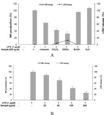
아마란스 종실 추출물의 항염증 효과를 찾기 위하여 마우스 대식세포주인 RAW 264.7 cell에서 LPS에 의해 유도된 NO 및 세포독성 효과를 확인하였다. 아마란스 분획층별로 시료의 농 도는 200 ㎍/㎖로, NO의 양은 Griess 시약을 사용하여 세포 배 양액 중에 존재하는 NO2-의 형태로 측정하였다. 측정결과 아마 란스 종실 EtOAc 분획층에서 가장 좋은 NO 억제 효능을 보여 EtOAc 분획층을 농도별로 측정하기 위하여 25, 50, 100, 200 ㎍/㎖
의 농도로 처리한 결과, LPS에 의해 유도된 NO의 증가를 농도 의존적으로 억제하는 것을 확인할 수 있었다(Fig.1). 같은 처리 조건에서 세포독성효과를 확인하기 위해 LDH assay를 동시에 측정한 결과 가장 높은 200 ㎍/㎖의 농도에서 2.51%로 세포 독 성이 처리 농도에서 거의 나타나지 않았다(Fig.1).
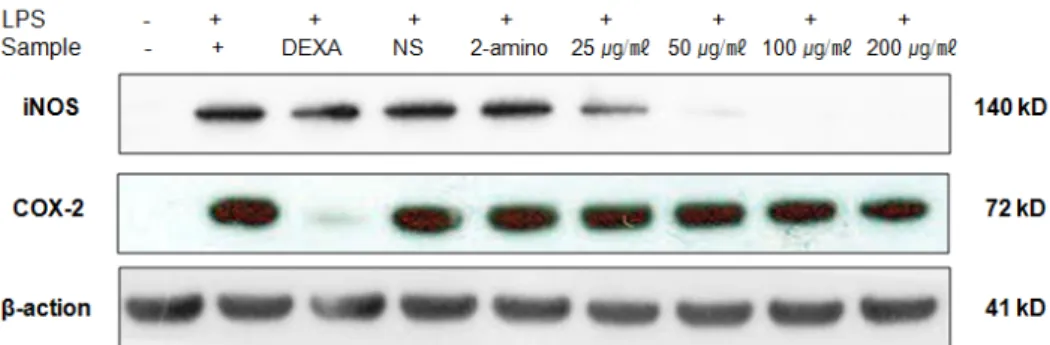
iNOS 및 COX-2 발현 억제 효능 평가(western-blotting) 아마란스 EtOAC 분획물에 의한 NO, PGE2 생성 억제 활성이 iNOS와 COX-2의 발현 억제로 인한 것인지 확인하기 위하여 단 백질 수준에서의 발현 억제 정도를 western blotting을 통하여 측정하였다. RAW 264.7 세포에 LPS (1 ㎍/㎖)를 사용하여 iNOS, COX-2 의 생성을 유도한 후 아마란스 종실 EtOAc 분획 층을 농도별로 처리하여 생성 억제 효능을 측정하였다. 대조군 으로는 소염제로 사용되는 dexamethason (DEXA), PGE2 inhibitor 인 NS-398 (NS), NO inhibitor인 2-amio-4-methyl pyridine (2-amino)를 각각 20 μM 처리하여 비교하였다. LPS에 의하여 iNOS와 COX-2는 현저하게 증가하였으며, 시료의 EtOAc 분획 물을 처리한 결과 iNOS 발현은 뚜렷하게, 그리고 COX-2는 약
A
B
Fig. 1. Inhibitory effect of 70% EtOH extract and solvent fractions of Amaranth seed on nitric oxide production and cell viability in RAW 264.7 cells.
A: Inhibitory effect of 70% EtOH extract and solvent fractions of Amaranth seed on nitric oxide production and cell viability in RAW 264.7 cells. B: Inhibitory effect of EtOAc fraction of Amaranth seed on nitric oxide production and cell viability in RAW 264.7 cells. The production of nitric oxide was assayed in the culture medium of cells stimulated with LPS (1 ㎍/㎖) for 24 h in the presence of samples. Cytotoxicity was determined using the LDH method.
Values are the mean ± SEM of triplicate experiments.
*p < 0.05, **p < 0.01.
하게 농도 의존적으로 억제함을 확인하였다(Fig. 2). 또한 대조 군으로 사용한 DEXA는 iNOS보다 COX-2를 강하게 억제함을 확인함으로써 항염 효능을 나타내고, NS와 2-amino인 경우는 iNOS와 COX-2의 발현 억제는 미비하지만 PGE2 와 NO에만 특 이적으로 작용하는 inhibitor로서 항염 효능이 있음을 확인할 수 있었다(KO et al., 2015).
Pro-inflammatory cytokine (TNF-α) 생성 억제 효능 TNF-α는 염증반응을 조절하는 대표적인 pro-inflammatory cytokine으로 급성 및 만성염증 질환 반응에서 중요한 역할을 담당한다. 이에 본 연구에서는 NO 억제 효과를 보이는 아마란 스의 EtOAc 분획물이 LPS에 의해 증가되는 TNF-α의 발현에 영향을 미치는 지를 확인하기 위하여 ELISA kit를 이용하여 조 사하였다. 그 결과 농도 의존적으로 TNF-α의 생성 억제 효과를 나타내었다(Fig. 3).
Prostaglandin E2 생성 억제 효과
PGE2는 염증매개인자로 통증, 혈관의 확장과 면역세포를 염증 부위로 이동시키는데 관여하는 것으로 알려져 있다. NO 억제 효능 을 보이는 아마란스 EtOAc 분획물이 염증매개인자인 PGE2 생성을 억제하는지 확인하기 위하여 LPS (1 ㎍/㎖)를 처리하여 염증반응 을 유도하여 PGE2 생성을 증가시키고, 농도별로 시료를 처리하여 세포배양액으로부터 PGE2 생성량을 측정하였다. 그 결과 농도 의 존적으로 PGE2 생성 억제 활성을 보임을 확인하였다(Fig. 4).
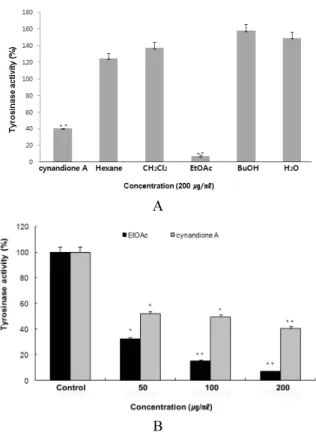
Tyrosinase 저해 활성
아마란스 종실 EtOAc 분획물이 NO와 PGE2 생성을 억제하는 것으로 보아 tyrosinase 활성 억제에도 관여하여 미백 효능에도
Fig. 2. Inhibitory effect of EtOAc fractions of Amaranth seed on the protein level of iNOS in RAW 264.7 cells.
RAW 264.7 cells (1.0 × 10⁶ cells/㎖) were pre-incubated for 18 h, and the cells were stimulated with LPS (1 ㎍/㎖) in the presence of EtOAc fractions (25, 50, 100, 200 ㎍/㎖) for 24 h. iNOS protein levels were determined using immunoblotting method. dex : dexamethason (20 μM), NS : NS-398 (20 μM), 2-amino : 2-amino-4-methyl pyridine (20 μM).
Fig. 3. Inhibitory effect of EtOAc fraction of Amaranth seed on TNF-α production in RAW 264.7 cells. Cells were pre-incubated for 18 h and them stimulated with LPS (1 ㎍/㎖) for 24 h in the presence of the EtOAc fraction of Amaranth seed. Supernatants were collected, and the TNF-α concentration in the supernatants was determined by ELISA. Values ane the mean ± SEM of triplicate experiments. *p < 0.05, **p < 0.01 versus LPS-treated group.
Fig. 4. Inhibitory effects of EtOAc fraction of Amaranth seed on PGE2 production in RAW 264.7 cells. The production of PGE2 was assayed in the culture medium of cells stimulated with LPS (1 ㎍/㎖) for 24 h in the presence of the EtOAc fraction of Amaranth seed. Supernatants were collected and the PGE2 concentration in the supernatants was determined by ELISA. Values ane the mean ± SEM of triplicate experiments.
*p < 0.05, **p < 0.01 versus LPS-treated group.
작용하는 지를 확인하기 위하여 mushroom tyrosinase 활성 억 제 효능 평가를 수행하였다. 아마란스 용매 분획물들을 200 ㎍/㎖
의 농도로 처리하여 tyrosinase 억제 효과를 확인한 결과 대조 군인 cynandione A보다 EtOAc 분획물이 훨씬 좋은 억제 효과 를 나타내었고(Fig. 5, A), EtOAc 분획물을 농도별로 처리한 결 과 농도 의존적으로 tyrosinase 효소 활성을 효과적으로 억제함 을 확인함(Fig. 5, B)으로써 염증에 의해 증가되는 PGE2와 같은 아라키돈산 대사산물의 억제와 함께 tyrosinase 활성도 억제됨 을 확인할 수 있었다.
적 요
본 연구는 아마란스(Amaranthus spp L.) 씨앗 추출물의 항 염 및 미백과 관련된 효능을 측정함으로써 식품, 의약품 및 화장
품 등에 유용한 소재로서 활용할 수 있는 가능성을 확인하고자 수행하였다. 아마란스(Amaranthus spp L.) 씨앗 추출물의 항 염증 활성 검색을 위하여 RAW 264.7 세포를 이용하고 실험을 진행하였고, 미백 효능 확인에 대하여서는 mushroom tyrosinase 를 사용하였다. RAW 264.7 세포를 이용하여 NO 생성 억제, iNOS 및 COX-2 단백질 발현 억제, 염증매개인자인 TNF-α 및 PGE2
생성 억제효과 등을 확인한 결과, 에틸아세테이트 분획물이 NO 생성과 iNOS 발현을 농도 의존적으로 강하게 억제함으로써 iNOS 발현 억제를 통한 NO 생성을 억제함을 확인할 수 있었다.
NO와 iNOS 억제 효과보다는 약하지만 PGE2와 COX-2 단백질 발현 역시 농도 의존적으로 억제함으로써 PGE2 억제 효능이 COX-2의 발현 억제를 통하여 이루어짐을 확인하였다. 또한 염 증에 의해 증가되는 아라키돈산 대사물들이 피부 멜라닌 합성 의 최초 속도결정단계에 작용하는 tyrosinase 활성을 증가시킨 다는 연구 결과에 근거하여 PGE2의 발현을 억제하는 아마란스 EtOAc 분획물이 tyrosinase 효소 억제 활성을 측정하고 미백효 능을 확인하였다. Mushroom tyrosinase 활성 억제 효능은 에 틸아세테이트 분획물이 우수한 효과를 보였으며, 농도 의존적 으로 tyrosinase 효소의 활성을 억제함을 확인할 수 있었다. 이 상의 결과들은 향후 아마란스 씨앗의 항염 효능을 갖는 유효성 분 탐색 및 멜라닌 세포를 이용하여 미백 관련 연구의 중요한 기 초자료로 활용할 수 있을 것이라 사료되며, 아마란스 씨앗이 우 수한 영양식품으로 알려져 있는 만큼 안전하면서도 항염과 미 백 효능이 있는 소재로서 기능성 식품, 의약품, 화장품 소재로 다양하게 응용할 수 있는 가능성을 확인하였다.
References
Assar, M.E. and J. Angulo. 2013. Oxidative stress and vascular inflammation in aging. Free Radic. Biol. Med. 65:380-401.
Cho, J.Y., D.M. Son, J.M. Kim, B.S. Seo, S.Y. Yang, B.W. Kim and B.G. Heo. 2008. Effects of LEDs on the germination, growth and physiological activities of amaranth sprouts.
Kor. J. Hort. Sci. Technol. 26:106-112 (in Korean).
Choi, C.R., J.J. Choio, S.R. Kim, J.H. Lee and M.S. Shin. 2000.
Comparisons of characteristics of amaranth starches isolated from five cultivars grown in Korea. Korean J. Food Sci.
Technol. 32:252-257 (in Korean).
Choi, H.S. 2011. Effect of adding amaranth powder on noodle quality. Koren J. Food & Nutr. 24:664-669 (in Korean).
Dubois, R., S.B. Abramson, L. Crofford, R.A. Gupta, L.S.
A
B
Fig. 5. Tyrosinase inhibitory effect of 70% EtOH extract and solvent fractions of Amaranth seed. Data were expressed in mean ± S.D. (N = 3). *p < 0.05, **p < 0.01, A: Inhibitory effect of 70% EtOH extract and solvent fractions of Amaranth seed against tyrosinase. Concentration of samples were 200 ㎍/㎖.
B: Inhibitory effect of EtOAc fractions of Amaranth seed against tyrosinase. Samples were treated with three concentra- tions (50, 100, and 200 ㎍/㎖).
Simon, L.B.A. Van De Putte and P.E. Lipsky. 1998. Cycloo- xygenase in biology and disease. FASEB J. 9:2045-2046.
Gins, M.S., M.P. Kolesnikov, P.F. Kononkov and V.K. Gins.
2010. Characteristics of the accumulation of phenolic com- pounds in amaranth leaves under the effect of growth stimu- lators. Russian Agricultural Sciences 36:349-352.
He, H.P., Y. Cai, M. Sum and H. Corke. 2002. Extraction and purification of squalene from amaranthus grain. J. Agric.
Food Chem. 50:368-372.
Hearing, V.J. and T.M. Ekel. 1976. Mammalian tyrosinase.
Biochem J. 157:549-557.
Hogquist, K.A., T.A. Baldwin and C. Jameson. 2005. Central tolence: learning self-control in the thymus. Nat. Rev. Immunol.
5:772-854.
Hong, S.Y., K.S. Cho, Y.I. Jin, Y.H. Yeon, S.J. Kim, J.H. Nam, J.C. Jeong, O.K. Kwon and H.B. Sohn. 2014. Comparison of growth characteristics, antioxidant activity and total phenolic contents of Amaranthus species according to the different cultivation regions and varieties in South Korea. Korean J.
Crop Sci. 59(1):16-21 (in Korean).
Hwang, D., B.C. Jang, G. Yu and B. Mary. 1997. Expression of mitogen-inducible cyclooxygenase induced by lipopolysacc- haride. Biochem. Pharmacol. 54:87-96.
Jo, H.J., J.W. Kim, J.A. Yoon, K.I. Kim, K.H. Chung, B.C.
Song and J.H. An. 2014. Antioxidant activities of amaranth (Amaranthus spp. L.) flower exracts. Korean J. Food & Nutr.
27(2):175-182 (in Korean).
Kim, J.S. and H.J. Ryoo. 2002. Application to the biscuits manu- facture of processed amaranth seeds. Korean J. Food & Nutr.
15(4):321-325 (in Korean).
Kim, J.Y., K.S. Jung and H.G. Jeong. 2004. Suppressive effects of the kaweol and cafestol on cyclooxygenase-2 expression in macrophases. FEBS Lett. 569:321-326.
Ko, C.S, W.C. Hyun, J.H. Kim, Y.J. Ko, S.M. Song, M.H. Ko, J.C. Lee, C.S. Kim and W.J. Yoon. 2015. Anti-inflammotory effects on 80% ethanol extract and ethyl acetate fraction of Acrosorium yendou Yamada in murine macrophage 264.7 cells. Korean J. Plant Res. 28(5):574-581 (in Korean).
Lee, H.J., Y.S. Jeong, S.Y. Ryu and J.H. Ryu. 1998. Inhibition of nitric oxide synthesis by 8-epi-xanthatin in activated RAW 264.7 cell. Yakhak Hoeji. 42(5):540-543 (in Korean).
Lee, J.H., H.I. Moon, J.I. Lee. C.W. Kang and S.T. Lee. 1996.
Isolation and identification of squalene and antineoplastic activity of its residue extract in amaranth. Korean J. Crop Sci. 41:450-455 (in Korean).
Lee, J.H., S.R. Kim, J.Y. Song, and M.S. Shin. 1999. Comparison on physicochemical properties of amaranth starch with other waxy cereal starches. Korean J. Food Sci. Technol. 31:612-618 (in Korean).
Matubara, H., K. Miharu, K. Kinoshita, K. Koyama, K.Y. Ye, K. Takahashi, I. Yoshimura, Y. Yamamoto, Y. Miura and Y.
Kinoshita. 1998. Inhibitory effect of lichen metabolites and their synthetic analogues on melanin biosynthesis in cultured B-16 mouse melanoma cells. Natural Product Sciences 4(3):
161-169.
Mishima, Y., S. Hatta, Y. Ohyama and M. Inazu. 1988.
Induction of melanogenesis suppression : cellular pharmacology and mode of differential action. Pigment Cell Res. 1(6):367- 374.
Morelli JG, J.J. Youn, M.B Lyons, R.C. Murphy and D.A.
Norris. 1989. Leukotriene C4 and C4 as potent mitogens for cultured human neonatal melanocytes. J. Invest. Dermatol.
93:719-22.
Mu, M.M., D. Chakravortty, T. Sugiyama, N. Koide, K.
Takahashi, I. Mori, T. Yoshida and T. Yokochi. 2001. The inhibitory action of quercetin on lipopoly-saccharide-induced nitric oxide production in RAW 264.7 macrophage cells. J.
Endotoxin. Res. 7:431-438.
Oh, S.H., S.Y. Choi, N.R. Lee, J.N. Lee, D.S. Kim, S.H. Lee and S.M. Park. 2014. Cell migration and anti-inflammatory effect of red ginseng extracts fermented with laetiporus sulphureus. J. Soc. Cosmet. Scientists Korea 40(3):297-305 (in Korean).
Ryu, J.H. H. Ahn, J.Y. Kim and Y.K. Kim. 2003. Inhibitory activity of plant extracts on nitric oxide synthesis in LPS-activated macrophage. Phytother. Res. 17:485-489.
Smith, W.L., G.R. Michael and D.L. De-Witt. 1996.
Prostaglandin endoperoxide H synthases (cyclooxygenases)-1 and 2. J. Biol. Chem. 271:33157-33160.
Takiwak,i H., S. Shirai, H. Kohno and H. Soh. 1994. The degrees of UVB-induced erythma and pigmentation correlate linearly and are reduced in a parallel manner by topical anti- inflammatory agents. J. Invest. Dermatol. 103:642-6.
Tomita, Y, K. Maeda and H. Tagami. 1992. Melanocyte-stimu- lating properties of arachidonic acid metabolites: possible role in post-inflammatory pigmentation. Pigment Cell Res.
5:357-61.
Weisz, A., L. Cicatiello and H. Esumi. 1996. Regulation of the
mouse inducible type nitric oxide synthase gene promoter by
interferon-gamma, bacterial lipopolysaccharide and NG-mono-
methyl-L-arginine. Biochem. J. 316:209-215.
Willeaume, V., V. Kruys, T. Mijatovic and G. Huez. 1995-1996.
Tummor necrosis factor-alpha production induced by viruses and by lipopolysaccharides in macrophages: similarities and differences. J. Inflamm. 46(1):1-12.
Yayeh, T., H. Jung, H.Y. Jeong, J.H. Park, B. Song, Y.S. Kwak, H.S. Kang, J.Y. Cho, J.W. Oh, S.K. Kim and M.H. Rhee.
2012. Korean red ginseng saponin fraction downregulates
proinfl ammatory mediators in LPS stimulated RAW264.7 cells and protects mice against endotoxic shock. J. Ginseng Res. 36:263-269 (in Korean).
Yoon, W.J., J.A. Lee, K.N. Kim, J.Y. Kim and S.Y. Park. 2007.
In vitro anti-inflammatory activity of the Artemisia fukudo extracts in murine macrophage RAW 264.7. Korean J. Food Sci. Technol. 39:464-469 (in Korean).
(Received 31 January 2017 ; Revised 12 April 2017 ; Accepted 14 April 2017)