273
Korean J Vet Serv 34(3) : 273~278 (2011)
*Corresponding author: Jeong-Hee Han, Tel. +82-33-250-8691, Fax. +82-33-256-3722, E-mail. [email protected]
개 세르토리세포종 케이스에서 면역조직화학적 마커를 통한 진단
김성재ㆍ한정희*
강원대학교 수의과대학 수의학과 병리학실 (접수 2011. 4. 8; 수정 2011. 7. 4; 게재승인 2011. 9. 14)
Diagnosis of immunohistochemical marker expressed by a canine Sertoli cell tumor case
Sung-Jae Kim, Jeong-Hee Han*
College of Veterinary Medicine and Institute of Veterinary Science, Kangwon National University, Chuncheon 200-701, Korea
(Received 8 April 2011; revised 4 July 2011; accepted 14 September 2011)
Abstract
Sertoli cell tumor (SCT) of the testicle arises from the supporting cells within the seminiferous tubules.
SCT is common in dogs, especially in cryptorchid testicles, but also has been reported in the stallion, ram, cat, and bull. Sertoli cell tumor sample was collected from 7-years male german shepherd. In this study, SCT arose from one testicle. Sample size is approximately 1.7 cm in diameter and it has a round form. In the microscopic, cells within the tumor variably resemble Sertoli cells (SCs) that nor- mally populate the seminiferous tubules and interstitial area. There is abundant stroma of dense, ma- ture fibrous connective tissue in SCT. In the immunohistochemical staining, cytokeratin AE1/AE3 was not expressed in the control and SCT. S-100 protein was expressed by SCs, germ cells and fibrous connective tissue of SCT. Melan A was expressed by leydig cells (LCs) of SCT. A study by using S-100 and melan A in canine SCT was almost never carried out. S-100 and melans A is considered to suggest for diagnosis and pathogenesis of canine SCTs. Inhibin-alpha and Vimentin were well known as the marekers of SCTs. Also, they were expressed by Sertoli cells and LSs of SCT in this study.
Key words : SCT, Cytokeratin, S-100, Melan A, Inhibin-alpha, Vimentin
서 론
고환 종양은 생식 세포나 성선 기질의 구성요소 로부터 발생하며, 일반적으로 네 종류로 구분할 수 있다(McLachlan과 Kennedy, 2002). 생식 세포 종양으 로는 seminoma, teratoma가 있으며, 성선 기질로부터 유래한 종양으로는 Sertoli cell tumor (SCT)와 Leydig
(interstitial) cell tumor가 있다(Peters 등, 2001; McLa- chlan과 Kennedy, 2002; Rosai, 2004). 주요한 종양은 seminoma와 SCT로 0.068∼4.6%의 발생률을 보이며, 나이가 든 성숙한 개에서 일반적이다(Peters 등, 2001; Meyts, 2007). Seminoma는 정세소관 내의 정자 발생상피를 구성하는 생식세포로부터 유래하며, 잠 복고환인 개에서 잘 발생한다. SCT는 정소세관 내 의 지지세포로부터 유래하며, sustentacular cell tumor 라고 부르기도 한다. 특히 SCT는 잠복고환일 때 잘
Fig. 1. Gross lesion of Sertoli cell tumor. The tumor (arrows) is rel- atively well circumscribed and has a solid, tan-yellowish cut surface.
Fig. 2.Histology of Sertoli cell tumor, SCT has abundant fibrous stroma (black arrows). The tumor cells (white arrows) have small, round nuclei and either vacuolated or dense eosino- philic cytoplasm, hematoxylin and eosin. Bar=350 μm.
발생하며, 뮐러관 잔존증이 있는 슈나우저에서 높은 발생률을 보인다고 보고되었다. 개에서 발생한 모든 SCT의 절반 정도가 잠복고환에서 발생했을 것으로 보이며, 잠복고환이 정상의 고환보다 SCT 발생률이 20배나 높다. SCT는 대부분 한쪽 고환에서만 일어 나지만, 양쪽 고환에서 일어나는 예도 있으며, 대략 20∼30%의 개에서 확연한 에스트로겐 과다증이 나 타나면서 여성화가 일어나는 특징이 있다(McLac- hlan과 Kennedy, 2002).
현재 사람에서는 고환종양의 정확한 조직학적 진 단과 병리기전을 연구하기 위해 많은 면역조직화학 적 마커들이 소개되고 있다(De Vico 등, 1994; Lau 등, 2007). Cytokeratins, C-Kit, CD30, epithelial mem- brane antigen, inhibin-alpha, OCT3/4, placental alkaline phosphatase (PLAP)와 alpha fetoprotein (AFP) 등이 사 람 고환종양에 특이성과 민감도가 있는 마커들이다 (De Vico 등, 1994). 개에서도 고환종양의 진단과 병 리기전을 밝혀내기 위해 면역조직학적 연구가 진행 되고 있지만, 사람보다 많이 알려진 것이 없고, 현재 종양의 진단과 병리기전을 연구하는데 종양 마커들 을 사용하는 것이 제한되어있다.
이번 실험에서 저먼 셰퍼드에서 발생한 SCT의 종 양에서의 면역조직학적 반응을 보기 위해 기존의 사 람과 개의 고환종양관련 실험에서 많이 연구된 cyto- keratin, vimentin, inhibin-alpha, S-100과 Melan A를 이 용하였다.
증 례
임상증상 및 시료채취
종양이 발견된 german shepherd는 노쇠하여 군부대 에서 폐견 처리되었으며, 7살 된 수컷이다. 활동적이 지 않고 우울한 상태였으며, 약간의 탈모증세가 있었 으나 외형적으로 특별한 병변은 없었다. 부검을 한 결과, 다른 장기에는 병변이 없었으며, 한쪽 고환에 서만 종양이 발견되었다. 종양이 발견된 고환은 잠복 고환 상태가 아니었고, 정상적으로 복강에서 하강하 여 음낭에 있었다. 정상 고환 시료는 중성화 수술을 한 2살 된 잡종 개에서 채취하였다.
조직병리학적 소견
육안소견: 종양은 한쪽 고환에만 있었으며, 직경이 약 1.7 cm정도 되었다. 종양이 있는 고환은 정상 고환 보다 더 비대해져 있었다. 종양은 고환 내에만 국한되 었으며 잿빛 노란색을 띠었다. 종양 조직은 정상조직 보다 단단했으며, 종양 조직의 압박을 받아 위축된 정 상 고환 조직들이 종양 주변에 경계를 형성하면서 종 양과 정상조직이 명확히 구분되어 있었다(Fig. 1).
조직학적 소견: 종양조직은 정세관이 비교적 잘 유 지되어 있었으며, 섬유소성 기질이 풍부하게 발달되 어있었다. 종양조직의 세포들은 정상적으로 정세관에 있는 SCs와 유사했으며, 정세관과 치밀하게 잘 발달
Table 1.Primary antibodies used for immunohistochemical staining
Antibody Clone Type Dilution
Cytokeratin AE1/AE3 AE1/AE3 Mouse 1:50
S-100 A1/A6/B Rabbit 1:400
Melan A A103 Mouse 1:50
Inhibin-α R1 Mouse 1:30
Vimentin V9 Mouse 1:75
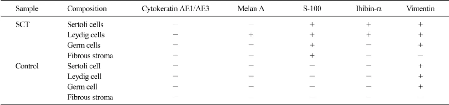
Table 2. Result of immunohistochemical staining in SCT and Control
Sample Composition Cytokeratin AE1/AE3 Melan A S-100 Ihibin-α Vimentin
SCT Sertoli cells − − + + +
Leydig cells − + + + +
Germ cells − − + − +
Fibrous stroma − − + − −
Control Sertoli cell − − − − +
Leydig cell − − − − +
Germ cell − − − − +
Fibrous stroma − − − − −
한 결합조직으로 구성된 기질에 의해 나누어진 공간 을 채우고 있었다. 종양 세포의 핵은 작고, 둥글며, 기저부에 위치하는 경향이 있었으며, 호산성 세포질 을 가지고 있었다. 세포의 유사분열 현상은 400배율 에서 드물게 관찰되었다(Fig. 2).
면역염색
육안 병리와 조직 병리를 바탕으로 Sertoli cell tumor 로 진단하였고, 확진과 면역조직화학적 반응을 보기 위해 면역염색을 하였다. 종양 및 정상 시료를 10% 중 성 포르말린에 고정했다. 시료 조직을 파라핀 왁스를 이용하여 블록을 만들었으며, 면역염색을 위해 절편기 로 5 μm 두께로 잘랐다. 일차 항체로는 Cytokeratin AE1/AE3 (Dako, USA), Vimentin (Dako, USA), Inhibin- alpha (Dako, USA), S-100 (Dako, USA), Melan A (Novo- castra, UK)를 사용하였다(Table 1).
각 절편은 xylene과 농도별 ethanol에서 탈파라핀 과정과 탈수 과정을 거쳤다. 비커에 담아서 전자레인 지에서 5분간 데운 10 mmol/L citrate buffer (pH 6.0)에 슬라이드를 담그고 랩을 씌워서 다시 전자레인지에 20분간 끓이는 항원 복원과정을 거쳤다. 끓은 용액을 얼음에서 식힌 후, TPBS (1L PBS, 5 μl Tween20)에 20분간, phosphate-buffer saline (PBS; pH 7.4, 137 mM NaCl, 2.7 mM KCl, 10 mM Na2HPO4, 2 mM KH2PO4) 에서 5분간 두 번 수세를 한 후, 20분간 실온에서 0.3% hydrogen peroxide로 처리하였다. 일차 항체와 이차 항체를 붙이는 과정은 ABC kit (Vector Laborato- ries, USA)의 프로토콜대로 진행하였다. DAB sub- strate kit (Zymed, USA)를 이용하여 현미경으로 보면 서 발색을 하였다. Harris hematoxylin을 이용하여 대 조 염색을 하였다.
면역염색의 결과는 Cytokeratin AE1/AE3는 Sertoli cell tumor (SCT)와 정상고환 조직에서 모두 음성이었
다. S-100은 정상 고환 조직에서는 반응을 보이지 않 았으나, SCT에서는 간질조직의 풍부하게 발달한 섬 유성 결합조직, SCs와 LCs에 약하지만 광범위하게 염색되었다. Melan A 역시 정상고환 조직에서는 반 응을 보이지 않았으며 SCT에서는 간질조직의 LCs에 강하게 염색되었다. Inhibin-alpha는 정상고환 조직에 서는 반응을 보이지 않았고 SCT에서는 SCs와 LCs에 강하게 염색되었으며, Vimentin은 SCT와 정상고환에 서 반응을 보였는데, SCs, LCs와 germ cells에 강하게 염색되는 양상을 보였다(Table 2, Fig. 3).
고 찰
원발성 고환종양은 늙고 성숙한 개에서 일반적이 며, 악성은 드물며 주로 양성종양이다. 개에서의 고 환종양은 seminoma와 SCT가 가장 많이 다발하며 이것 은 잠복고환과 연관성이 높으나(McLachlan과 Kennedy, 2002), 이번 사례는 정상적으로 고환이 음낭 내부로 하강한 상태였다. SCT에서 종양의 크기가 클 때, 전 이 될 확률이 높으며 2 cm 이하의 작은 종양은 전이 될 확률이 매우 낮다(McLachlan과 Kennedy, 2002). 부 검 결과, 종양의 크기가 작고, 다른 장기에 전이한 흔 적은 없었으며, 현미경상 유사분열 빈도가 낮은 것으 로 보아 양성종양일 가능성이 높았다.
조직학적으로 SCT는 tubular form과 diffuse form으
Fig. 3. Immunohistochemical markers in Sertoli cell tumor. (A) S-100. (B) Melan A. (C) Inhibin-alpha. (D) Vimentin. Immunostain and counter- stain with Harris hematoxylin. Bar=350 μm.
로 나누어진다. tubular form은 정세관의 형태가 잘 유 지되었으며, 종양세포들이 정세관의 기저막을 따라 층을 형성하고 있으며, diffuse form은 치밀한 섬유성 기질에 종양 세포들이 침투해 얇은 층이나 덩어리를 형성한다. 또한 종양세포들은 tubular type에 비해 크 기가 다양하고 불규칙하다(McLanchlan과 Kennedy, 2002). 이 케이스는 정세관의 형태가 잘 유지되어 있 었으며, 그 기저막을 따라 종양세포(SCs)들이 층을 형성한 tubular type이였다.
정상 고환 조직 모두에서 Cytokeratin (CK)은 면역 반응을 나타내지 않았다. CK는 상피세포에 특이성을 가지고 있는 악성 상피종의 마커로서(Braun 등, 2000), 중신의 표면에 SCs가 응축되고 상피로 분화되 는 단계에서 일시적으로 SCs이 CK에 면역반응을 보 이나(Stosiek 등, 1990; Rogatsch 등 1996), 그 후 고환 의 발달을 통해 정세관 내에 SCs가 자리잡는 과정에 서 CK에 대한 반응을 잃게 되며(Wilhelm 등, 2007), 정상적으로 성숙한 정세관의 상피는 다른 조직의 상 피와 달리 CK filament로 구성되어있지 않은 것이 특 징이다(Maymon 등, 2000). 그러므로 정상의 성숙한
개의 고환은 CK에 대한 면역반응이 없으며, 이 실험 의 정상고환에서도 CK에 대한 면역반응이 없었다.
또한 SCT에서도 면역반응이 없었는데 이것은 SCT 세포들의 분화도와 관련이 있으며, 분화도가 높은 SCT 타입이라는 것을 의미한다(Banco 등, 2010).
S-100은 calcium binding protein으로서, 세포 내 및 세포 외 기능을 한다고 보고되었으며, 췌장암, 유방 암, 전립선암, 폐암 등 다양한 종양에서 반응을 나타 내며(Arumugam과 Logsdon, 2010), 사람 고환 종양에 서의 S-100에 관한 연구는 많이 이루어져 왔다 (McCluggage 등, 1998; Tanaka 등, 2002; Sato 등, 2005; Angela 등, 2006). 사람의 Large cell calcifying Sertoli cell tumor (LCSCT)는 SCT의 분화도가 높은 타 입으로 종양조직의 SCs에 S-100이 면역반응을 나타 내는데, 이것은 SCs의 세포질 내 칼슘 침착과 깊은 연관이 있는 것으로 보인다(Tanaka 등, 2002). 이번 사례에서는 종양 조직의 SCs 및 germ cell에 전체적 으로 면역반응을 보였으며, 일부 강하게 염색된 부분 도 있었다. 또한, LCSCT와 달리 종양 세포를 둘러싼 섬유성 결합조직에도 일부 강하게 염색이 되었다. 쥐
에서 유래한 fibroblast 및 fibrosarcoma와 전이성 상피 세포가 S-100에 면역반응을 보였다는 보고가 있는데 (Shiras 등, 2001), 종양조직에 발달한 결합조직에 면 역반응을 보인 것은 S-100이 fibroblast에 반응을 나타 낸 것으로 생각한다.
Melan-A는 자가조직의 세포면역성 T세포에 의해 인식되는 악성 흑색종 항원이며, A103은 melan-A 단 백질에 특이성을 가진 유전자 재조합형의 단클론 항 체이다(Chen 등, 1996; Busam 등, 1998). Melan-A는 사람의 SCT에서 지지세포와 간질세포에 면역반응을 보이며, 지지세포보다 간질세포 영역에 더 강한 면역 반응을 보인다(Busam 등, 1998). 정상 고환에서도 지 지세포와 간질세포의 세포질이 약하게 면역반응을 보였지만(Sato 등, 2005), 이번 사례에서는 정상 고환 은 면역반응이 없었으며, SCT의 경우, 간질세포는 사 람과 마찬가지로 면역반응이 있었지만, 지지세포는 면역반응이 없었다. 종양 세포 세포질의 면역반응은 종양 세포의 분화도 정도와 관련이 있는데, 분화도가 높을수록 강하게 염색된다(Stewart 등, 2000). 이번 개 SCT 케이스는 잘 분화된 SCT 형태라는 것을 알 수 있다. 하지만 종양조직의 SCs에서 면역 반응을 보이 지 않은 것은 개 SCT의 특징인지 이번 케이스의 특 징인지는 더 많은 연구가 필요할 것으로 생각된다.
15개의 개 SCT 시료를 가지고 면역염색을 한 실험 에서 vinmentin과 inhibin-alpha가 가장 뛰어난 마커라 고 밝혔으며, 두 개의 마커 모두 93.3%의 면역반응을 보였다고 보고했다(Yu 등, 2009). Inhibin-alpha는 SCs 와 LCs에서 분비되는 호르몬으로 고환의 수출관과 부고환의 기시부위에서 재흡수되며, 혈류에 도달하여 뇌하수체의 FSH 분비에 네거티브 피드백 작용을 한 다(Dellman과 Eurell, 2005). Inhibin-alpha는 신생아의 SCs에서는 면역반응을 보이지만 정상의 성숙한 개에 서는 반응을 보이지 않으며, 개의 경우, SCs보다 LCs 에서 많은 양이 분비된다고 하였다(Banco 등, 2010).
또한, SCT를 가진 개에서 면역학적으로 감지할 수 있는 inhibin의 고환조직의 농도가 정상의 개보다 높 으며, 혈중의 농도 역시 짙다는 것을 밝혀냈으며 (Kawakami 등, 2007), 이것은 사람도 마찬가지다 (Iczkowski와 Butler, 2006). 이번 케이스에서는 정상고 환 조직에서는 면역반응이 없었으며, SCT의 SCs와 LCs에 inhibin-alpha가 반응을 보였다. 이 결과는 SCT 의 SCs와 LCs가 정상조직의 세포들보다 짙은 농도의 inhibin-alpha를 분비한다는 것을 보여준다.
Vimetin은 결합조직, 상피, 조혈세포, 근육에 포함
된 중배엽 유래의 종양마커로 알려졌으며, 사람의 SCT에서 면역염색 양성 반응을 보인다(Sung 등, 2006). 이번 케이스에서 vimentin은 정상고환조직과 SCT의 SCs와 LCs의 세포질이 강하게 염색되었다. 이 것은 vimentin이 정상 고환의 세포들을 형성하는 in- termediate filamen이며, 종양으로 발전된 후에도, 여전 히 개 SCT 세포들의 기본 구조를 형성하는 inter- mediate filament라는 것을 보여준다(McCluggage 등, 1998).
감사의 글
본 연구는 강원대학교 동물의학종합연구소의 지원 에 의해 수행되어 이에 감사드립니다.
참 고 문 헌
Angela LD, Julie AY, Susan JB, and Robert AF. 2006. Extrates- ticular interstitial and Sertoli cell tumors in previously neutered dogs and cats: A report of 17 cases. Can Vet J 47: 763-766.
Arumugam T, Logsdon CD. 2010. S100P: a novel therapeutic tar- get for cancer. Amino Acids: 1-7.
Banco B, Giudice C, Veronesi MC, Gerosa E, Grieco V. 2010.
An Immunohistochemical study of normal and neoplas- tic canine Sertoli cells. J Comp Path 143: 239-247.
Braun S, Pantel K, Müller P, Janni W, Hepp F, Kentenich CR, Gastroph S, Wischnik A, Dimpfl T, Kindermann G, Rie- thmüller G, Schlimok G. 2000. Cytokeratin-positive cells in the bone marrow and survival of patients with stage I, II, or III breast cancer. N Engl J Med 342: 525-533.
Busam KJ, Iversen K. Coplan KA, Old LJ, Stockert E, Chen YT, McGregor D, Jungbluth A. 1998. Immunoreactivity for A103, an antibody to melan-A (Mart-1), in adrenocort- ical and other steroid tumors. Am J Surg Pathol 22:
57-63.
Chen YT, Stockert E, Jungbluth A, Tsang S, Coplan KA, Scanlan MJ, Old LJ. 1996. Serological analysis of Melan-A (MART-1), a melanocyte-specific protein homogeneou- sly expressed in human melanomas. Proc Natl Acad Sci USA 93: 5915-5919.
De Vico G, Papparella S, Di Guardo G. 1994. Number and size of silver-stained nucleoli (Ag-NOR clusters) in canine seminomas: correlation with histological features and tu- mour behaviour. J Comp Pathol 110: 267-273.
Dellman DH, Eurell JA. 2005. Male reproduct system. pp.
230-233. In: Dellman DH, Eurell JA(ed). Textbook of veterinary histology. 5th ed. LIPWW, United states.
Iczkowski KA, Butler SL. 2006. New immunohistochemical markers in testicular tumors. Anal Quant Cytol Histol 28: 181-187.
Kawakami E, Hirano T, Hori T, Tsutsui T. 2007. Testicular su- peroxide dismutase activity, heat shock protein 70 con- centration and blood plasma inhibin-α concentration of dogs with a Sertoli cell tumor in a unilateral cryptorchid testis. J Vet Sci 69: 1259-1262.
Lau SK, Weiss LM, Chu PG. 2007. D2-40 Immunohistochemistry in the differential diagnosis of seminoma and embryo- nal carcinoma: a comparative immunohistochemical Study with KIT (CD117) and CD30. Mod pathol 20:
320-325.
Maymon BB, Paz G, Elliott DJ, Hammel I, Kleiman SE, Yogev L, Hauser R, Botchan A, Yavetz H. 2000. Maturation phenotype of Sertoli cells in testicular biopsies of azoo- spermic men. Human Reproduction 15: 1537-1542.
McCluggage WG, Shanks JH, Whiteside C, Maxwell P, Banerjee SS, Biggart JD. 1998. Immunohistochemical study of testicular sex cord-stromal tumors, including staining with anti-inhibin antibody. Am J Surg Pathol 22:
615-619.
McLachlan NJ, Kennedy PC. 2002. Tumors of the genital system.
pp. 547-573. In: Meuten DJ(ed.). Tumors in Domestic Animals. 4th ed. Blackwell. Iowa state press. Ames.
Meyts RD. 2007. Recent advances and future directions in re- search on testicular germ cell cancer. Int J Androl 30:
192-197.
Peters M, Teerds K, Van Der Gaag I, De Rooij D, van Sluijs F.
2001. Use of antibodies against LH receptor, 3beta-hy- droxysteroid dehydrogenase and vimentin to characterize different types of testicular tumour in dogs. Reproduc- tion 121: 287-296.
Rogatsch H, Jezek D, Hittmair A, Mikuz G, Feichtinger H. 1996.
Expression of vimentin, cytokeratin, and desmin in Sertoli cells of human fetal, cryptorchid, and tumour-ad- jacent testicular tissue. Virchows Archiv 427: 497-502.
Rosai J. 2004. Rosai and Ackerman's Surgical Pathology. 4th ed.
pp. 1412-1456. Mosby, Edinburgh.
Sato K, Ueda Y, Sakurai A, Ishikawa Y, Okamoto S, Ikawa H, Katsuda S. 2005. Large cell calcifying Sertoli cell tumor of the testis: comparative immunohistochemical study with Leydig cell tumor. Pathol Int 55: 366-371.
Shiras A, Shepal V, Bhonde M, Shastry P. 2001. S100 expression in primary mouse fibroblast cultures. In Vitro Cell Dev Biol Anim 37: 172-174.
Stewart CJR, Nandini CL, Richmond JA. 2000. Value of A103(melan-A) immunostaining in the differential diag- nosis of avarian sex cord stromal tumours. J Clin Pathol 53: 206-211.
Stosiek P, Kasper M, Karsten U. 1990. Expression of cytokeratins 8 and 18 in human Sertoli cells of immature and atro- phic seminiferous tubules. Differntiation 43: 66-70.
Sung MT, Jones TD, Beck SD, Foster RS, Cheng L. 2006. OCT4 is superior to CD30 in the diagnosis of metastatic em- bryonal carcinomas after chemotherapy. Hum Pathol 37:
662-667.
Tanaka Y, Carney JA, Ijiri R, Kato K, Miyake T, Nakatani Y, Misugi K. 2002. Utility of immunostaining for S-100 protein subunits in gonadal sex cord-stromal tumors, with emphasis on the large-cell calcifying Sertoli cell tu- mor of the testis. Hum Pathol 33: 285-289.
Wilhelm D, Palmer S, Koopman P. 2007. Sex determination and gonadal development in mammals. Physiol Rev 87: 1-28.
Yu CH, Hwang DN, Yhee JY, Kim JH, Im KS, Nho WG, Lyoo YS, Sur JH. 2009. Comparative immunohistochemical characterization of canine seminomas and Sertoli cell tumors. J Vet Sci 10: 1-7.