INTRODUCTION
Involvement of the inferior vena cava (IVC) has been re- ported in up to 35% of patients with renal cell carcinoma (RCC) (1-6). Although complete removal of the extension of the tumor thrombus into the IVC does not affect patient prognosis (7, 8), non-removal has been associated with poor survival rates (7). The most effective therapeutic option in patients with RCC and IVC tumor thrombus is aggressive surgical resection, including radical nephrectomy with IVC thrombectomy, even in patients with distant metastases (8- 11). Five-year survival rates following surgery have been re- ported to range from 32% to 64% (2, 6, 7, 9).
Even with improvements in preoperative diagnostic modal- ities, anesthesiology, and perioperative care, there is still con- siderable morbidity and mortality in this type of surgery, ranging from 2.7% to 40% (3, 8, 12), arising primarily from massive pulmonary embolism and hemorrhage (7, 13). These complications are closely related to the cephalic extension of the IVC tumor thrombus, suggesting that radical surgery should be reserved for patients with tumor thrombi extend-
ing beyond the level of the diaphragm (12). We have design- ed a safe and successful surgical strategy through a review of our surgical experience.
MATERIALS AND METHODS
From January 1997 to December 2006, 35 patients at the Asan Medical Center, Seoul, Korea, underwent IVC tumor thrombectomy with radical nephrectomy, performed by uro- logical and vascular surgeons. Preoperative assessment of dis- tant metastases and extents of thrombus was determined main- ly by computed tomography (CT) of the thorax, abdomen, and pelvis. Nineteen patients underwent magnetic resonance image (MRI). After discharge, patients were followed up every 3 months and CT scans were performed every 6 months.
Tumors were classified according to the TNM classifica- tion of the UICC International Union against Cancer (IUAC) (14). Extent of the thrombus was graded at level I or renal (i.e., extending ≤2 cm above the ostium of the renal vein into the IVC); level II or infrahepatic (i.e., extending >2 cm
104
Tae-Won Kwon1, Hyangkyoung Kim1, Ki-Myung Moon1, Yong-Pil Cho1, Cheryn Song2, Chung-Soo Kim2, and Hanjong Ahn2
Division of Vascular Surgery, Departments of Surgery1 and Urology2, University of Ulsan College of Medicine and Asan Medical Center, Seoul, Korea
Address for Correspondence Tae-Won Kwon, M.D.
Division of Vascular Surgery, Department of Surgery, University of Ulsan College of Medicine and Asan Medical Center, 86 Asanbyeongwon-gil, Songpa-gu, Seoul 138-736, Korea
Tel : +82.2-3010-3492, Fax : +82.2-474-9027 E-mail : [email protected]
Surgical Treatment of Inferior Vena Cava Tumor Thrombus in Patients with Renal Cell Carcinoma
Radical nephrectomy with inferior vena cava (IVC) thrombectomy remains the most effective therapeutic option in patients with renal cell carcinoma and IVC tumor throm- bus. Cephalic extension of the thrombus is closely related to perioperative morbidi- ty. We purposed to design a safe and successful surgical strategy through a review of our surgical experience and treatment results in 35 patients (male:female=28:7, mean age=56 yr [32-77]) who underwent IVC thrombectomy with radical nephrec- tomy between January 1997 and December 2006. The limit of tumor extension was level I in 10 patients (28.6%), level II in 17 (48.6%), and level III and IV in 4 patients each (11.4%). Liver mobilization with hepatic vascular exclusion was performed in 12 patients and cardiopulmonary bypass in 7. Thirty-two primary closures, 2 patch closures, and 1 graft interposition were performed. One patient underwent simulta- neous pulmonary embolectomy because of an operative pulmonary embolism. There was no operative mortality, and the overall survival at 5-yr was 50.8%. Complete thrombus removal without tumor fragmentation under long venotomy on fully ex- posed involved IVC is recommended for successful result in a bloodless operative field. The applicability of liver mobilization, hepatic vascular exclusion, and cardiopul- monary bypass, can be determined by the level of thrombus.
Key Words : Vena Cava, Inferior; Thrombectomy; Kidney Neoplasms
ⓒ 2010 The Korean Academy of Medical Sciences.
This is an Open Access article distributed under the terms of the Creative Commons Attribution Non-Commercial License (http://creativecommons.org/licenses/by-nc/3.0) which permits unrestricted non-commercial use, distribution, and reproduction in any medium, provided the original work is properly cited.
Received : 18 September 2008 Accepted : 17 February 2009
above the ostium of the renal vein and below the intrahep- atic vena cava); level III or intrahepatic (i.e., extending into the intrahepatic portion of the vena cava, but below the dia- phragm); or level IV or atrial (i.e., extending into the right atrium) (9).
Statistical analysis was performed using the statistical pack- age SPSS for Windows (Version 12.0, SPSS Inc., Chicago, IL, USA). Statistical significance was defined when a P value of
<0.05 was obtained. Overall survival rate was calculated using the Kaplan-Meier method.
Surgical procedure
Under general anesthesia, a central venous line and radial artery catheter were placed to allow fluid administration and blood pressure monitoring. Intraoperative continuous TEE surveillance was used to detect tumor or gas emboli and to confirm complete thrombus removal.
Laparotomy was usually performed using an inverted T incision or a chevron incision, both of which can be extended to the thorax when cardiopulmonary bypass (CPB) is requir- ed. The colon was retracted medially and the parietal peri- toneum was incised over the kidney near the colon. In pati- ents who did not undergo preoperative renal artery emboliza- tion, the renal artery was ligated first. The colon was reflect- ed medially to expose the IVC and aorta. The IVC was freed from the lumbar veins, both renal veins were exposed, and slings were passed around these structures, taking care not to dislodge the thrombus. Following administration of hep- arin, clamps were applied successively to the distal IVC, the renal vein contralateral to the tumor, and the upper IVC prox- imal to the thrombus. A lateral venotomy was performed from the ostium of the tumoral renal vein to the IVC beyond
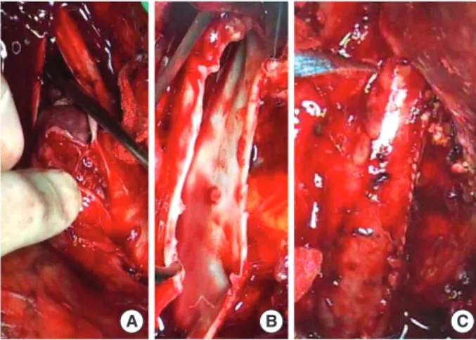
the proximal limit of the thrombus and the thrombus was completely removed under direct vision (Fig. 1A, B). We avoided blind digital extraction, milking, or Fogarty throm- bectomy of IVC thrombus. The ostium of renal vein was sent for frozen biopsy. Primary repair of the cavotomy was done if the margin was free of the tumor, and when gross invasion into the IVC wall was present cavectomy with prosthetic graft interposition or patch angioplasty was performed (Fig. 1C).
After closure of the IVC, radical nephrectomy was performed.
Following surgery, patients were transferred to the intensive care unit. Anticoagulation therapy, using low molecular-weight heparin followed by antiplatelet agent(s), was administered for 6 months after the procedure.
Surgical strategy based on the limit of thrombus extension
Operative maneuvers based on the limit of thrombus exten- sion are summarized at Table 1.
Level I (renal) or II (infrahepatic) thrombus
Liver mobilization was individualized depending on throm- bus extent. If preoperative imaging showed sufficient space to apply a proximal venous clamp, exposure of the IVC was limited to the infrahepatic portion. Venous clamps were suc- cessively positioned on the infrarenal IVC, the renal vein con- tralateral to the tumor, and the IVC between the origin of the hepatic veins and the limit of thrombus extension.
Level III (intrahepatic) thrombus
The liver was mobilized completely by the piggyback tech- nique, dividing the falciform, triangular, and coronary liga- ments, and the IVC was completely dissected from the renal vein level to the hepatic veins. After complete hepatic mobi- lization and IVC cross-clamping, hepatic vascular exclusion was performed to prevent bleeding from the hepatic vein and hepatic congestion. The suprahepatic IVC, the infrarenal IVC, the contralateral renal vein, and the hepatic vein were occlud- ed in sequence. Hepatic venous thrombi were removed with a Fogarty catheter.
Level IV (intraatrial) thrombus
Laparotomy was associated with sternotomy for CPB. CPB was started after cannulation of the aorta, superior vena cava, and right femoral vein or infrarenal IVC. Deep hypothermia was usually not used. After making a long venotomy below the diaphragm, a right atriotomy for tumor mobilization and
Fig. 1. Operative pictures. After full exposure of the involved seg- ment of the inferior vena cava (IVC), upper margin of thrombus (A) and presence of remaining thrombus attached to the venous wall is identified under direct vision (B). Since invasion into the vein often starts from renal vein level, the venotomy site might be suc- cessfully repaired without narrowing in case of resection of IVC wall when incision was extended from renal vein ostium (C).
A B C
Liver mobilization
Hepatic vascular exclusion
Cardiopulmo- nary bypass
Level I, II ± - -
Level III + + -
Level IV + + +
Table 1. Operative maneuvers
extraction was performed. Complete removal of the throm- bus was confirmed by inspecting and palpating the right atri- um and the wall of IVC.
RESULTS
The 35 patients consisted of 28 men and 7 women, of mean age 56 yr (range, 32-77 yr). Eleven patients had tumors in the right kidney and 24 in the left kidney. Median follow up period was 28 months (range, 2-80 months). Preoperative renal artery embolization was performed in 10 patients.
Based on histological findings, the tumors were classified as pT3b in 22 patients (62.9%), pT3c in 6 (17.1%), and pT4 in 7 (20.0%) (Table 2). Pathological examination revealed 28 clear cell carcinomas (80.0%), 5 papillary cell carcinomas (14.3%), 1 sarcomatoid carcinoma (2.9%), and 1 chromo- phobe carcinoma (2.9%). Four patients (11.4%) had lymph node metastases and 8 (22.9%) had distant metastases at the time of diagnosis.
The limit of tumor extension was level I in 10 patients (28.6%), level II in 17 (48.6%), level III in 4 (11.4%), and level IV in 4 (11.4%) (Table 2). Four patients had concomi- tant iliac vein thrombosis.
Right liver mobilization with hepatic vascular exclusion was performed in 12 patients, 4 of 17 with level II thrombi, 4 of 4 with level III and 4 of 4 with level IV. CPB was used in 7 patients, including 2 with level III thrombi, 4 with intraa- trial thrombi and 1 with intraoperative pulmonary embolism.
After thrombectomy, primary closure was possible in 32 patients (91.4%) without significant diameter loss, because of dilatation of the IVC. Because of involvement of the IVC wall, 2 patients required patch closure and 1 required graft interposition (Table 2). Except three patients with recurrent tumor of the IVC, all patients showed good patency of the IVC (Fig. 2).
There was no operative mortality or hepatic insufficiency.
However, simultaneous pulmonary embolectomy was per- formed in 2 patients, both with infrahepatic thrombi, be- cause of operative pulmonary embolism during dissection and manipulation of the kidney and IVC. Patients with level III and IV thrombosis required larger amount of mean blood transfusion compared to patients with level I and II (14.8 vs.
5.9 packs, P<0.05). Operation time was significantly longer in patients with level III and IV thrombosis than level I and II (594.0 vs. 325.7 min, P<0.05). Likewise, postoperative intensive care unit stay was longer (3.8 vs. 1.7 days, P<0.05).
The overall 5-yr survival rate was 50.6% and median sur- vival was 54 months (Fig. 3A). Seventeen patients presented with tumor recurrence, most commonly in the lungs (58.8
%). There were 11 (31.4%) late deaths during the follow- up period. The 2-yr survival rate of patients with renal, infra- hepatic, intrahepatic, and atrial tumor extension was 90.0%, 79.5%, 66.7%, and 50.0%, respectively (Fig. 3B). The 2-yr survival rate of stage 3 and stage 4 was 82.5% and 64.3%
(Fig. 3C). Extent of tumor thrombus was not significantly associated with survival, nor was stage, lymph node involve-
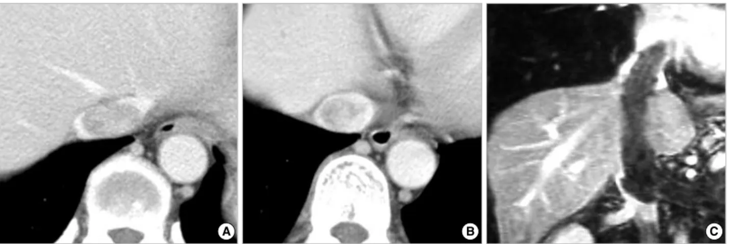
Fig. 2. Computed tomography of the patients with level IV thrombosis. Thrombus is extended to the suprahepatic IVC (A) and right atrium (B). Coronal reconstruction view demonstrates IVC thrombus extended from left renal vein to the right atrium (C).
A B C
Number (%) Level
I 10 (28.6)
II 17 (48.6)
III 4 (11.4)
IV 4 (11.4)
Stage
T3b 22 (62.9)
T3c 6 (17.1)
T4 7 (20.0)
Operation
Primary closure 32 (91.4)
Patch angioplasty 2 (5.7)
Graft interposition 1 (2.9)
Table 2. Number of patients according to level of tumor throm- bus extension, TNM stage, and operation
ment, visceral involvement, or invasion of the IVC wall (P>
0.05 for each association).
Perioperative IVC filters were used in 8 patients, including preoperative IVC filters in 3, intraoperative in 3, and post- operative in 2. Four IVC filters were inserted for preventing pulmonary embolism, 2 after pulmonary embolectomy, and 2 with pulmonary embolisms during follow-up. Intraopera- tive IVC filter placement was performed through femoral vein or internal jugular vein after IVC repair. One IVC fil- ter which was inserted prophylactically, was retrieved dur- ing surgery because it hindered proximal venous clamp. No postoperative IVC occlusion was related to IVC filter use dur- ing follow-up period.
DISCUSSION
Treatment strategies of RCC has continued to evolve from understanding of molecular and genetic characteristics of this disease (15). However, patients with RCC and IVC throm- bosis have major concern on immediate postoperative compli- cation mainly from massive pulmonary embolism and bleed- ing during surgery before getting benefit on outcome with adjuvant therapies (1, 6, 7, 9). To proceed safe and success- ful operation regarding IVC thrombectomy, first of all, inten- sive intraoperative hemodynamic monitoring was required.
We found that patients with intraoperative pulmonary embo- lisms showed sudden decreases in O2saturation and blood pressure, suggesting a need for continuous surveillance with intraoperative bidimensional TEE. TEE is useful in the pre- operative evaluation of thrombus extent, in detecting tumor or gas emboli, and in intraoperative confirmation of complete thrombus removal (6, 16, 17). In our experience, intraoper- ative pulmonary embolism developed during dissection and manipulation of the IVC and kidney, and we recommend using TEE routinely at every stage of the procedure.
Pulmonary embolisms can be prevented by sufficient expo- sure of the IVC for proximal control and direct inspection of involved IVC wall. Complete thrombus extraction without
tumor fragmentation can be achieved through liver mobiliza- tion using liver transplantation techniques (18-22). Piggy- back liver mobilization can ensure direct visualization of the entire length of the IVC, thus permitting a cavotomy to be extended when necessary. Reported complications of liver mobilization include duodenal injury, liver laceration, and pulmonary embolism, suggesting that liver mobilization be limited to level III and IV tumor extensions and selected pati- ents with level II extensions (13, 18, 21). These surgical strate- gies also related with the long term outcome. Blind removal of a thrombus can cause postoperative embolisms and early disease recurrence because of tumor fragmentation and the intraluminal persistence of part of a thrombus (21). Patients with incomplete resection have a significantly worse prog- nosis, and their five-year survival rates following surgery have been reported to range from 0% to 17.5% (2, 7, 9). Our over- all survival rate of 50.8% at 5-yr was similar to other reports of tumor resection through long cavotomy under full expo- sure of the involved IVC (3, 6).
We found that lateral venotomy, starting from the ostium of the affected renal vein, was convenient to effect repairs. In case of tumor invasion of the caval wall, medial venotomy car- ries a higher risk of narrowing after caval wall excision and renal vein repair, and often requires caval wall reconstruction.
Other reported strategies that may prevent pulmonary embolism include suprarenal IVC control using suprarenal IVC clips, sternotomy with direct clamping, IVC interrup- tion by suture/staple ligation, and placement of a suprarenal IVC filter (23-27). Good technical success has been seen after various types of filters were used, with venography at the time of filter placement providing a preoperative assessment of the extent of the tumor thrombus (26). Sosa et al. (28) reported that preoperatively placed Mobin-Uddin vena caval umbrel- las trapped tumor thrombi and/or clots in 5 of 7 patients.
Intraoperative deployment of a filter may protect the propa- gation of bland thrombi in patients with distal thrombi as well as tumor thrombi (27). In contrast, the overall rate of vena caval thrombosis or occlusion and renal dysfunction asso- ciated with infrarenal filter placement was 3-5%, suggesting
Fig. 3. Cumulative survival rate of RCC with IVC thrombosis patients. Overall survival rate (A) and stratified data according to extent of tumor thrombus (B, P >0.05) and stage (C, P >0.05) are depicted.
Cumulative survival rate
1.0
0.8
0.6
0.4
0.2
0.0
0 20 40 60 80
Follow up (months) A
Cumulative survival rate
1.0
0.8
0.6
0.4
0.2
0.0
0 20 40 60 80
Follow up (months) n=25
n=9
3 Stage
B
Cumulative survival rate
1.0
0.8
0.6
0.4
0.2
0.0
0 20 40 60 80
Follow up (months) C 4
1 Extent
2 3 4 n=4
n=4 n=10
n=17
that filter placement may increase significant overall morbidi- ty in the setting of recent nephrectomy (24). Moreover, incor- poration of the tumor in the filter may increase the difficul- ty of complete resection (27). In our experience, use of pre- operative IVC filters granted little room for applying venous clamps and we had to remove one during operation. Because direct inspection of involved IVC wall alone might decrease chance of pulmonary embolism, we do not recommend the prophylactic use of IVC filters. If IVC filter is required, care- ful selection of the IVC filter location is crucial, based on the extent of dissection.
Cardiopulmonary bypass or Pringle’s maneuver may min- imize bleeding from the operative field, thus achieving hemo- dynamic stability (13, 19, 29). CPB with or without hypo- thermia and circulatory arrest may achieve stable hemody- namic status during clamping of the IVC, thus providing a bloodless operative field for tumor resection and venous recon- struction in patients with level III or IV thrombi. However, half of IVC thrombi are infrahepatic and only 10% are located in the right atrium (11). Generally, CPB is the main cause of a complex systemic inflammatory response, which signif- icantly contributes to several adverse postoperative outcomes, including renal, pulmonary, and neurological complications, bleeding, and even multiple organ dysfunctions (5, 30). Post- operative bleeding and coagulation disturbances were the most frequently reported complications in patients who under- went IVC thrombectomy with nephrectomy. Our findings suggest that CPB is not always necessary to achieve nephrec- tomy with thrombectomy of the IVC in patients without intraatrial thrombi. We usually use CPB without hypother- mia or circulatory arrest. Furthermore, when infrarenal IVC is confirmed free from thrombus, we cannulate the infrarenal IVC instead of the femoral vein. This does not require fur- ther dissection of the femoral vein and may facilitate IVC manipulation, reducing bleeding from the IVC and assur- ing venous drainage of the lower extremities. Oblique appli- cation of a vascular clamp across the infrarenal IVC and con- tralateral renal vein can also assure renal venous drainage.
Another reported surgical technique that may achieve hemodynamic stability is aortic cross-clamping during IVC thrombectomy. Partial or total cross-clamping of the infra- renal or supraceliac segment of the abdominal aorta can main- tain systemic blood pressure above 100 mmHg by maintain- ing venous return (13). However, in our experience, even patients without venous occlusion tolerated this procedure without significant hemodynamic instability, and transient hypotension could be managed by fluid and blood adminis- tration.
The role of anticoagulation after operation for preventing recurrent IVC thrombosis is not well described in many arti- cles. The brief use of low molecular weight heparin or dex- tran was reported during the immediate postoperative peri- od to prevent deep vein thrombosis (19, 26).
In conclusion, our findings suggest that safe IVC thrombec-
tomy can be accomplished by vigilance on hemodynamic status and by complete thrombus removal without fragmen- tation through long venotomy on fully exposed involved IVC. Individual surgical planning is necessary, including liver mobilization, hepatic vascular exclusion, and possible CPB, depending on the level of thrombus.
REFERENCES
1. Kearney GP, Waters WB, Klein LA, Richie JP, Gittes RF. Results of inferior vena cava resection for renal cell carcinoma. J Urol 1981;
125: 769-73.
2. Hatcher PA, Anderson EE, Paulson DF, Carson CC, Robertson JE.
Surgical management and prognosis of renal cell carcinoma invad- ing the vena cava. J Urol 1991; 145: 20-3.
3. Nesbitt JC, Soltero ER, Dinney CP, Walsh GL, Schrump DS, Swan- son DA, Pisters LL, Willis KD, Putnam JB Jr. Surgical management of renal cell carcinoma with inferior vena cava tumor thrombus. Ann Thorac Surg 1997; 63: 1592-600.
4. Tsuji Y, Goto A, Hara I, Ataka K, Yamashita C, Okita Y, Kamidono S. Renal cell carcinoma with extension of tumor thrombus into the vena cava: surgical strategy and prognosis. J Vasc Surg 2001; 33:
789-96.
5. Bissada NK, Yakout HH, Babanouri A, Elsalamony T, Fahmy W, Gunham M, Hull GW, Chaudhary UB. Long-term experience with management of renal cell carcinoma involving the inferior vena cava.
Urology 2003; 61: 89-92.
6. Blute ML, Leibovich BC, Lohse CM, Cheville JC, Zincke H. The Mayo Clinic experience with surgical management, complications and outcome for patients with renal cell carcinoma and venous tu- mour thrombus. BJU Int 2004; 94: 33-41.
7. Skinner DG, Pritchett TR, Lieskovsky G, Boyd SD, Stiles QR. Vena caval involvement by renal cell carcinoma. Surgical resection pro- vides meaningful long-term survival. Ann Surg 1989; 210: 387-92.
8. Moinzadeh A, Libertino JA. Prognostic significance of tumor throm- bus level in patients with renal cell carcinoma and venous tumor thrombus extension. Is all T3b the same? J Urol 2004; 171: 598-601.
9. Neves RJ, Zincke H. Surgical treatment of renal cancer with vena cava extension. Br J Urol 1987; 59: 390-5.
10. Haferkamp A, Bastian PJ, Jakobi H, Pritsch M, Pfitzenmaier J, Albers P, Hallscheidt P, Muller SC, Hohenfellner M. Renal cell carcinoma with tumor thrombus extension into the vena cava: prospective long- term followup. J Urol 2007; 177: 1703-8.
11. Kirkali Z, Van Poppel H. A critical analysis of surgery for kidney can- cer with vena cava invasion. Eur Urol 2007; 52: 658-62.
12. Staehler G, Brkovic D. The role of radical surgery for renal cell car- cinoma with extension into the vena cava. J Urol 2000; 163: 1671-5.
13. Jibiki M, Iwai T, Inoue Y, Sugano N, Kihara K, Hyochi N, Sunamori M. Surgical strategy for treating renal cell carcinoma with thrombus extending into the inferior vena cava. J Vasc Surg 2004; 39: 829-35.
14. Sobin LH, Wittekind CH. UICC International Union Against Can- cer. In: TNM classificatoin of malignant tumors. 6th ed. New York:
Wiley-Liss, 2002; 193-5.
15. Rini BI, Rathmell WK, Godley P. Renal cell carcinoma. Curr Opin Oncol 2008; 20: 300-6.
16. Oikawa T, Shimazui T, Johraku A, Kihara S, Tsukamoto S, Miyana- ga N, Hattori K, Kawai K, Uchida K, Takeshima H, Saito S, Toy- ooka H, Akaza H. Intraoperative transesophageal echocardiogra- phy for inferior vena caval tumor thrombus in renal cell carcinoma.
Int J Urol 2004; 11: 189-92.
17. Zini L, Haulon S, Decoene C, Amara N, Villers A, Biserte J, Leroy X, Koussa M. Renal cell carcinoma associated with tumor thrombus in the inferior vena cava: surgical strategies. Ann Vasc Surg 2005;
19: 522-8.
18. Kaplan S, Ekici S, Dogan R, Demircin M, Ozen H, Pasaoglu I. Sur- gical management of renal cell carcinoma with inferior vena cava tumor thrombus. Am J Surg 2002; 183: 292-9.
19. Vaidya A, Ciancio G, Soloway M. Surgical techniques for treating a renal neoplasm invading the inferior vena cava. J Urol 2003; 169:
435-44.
20. Delis S, Dervenis C, Lytras D, Avgerinos C, Soloway M, Ciancio G. Liver transplantation techniques with preservation of the natural venovenous bypass: effect on surgical resection of renal cell carci- noma invading the inferior vena cava. World J Surg 2004; 28: 614-9.
21. Gallucci M, Borzomati D, Flammia G, Alcini A, Albino G, Carica- to M, Esposito A, Vincenzi B, Rossi M, Coppola R, Berloco P. Liver harvesting surgical technique for the treatment of retro-hepatic caval thrombosis concomitant to renal cell carcinoma: perioperative and long-term results in 15 patients without mortality. Eur Urol 2004;
45: 194-202.
22. Ciancio G, Livingstone AS, Soloway M. Surgical management of renal cell carcinoma with tumor thrombus in the renal and inferior vena cava: the University of Miami experience in using liver trans- plantation techniques. Eur Urol 2007; 51: 988-94.
23. Rosenthal D, Gershon CR, Rudderman R. Renal cell carcinoma invad- ing the inferior vena cava: the use of the Greenfield filter to prevent tumor emboli during nephrectomy. J Urol 1985; 134: 126-7.
24. Brenner DW, Brenner CJ, Scott J, Wehberg K, Granger JP, Schell- hammer PF. Suprarenal Greenfield filter placement to prevent pul- monary embolus in patients with vena caval tumor thrombi. J Urol 1992; 147: 19-23.
25. Hirota S, Matsumoto S, Ichikawa S, Tomita M, Koshino T, Sako M, Kono M. Suprarenal inferior vena cava filter placement prior to tran- scatheter arterial embolization (TAE) of a renal cell carcinoma with large renal vein tumor thrombus: prevention of pulmonary tumor emboli after TAE. Cardiovasc Intervent Radiol 1997; 20: 139-41.
26. Wellons E, Rosenthal D, Schoborg T, Shuler F, Levitt A. Renal cell carcinoma invading the inferior vena cava: use of a ‘‘temporary’’
vena cava filter to prevent tumor emboli during nephrectomy. Urol- ogy 2004; 63: 380-2.
27. Blute ML, Boorjian SA, Leibovich BC, Lohse CM, Frank I, Karnes RJ. Results of inferior vena caval interruption by greenfield filter, ligation or resection during radical nephrectomy and tumor throm- bectomy. J Urol 2007; 178: 440-5.
28. Sosa RE, Muecke EC, Vaughan ED Jr, McCarron JP Jr. Renal cell carcinoma extending into the inferior vena cava: the prognostic sig- nificance of the level of vena caval involvement. J Urol 1984; 132:
1097-100.
29. Hedderich GS, O'Connor RJ, Reid EC, Mulder DS. Caval tumor thrombus complicating renal cell carcinoma: a surgical challenge.
Surgery 1987; 102: 614-21.
30. Elahi MM, Khan JS. Living with off-pump coronary artery surgery:
evolution, development, and clinical potential for coronary heart dis- ease patients. Heart Surg Forum 2006; 9: E630-7.