INTRODUCTION
Giant cell tumor of the pancreas is a very rare neoplasm and appears as two different phenotypes: undifferentiated carcino- ma and osteoclast-like giant cell tumor (OGTP) (1-3). OGTP, mimicking giant cell tumor of bone should be distinguished from undifferentiated carcinoma. In contrast to OGTP, undif- ferentiated carcinoma exhibit more pleomorphic and anaplas- tic features. OGTP is the more rare type and has a better prog- nosis than undifferentiated carcinoma (1-3). An implication of these differences in histological and clinical characteristics is that these tumors have a different histogenesis. To date, generally, undifferentiated carcinoma is believed to have an epithelial origin without debating. However, the histogene- sis of OGTP remains controversial either epithelial or mes- enchymal derivation in the literature (4-16).
Here, we report a case of OGTP, performed the histopatho- logical, immunohistochemical, ultrastructural, and molecu- lar biological studies and review the literature pertaining to this condition.
CASE REPORT
A 63-yr-old woman was admitted to Chonnam National
University Hospital with a three month history of epigas- tric pain. Physical examination showed a smooth palpable mass in the epigastrium. The laboratory examinations were within normal range. An abdominal computerized tomog- raphy revealed the presence of a large cystic mass, arising from the tail of pancreas (Fig. 1A). A distal pancreatectomy with splenectomy was performed. The resected tumor was a 8×7×6-cm hemorrhagic and cystic mass in the tail of the pancreas, mimicking pancreatic pseudocyst (Fig. 1B). The histology confirmed OGTP with ductal adenocarcinoma. She received third courses of chemotherapy with gemcitabine.
She has been well, without evidence of tumor recurrence 6 months after surgery.
Microscopic findings
Histologically, the tumor was composed of typical ductal adenocarcinoma and surrounding mononuclear stromal cells intermingled with osteoclast-like giant cells and pleomorphic large cells (Fig. 2). The mononuclear cells demonstrated irreg- ular, pleomorphic nuclei with varying degrees of cytologic atypia. In contrast, osteoclast-like giant cells had multiple small uniform nuclei without atypia. Only some pleomor- phic large cells showed atypical mitoses. In addition, there was a small area of moderately to well differentiated ductal
Young-Eun Joo, Tag Heo*, Chang-Hwan Park, Wan-Sik Lee, Hyun-Soo Kim, Jung-Chul Kim�, Yang-Seok Koh�, Sung-Kyu Choi, Chol-Kyoon Cho�, Jong-Sun Rew, Sei-Jong Kim
Departments of Internal Medicine, Emergency Medicine*, and Surgery�, Chonnam National University Medical School, Gwangju, Korea
Address for correspondence Young-Eun Joo, M.D.
Department of Internal Medicine, Chonnam National University Medical School, 8 Hak-dong, Dong-ku, Gwangju 501-757, Korea
Tel : +82.62-220-6296, Fax : +82.62-225-8578 E-mail : [email protected]
516
A Case of Osteoclast-like Giant Cell Tumor of the Pancreas with Ductal Adenocarcinoma: Histopathological, Immunohistochemical, Ultrastructural and Molecular Biological Studies
Osteoclast-like giant cell tumor of the pancreas is a very rare neoplasm, of which the histiogenesis remains controversial. A 63-yr-old woman was hospitalized for evaluation of epigastric pain. An abdominal computerized tomography revealed the presence of a large cystic mass, arising from the tail of pancreas. A distal pan- createctomy with splenectomy was performed. Histologically, the tumor was com- posed of mononuclear stromal cells intermingled with osteclast-like giant cells. In addition, there was a small area of moderately to well differentiated ductal adeno- carcinoma. The final pathologic diagnosis was osteoclast-like giant cell tumor of the pancreas with ductal adenocarcinoma. Here, we describe the histopathologi- cal, immunohistochemical, ultrastructural and molecular biological findings of this tumor with review of the literature pertaining to this condition.
Key Words : Giant Cell Tumors; Pancreas Neoplasms; Osteoclasts; Histiogenesis
Received : 13 January 2004 Accepted : 20 May 2004
adenocarcinoma. Frequently cystic degeneration was found in this tumor.
Immunohistochemical findings
All procedures for immunohistochemical staining were done by the Micro-Probe staining system (Fisher Scientific, Pitts-
burgh, PA, U.S.A.) based on capillary action (17). Paraffin sections, of 4 m in thickness with mounted probe on slides, were immunostained with the panel of primary antibodies listed Table 1, by the avidin-biotin peroxidase complex method.
The results of immunohistochemical stainings are summa- rized in Table 2. Immunohistochemically, cytokeratin (CK) and epithelial membrane antigen (EMA) are strongly posi- tive for ductal carcinoma cells, whereas both osteoclast-like giant cells and mononuclear cells were negative for CK (Fig.
3A) and EMA. Osteoclast-like giant cells and mononuclear cells were positive for histiocytic markers, CD-68 (Fig. 3B) and lysozyme. In general, immunoreactivity of vimentin was more intense in pleomorphic large cells than that of osteoclast- like giant cells and mononuclear cells. Most of the nuclei in ductal carcinoma cells expressed PCNA, demonstrating intense proliferative activity, while no nuclei were stained in osteo- clast-like giant cells and mononuclear cells. Anti-p53 anti- body did react with some atypical mononuclear cells of weak intranuclear staining, but did not react with either ductal car-
Fig. 1.(A) An abdominal computerized tomography reveals the presence of a large cystic mass, arising from the tail of pancreas. (B) The resected tumor is a 8×7×6-cm hemorrhagic and cystic mass in the tail of the pancreas.
A B
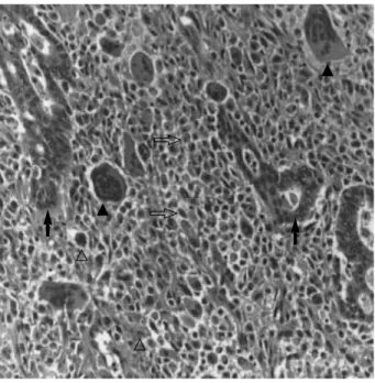
Fig. 2.Histologically, the tumor is composed of typical ductal ade- nocarcinoma (closed arrow) and surrounding mononuclear stro- mal cells (open arrow) intermingled with osteoclast-like giant cells (closed arrow head) and pleomorphic large cells (open arrow head) (H&E ×200).
CK, cytokeratin; EMA, epithelial membrane antigen; PCNA, proliferating cell nuclear antigen; M, monclonal antibody; P, polyclonal antibody.
Primary antibody Type Source Dilution
CK M Zymed 1:150
EMA M DAKO 1:75
CD-68 M DAKO 1:50
Lysozyme M Zymed 1:100
Vimentin M Zymed 1:100
PCNA M DAKO 1:100
p53 M Zymed 1:50
Table 1.Primary antibodies used in the immunohistochemical studies
cinoma cells or osteoclast-like giant cells.
Electron microscopic findings
For electron microscopy, the tissue specimen was fixed in 2.5% glutaraldehyde, fixed in osmium tetroxide, and embed- ded in epon mixture. Ultrathin sections, 80 nm in thickness, were made by LKB-V ultramicrotome with diamond knife, stained with uranyl acetate and lead citrate, and examined
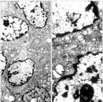
with a JEM 100CX type II electron microscope. The nucleus of the mononuclear cell contained prominent nucleoli, with dispersed clumps of heterochromatin. The cytoplasm con- tained ample mitochondria, and moderate amount of rough endoplasmic reticulum (Fig. 4) (Table 2). But we did not find the evidence of epithelial differentiation including microvil-
Fig. 3.Immunohistochemical staining of cytokeratin (A) and CD-68 (B). Cytokeratin (CK) is strongly positive for ductal carcinoma cells, whereas both osteoclast-like giant cells and mononuclear cells are negative for CK. However, osteoclast-like giant cells and mononuclear cells are positive for histiocytic markers, CD-68 (×200).
A B
Fig. 4.Electron microscopically, the nucleus of the mononuclear cell contains prominent nucleoli, with dispersed clumps of hetero- chromatin. The cytoplasm contained ample mitochondria, and moderate amount of rough endoplasmic reticulum (×4,000).
CK, cytokeratin; EMA, epithelial membrane antigen; PCNA, proliferat- ing cell nuclear antigen.
Pleomorphic large cell Osteoclast-
like giant cell Mononu-
clear cell Ductal car- cinoma cell Study
Immunohistochemistry
CK + - - -
EMA + - - -
CD-68 - + + -
Lysozyme - + + -
Vimentin - + + +
PCNA + - - -
p53 - + - -
Electron microscopy
Mitochondria Abundant Abundant
Endoplasmic reticulum Moderate Empty
Microvilli None None
Desmosome None None
K-ras gene analysis
Codon 12 - - - -
Codon 13 - - - -
Table 2.Results of immunohistochemical, ultrastructural and molecular biological studies
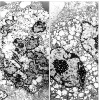
li and desmosomes. In the ultrastructure of multinucleated osteoclast-like giant cell, there were multiple nuclei with dispersed chromatin and a fine chromatin rim. The cytoplasm was characterized by abundant mitochondria of varying size, free ribosomes, and dilated empty rough endoplasmic retic- ulum cisternae (Fig. 5) (Table 2).
Molecular biologic findings
To analyze K-ras gene mutations, we microdissected in both ductal carcinoma and giant cell tumor components separate- ly. After extracting DNA, polymerase chain reaction (PCR) amplification and direct sequencing were done to detect K- ras gene mutation using ABI 3700DNA analyzer. The size of amplified PCR product was 223 bp. All mutations were verified in both the sense and anti-sense directions. DNA from ductal carcinoma with the known K-ras mutation was used as a positive control. A microdissection method was applied to allow analyses of K-ras gene mutations as performed by Sakai et al. (15). However, none of the microdissected osteo- clast-like giant cell, mononuclear cell, and ductal carcinoma cell contained K-ras gene mutations at codon 12 (GGT) and 13 (AGT) in our case (Table 2).
DISCUSSION
OGTP is very rare and comprises less than 1% of nonen- docrine pancreatic tumor (1-3). To date, only two cases have
been reported in Korea (18, 19). In the review of English liter- ature (20-22), OGTP tends to present in the 7th decade of life, although younger patients have been reported. There was no sex preference. The main symptoms and signs were abdomi- nal pain, palpable mass, weight loss and jaundice. OGTP commonly exhibited as large cystic neoplasms with hemor- rhage and necrosis of variable extent. There was predilection to involve head and body portion of the pancreas. The prog- nosis of OGTP is better than that of pancreatic ductal ade- nocarcinoma and undifferentiated carcinoma, suggesting that OGTP grows more slowly and is less likely to metastasize than pancreatic ductal adenocarcinoma and undifferentiated carcinoma. However, the outcome of OGTP is extremely vari- able in the literature. Although it is difficult to decide treat- ment modality because of its rarity, the choice of treatment of OGTP may be surgical resection if possible. Also, in pre- vious reports, experience of adjuvant treatments including chemotherapy or radiotherapy was not described, so no detailed information was provided. However, because the outcome of OGTP is extremely variable, it may be reasonable to con- sider adjuvant treatment for prevention of tumor recurrence.
In our case, we did attempt chemotherapy with gemcitabine, according to treatment protocol established for pancreatic ductal adenocarcinoma. We think that this attempt is impor- tant and necessary to detail the treatment modalities and clini- cal outcome.
Although the histiogenesis of OGTP has been previously investigated by immunohistochemical, ultrastructural, and molecular biologic studies, the origin of these cells is still con- troversial.
In our case, osteoclast-like giant cells and mononuclear cells were positive for histiocytic markers, CD68 and lysozyme in contrast to negative for epithelial markers, CK and EMA.
In the ultrastructure of multinucleated, osteoclast-like giant cells and mononuclear cells, we did not find either microvilli or desmosomes for the clue of epithelial differentiation.
Tumor progression is regulated by many biological pro- cesses including cell proliferation and genetic alterations of tumor suppressor gene and oncogene. PCNA reflects cell pro- liferation and mutation of the p53 tumor suppressor gene represents one of the common genetic alterations in pancre- atic ductal carcinoma. In our case, ductal carcinoma cells ex- pressed PCNA while no nuclei were stained in osteoclast- like giant cells and mononuclear cells. p53 immunoreactivi- ty showed only weak intranuclear staining in some atypical mononuclear cells, but did not show in ductal carcinoma cells and osteoclast-like giant cells.
K-ras gene mutations are a hallmark of pancreatic ductal carcinoma and appear to be present in 80% to 90% of tumors.
Evidence of K-ras gene mutations indicates that its origin is an epithelial nature. But in our case, none of the microdis- sected osteoclast-like giant cell, mononuclear cell, and duc- tal carcinoma cell contained K-ras gene mutations.
These results may support that both osteoclast-like giant
Fig. 5.Electron microscopically, osteoclast-like giant cell has mul- tiple nuclei with dispersed chromatin and a fine chromatin rim. The cytoplasm is characterized by abundant mitochondria of varying size, free ribosomes, and dilated empty rough endoplasmic retic- ulum cisternae (×4,000).
cells and mononuclear cells in OGTP originate from mes- enchymal rather than epithelial cell. However, although most reports have debated the histiogenesis of OGTP from immuno- histochemical, ultrastructural, and molecular biologic as- pects, it is still obscure whether the histiogenesis of OGTP is epithelial or mesenchymal origin (4-16). There are several possible explanations for these discrepancies. First, OGTP is too rare to discern differences in the origin of these cells. Sec- ond, immunohistochemical staining methods and primary antibodies used are different in different cases (4). Third, OGTP can express different phenotypes with variable stages and degrees of differentiation during tumor progression (4, 23). Fourth, circulating precursor cell may be stimulated by tumor derived factors such as cytokines, to proliferate and differentiate into different cell types (4, 16, 20, 21).
Clinical characteristics and histiogenesis of OGTP are still now variable and controversial in the literature. Therefore, further studies based on a large populations are necessary to clarify its histiogenesis.
REFERENCES
1. Rosai J. Carcinoma of pancreas simulating giant cell tumor of bone:
electron-microscopic evidence of its acinar cell origin. Cancer 1968;
22: 333-44.
2. Alguacil-Garcia A, Weiland LH. The histologic spectrum, progno- sis, and histogenesis of the sarcomatoid carcinoma of the pancreas.
Cancer 1977; 39: 1181-9.
3. Lewandrowski KB, Weston L, Dickersin GR, Rattner DW, Comp- ton CC. Giant cell tumor of the pancreas of mixed osteoclastic and pleomorphic cell type: evidence for a histogenetic relationship and mesenchymal differentiation. Hum Pathol 1990; 21: 1184-7.
4. Sun AP, Ohtsuki Y, Liang SB, Sonobe H, Iwata J, Furihata M, Take- uchi T, Qiu Y, Chen BK, Watanabe R, Ohmori K. Osteoclast-like giant cell tumor of the pancreas with metastases to gallbladder and lymph nodes. A case report. Pathol Res Pract 1998; 194: 587-94.
5. Machado MA, Herman P, Montagnini AL, Jukemura J, Leite KR, Machado MC. Benign variant of osteoclast-type giant cell tumor of the pancreas: importance of the lack of epithelial differentiation.
Pancreas 2001; 22: 105-7.
6. Hansen T, Burg J, Kirkpatrick CJ, Kriegsmann J. Osteoclast-like giant cell tumor of the pancreas with ductal adenocarcinoma: case report with novel data on histogenesis. Pancreas 2002; 25: 317-20.
7. Berendt RC, Shnitka TK, Wiens E, Manickavel V, Jewell LD. The osteoclast-type giant cell tumor of the pancreas. Arch Pathol Lab Med 1987; 111: 43-8.
8. Dizon MA, Multhaupt HA, Paskin DL, Warhol MJ. Osteoclastic giant cell tumor of the pancreas: an immunohistochemical study.
Arch Pathol Lab Med 1996; 120: 306-9.
9. Dworak O, Wittekind C, Koerfgen HP, Gall FP. Osteoclastic giant
cell tumor of the pancreas. An immunohistological study and review of the literature. Pathol Res Pract 1993; 189: 228-31.
10. Watanabe M, Miura H, Inoue H, Uzuki M, Noda Y, Fujita N, Yamaza- ki T, Sawai T. Mixed osteoclastic/pleomorphic-type giant cell tumor of the pancreas with ductal adenocarcinoma: histochemical and immunohistochemical study with review of the literature. Pancreas 1997; 15: 201-8.
11. Gatteschi B, Saccomanno S, Bartoli FG, Salvi S, Liu G, Pugliese V.
Mixed pleomorphic-osteoclast-like tumor of the pancreas. Light micro- scopical, immunohistochemical, and molecular biological studies.
Int J Pancreatol 1995; 18: 169-75.
12. Imai Y, Morishita S, Ikeda Y, Toyoda M, Ashizawa T, Yamamoto K, Inoue T, Ishikawa T. Immunohistochemical and molecular anal- ysis of giant cell carcinoma of the pancreas: a report of three cases.
Pancreas 1999; 18: 308-15.
13. Martin A, Texier P, Bahnini JM, Diebold J. An unusual epithelial pleomorphic giant cell tumour of the pancreas with osteoclast-type cells. J Clin Pathol 1994; 47: 372-4.
14. Westra WH, Sturm P, Drillenburg P, Choti MA, Klimstra DS, Albores- Saavedra J, Montag A, Offerhaus GJ, Hruban RH. K-ras oncogene mutations in osteoclast-like giant cell tumors of the pancreas and liver: genetic evidence to support origin from the duct epithelium.
Am J Surg Pathol 1998; 22: 1247-54.
15. Sakai Y, Kupelioglu AA, Yanagisawa A, Yamaguchi K, Hidaka E, Matsuya S, Ohbuchi T, Tada Y, Saisho H, Kato Y. Origin of giant cells in osteoclast-like giant cell tumors of the pancreas. Hum Pathol 2000; 31: 1223-9.
16. Goldberg RD, Michelassi F, Montag AG. Osteoclast-like giant cell tumor of the pancreas: immunophenotypic similarity to giant cell tumor of bone. Hum Pathol 1991; 22: 618-22.
17. Reed JA, Manahan LJ, Park CS, Brigati DJ. Complete one-hour im- munocytochemistry based on capillary action. Biotechniques 1992;
13: 434-43.
18. Sung SH, Han WS. Fine needle aspiration cytology of osteoclastic giant cell yumor of the pancreas. Korean J Cytopathol 1998; 9: 89- 94.
19. Song HG, Kim YI, Yu ES, Lee HS. Two histologic variants of giant cell carcinoma of the pancreas. Korean J Pathol 1987; 21: 192-8.
20. Shiozawa M, Imada T, Ishiwa N, Rino Y, Hasuo K, Takanashi Y, Nakatani Y, Inayama Y. Osteoclast-like giant cell tumor of the pan- creas. Int J Clin Oncol 2002; 7: 376-80.
21. Leighton CC, Shum DT. Osteoclastic giant cell tumor of the pan- creas: case report and literature review. Am J Clin Oncol 2001; 24:
77-80.
22. Oehler U, Jurs M, Kloppel G, Helpap B. Osteoclast-like giant cell tumour of the pancreas presenting as a pseudocyst-like lesion. Vir- chows Arch 1997; 431: 215-8.
23. Suster S, Phillips M, Robinson MJ. Malignant fibrous histiocytoma (giant cell type) of the pancreas. A distinctive variant of osteoclast- type giant cell tumor of the pancreas. Cancer 1989; 64: 2303-8.