aResearch Assistant, eAssociate Professor, Department of Orthodontics, Faculty of Dentistry, Dicle University, Diyarbakır, Turkey.
bProfessor and Chair, Department of Orthodontics, Faculty of Dentistry, Izmir Katip Celebi University, Izmir, Turkey.
cProfessor, Department of Pediatric Dentistry and Orthodontics, College of Dentistry, King Saud University, Riyadh, Saudi Arabia.
dResearch Assistant, Department of Orthodontics, Faculty of Dentistry, Erciyes University, Kayseri, Turkey,
Corresponding author:Tancan Uysal.
İzmir Katip Çelebi Üniversitesi Dişhekimliği Fakültesi, Ortodonti Ortodonti Anabilim, Dalı, İzmir 35630, Turkey.
+902323293999; e-mail, [email protected].
Received April 22, 2011; Last Revision July 27, 2011; Accepted August 1, 2011.
http://dx.doi.org/10.4041/kjod.2011.41.6.431
Cone-beam computed tomography assessment of mandibular asymmetry in unilateral cleft lip and palate patients
Ilknur Veli, DDS,a Tancan Uysal, DDS, PhD,b,c Faruk Izzet Ucar, DDS,d Murat Eruz, DDS,d Torun Ozer, DDS, PhDe
Objective: To determine whether there is any difference between the cleft and non-cleft sides of the man- dible in unilateral cleft lip and palate (UCLP) patients, or the right and left sides in control patients; and to determine if there is any difference between the mandibular asymmetry of UCLP patients and that of control patients. Methods: We examined cone-beam computed tomography (CBCT) scans of 15 patients with UCLP and 15 age- and gender-matched control patients. We evaluated 8 linear, 3 surface, and 3 volu- metric measurements and compared the cleft/non-cleft sides of UCLP patients and the right/left sides of controls. Results: There were no statistically significant gender differences in any linear, surface, or volu- metric measurement. The single significant side-to-side difference in UCLP patients was a longer coronoid unit on the cleft side than on the non-cleft side (p = 0.046). Body volume was significantly lower in the UCLP group than in the control group (p = 0.008). Conclusions: In general, UCLP patients have sym- metrical mandibles, although the coronoid unit length is significantly longer on the cleft side than on the non-cleft side. UCLP patients and controls differed only in body volume. (Korean J Orthod 2011;41(6):
431-439)
Key words: Asymmetry, Cone-beam computed tomography, Cleft lip/palate
INTRODUCTION
Facial asymmetry, defined as a difference in size be- tween the left and right hemifaces, is a natural phe- nomenon1 that is caused primarily by mandibular
asymmetry.2 The etiology of mandibular asymmetry is multifactorial,3 including genetic or congenital malfor- mations such as cleft lip and palate.4
The development of mandibular asymmetry in uni- lateral cleft lip and palate (UCLP) patients may be caused by the following etiologic factors:5 (1) true skeletal mandibular asymmetry, (2) positional adapta- tion of the lower jaw to asymmetric mandibular fossae, and (3) functional adaptation to dentoalveolar and oc- clusal disharmonies. In the literature, some authors re- port significant mandibular asymmetries in cleft lip and palate patients,5,6 whereas others have found no such asymmetry.7,8
A number of tools have been used to assess man- dibular asymmetry, including clinical examination; fron- tal- and side-view photographs; and 2-dimensional (2D) radiographs, such as lateral and posteroanterior cephalograms, oblique radiographs of the mandible tak-
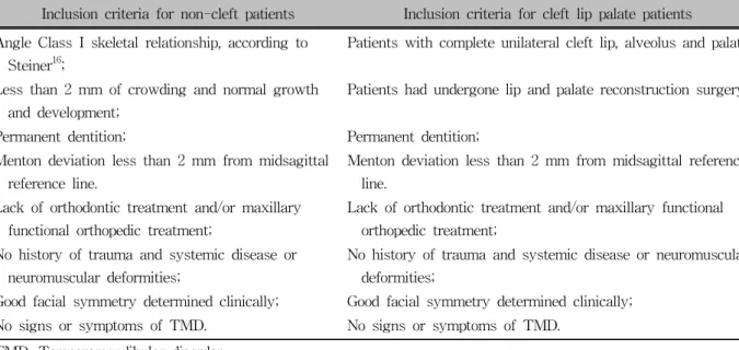
Table 1. Criteria for sample selection
Inclusion criteria for non-cleft patients Inclusion criteria for cleft lip palate patients Angle Class I skeletal relationship, according to
Steiner16;
Patients with complete unilateral cleft lip, alveolus and palate
Less than 2 mm of crowding and normal growth and development;
Patients had undergone lip and palate reconstruction surgery;
Permanent dentition; Permanent dentition;
Menton deviation less than 2 mm from midsagittal reference line.
Menton deviation less than 2 mm from midsagittal reference line.
Lack of orthodontic treatment and/or maxillary functional orthopedic treatment;
Lack of orthodontic treatment and/or maxillary functional orthopedic treatment;
No history of trauma and systemic disease or neuromuscular deformities;
No history of trauma and systemic disease or neuromuscular deformities;
Good facial symmetry determined clinically; Good facial symmetry determined clinically;
No signs or symptoms of TMD. No signs or symptoms of TMD.
TMD, Temporomandibular disorder.
en at 45o, and panoramic radiographs.9,10 These 2D ra- diographs can be misleading, since complex 3-dimen- sional (3D) structures are projected onto flat 2D surfa- ces, creating distortion and magnification errors.11,12 Cone-beam computed tomography (CBCT), a 3D imaging technique designed specifically to create im- ages of the maxillofacial region, allows 3D re- constructions of craniofacial structures from acquired volumetric data.13 CBCT provides high-resolution im- ages (i.e., with an isotropic resolution ranging between 0.125 mm and 0.4 mm) with short scanning times (10 - 70 seconds), and requires low doses of radiation (up to 15 times lower than that of medical computed to- mography scans).14 CBCTs therefore provide an oppor- tunity for multiplanar imaging and assessment of 3D information.
However, whereas many researchers have used 2D radiographs to assess mandibular asymmetry in cleft lip and palate patients,5-7 few have used 3D imaging to investigate this phenomenon. Indeed, we could find no published studies that have evaluated mandibular asym- metry in cleft lip and palate patients using CBCT.
Therefore, we undertook this study to determine (1) whether there are any differences in mandibular meas- urements between the cleft and non-cleft sides of UCLP patients or the right and left sides of control pa-
tients; and (2) whether there are any significant differ- ences in mandibular asymmetry between UCLP and control patients.
MATERIAL AND METHODS
We examined the CBCT scans of 15 patients (8 males and 7 females) with UCLP (8 right and 7 left;
mean age: 21.2 ± 2.1 years, range: 17.3 - 24.4 years) and 15 control patients (mean age: 22.6 ± 3.2 years, range: 17.1 - 25.2 years) that were selected from the archives of the Oral and Maxillofacial Radiology Department of Faculty of Dentistry, Dicle University.
The CBCT scans were taken as part of a set of clin- ically necessary radiographs. Therefore, patients were not unnecessarily subjected to additional radiation, and consequently ethical committee approval was not nee- ded. All patients attending the dental clinic of Dicle University sign an informed consent form indicating their agreement to CBCT scans.
We used CBCT scans from patients without cleft palate as controls. These patients were matched by age and gender to the UCLP patients in the study. Selec- tion criteria for both cleft and control patients are pro- vided in Table 1. Only cases of complete UCLP were included in the present investigation because indivi-
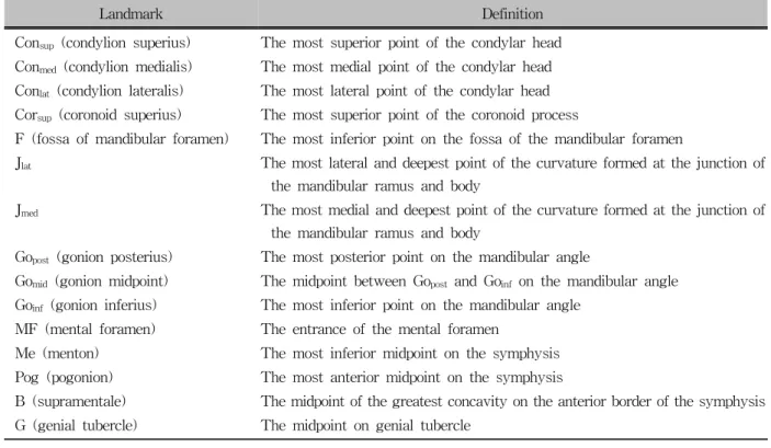
Table 2. Description of mandibular landmarks used in the study
Landmark Definition
Consup (condylion superius) The most superior point of the condylar head Conmed (condylion medialis) The most medial point of the condylar head Conlat (condylion lateralis) The most lateral point of the condylar head Corsup (coronoid superius) The most superior point of the coronoid process
F (fossa of mandibular foramen) The most inferior point on the fossa of the mandibular foramen
Jlat The most lateral and deepest point of the curvature formed at the junction of the mandibular ramus and body
Jmed The most medial and deepest point of the curvature formed at the junction of the mandibular ramus and body
Gopost (gonion posterius) The most posterior point on the mandibular angle
Gomid (gonion midpoint) The midpoint between Gopost and Goinf on the mandibular angle Goinf (gonion inferius) The most inferior point on the mandibular angle
MF (mental foramen) The entrance of the mental foramen
Me (menton) The most inferior midpoint on the symphysis Pog (pogonion) The most anterior midpoint on the symphysis
B (supramentale) The midpoint of the greatest concavity on the anterior border of the symphysis G (genial tubercle) The midpoint on genial tubercle
duals with UCLP have unilateral malformation, allow- ing us to use the measurements of the contralateral non-cleft side of each individual as an internal con- trol.15 We included patients with non-significant facial asymmetry in order to evaluate isolated asymmetry of the mandible in UCLP individuals.
Facial asymmetry was determined by the degree of menton deviation (MD) from the midsagittal reference line, as defined by Grummons and Kappeyne van de Coppello.17
All CBCT images were acquired using an iCAT 3D imaging device (Imaging Sciences International, Hat- field, PA, USA), set at 5.0 mA and 120 kV. Scans with a voxel size of 0.3 mm were made with a single 360-degree rotation, 9.6-second scan. According to rou- tine image exposure protocol, patients’ heads were ori- ented by adjusting the Frankfort plane parallel to the horizontal plane, lateral scout radiographs were taken, and small adjustments were made. This ensures in- clusion of all areas of interest and minimizes head ori- entation errors.
For better evaluation and a precise 1-to-1 ratio, mea- surements of anatomic surface landmarks and recon-
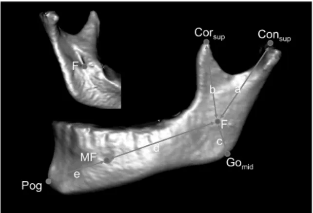
structed 3D models of UCLP patients were used in this study. DICOM files obtained from the CBCT scans were reconstructed using Mimics 10.0 (Materialise NV, Leuven, Belgium). This software allows the use of both Hounsfield and gray values to separate the area of interest from its surrounding structures, enabling the visualization of areas that are superimposed by other structures in the intact model. One important structure in the diagnosis of facial asymmetry, the condyle, can be evaluated separately after the mandible has been isolated from the rest of the image.11 We used the au- to-segmentation function of the software to isolate the mandibles from the images and removed the teeth above the alveolar bone of the mandibles. All land- mark identifications and measurements were made us- ing the Mimics 10.0 software. We used the landmarks described by You et al.18 in their examination of asym- metric mandibles based on condylar, coronoid, angular, body, and chin units (Table 2). These authors used the mandibular and mental foramina as important reference points at the junction of the skeletal units. Point F was proposed as a good reference point for the mandibular and mental foramina in 3D images of the mandible19
Fig 3. An example of surface and volumetric measurements.
Fig 1. Landmarks and measurements used in this study. A, condylar unit length; b, coronoid unit length;
c, angular unit length; d, body unit length; e, chin unit length; Corsup, coronoid superius; Consup, condylion su- perius; F, fossa of mandibular foramen; Gomid, gonion midpoint; MF, mental foramen; Pog, pogonion.
Fig 2. Landmarks and measurements used in this study. f, Condylar width; g, ramal height; h, body length; Jlat, the most lateral and deepest point of the curvature formed at the junction of the mandibular ra- mus and body; Jmed, The most medial and deepest point of the curvature formed at the junction of the mandibular ramus and body; Gomid, gonion midpoint;
Consup, condylion superius; Conmed, condylion medialis;
Conlat, condylion lateralis; Me, menton.
(Fig 1). Because primary intramembranous ossification begins in the mental foramen, it is generally accepted as a good point for the division of the mandibular cor- pus into body and chin units.18 Therefore, we used point F as a guide to measure the skeletal unit lengths.
All linear measurements were performed by control- ling the localization of the landmarks in all dimensions
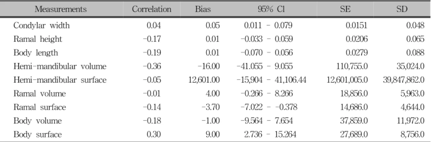
Table 3. Bland and Altman Plot to assess the repeatability
Measurements Correlation Bias 95% Cl SE SD
Condylar width 0.04 0.05 0.011 - 0.079 0.0151 0.048
Ramal height -0.17 0.01 -0.033 - 0.059 0.0206 0.065
Body length -0.19 0.01 -0.070 - 0.056 0.0279 0.088
Hemi-mandibular volume -0.36 -16.00 -41.055 - 9.055 110,755.0 35,024.0
Hemi-mandibular surface -0.05 12,601.00 -15,904 - 41,106.44 12,601,005.0 39,847,862.0
Ramal volume -0.01 4.00 -0.266 - 8.266 18,856.0 5,963.0
Ramal surface -0.14 -3.70 -7.022 - -0.378 14,686.0 4,644.0
Body volume -0.18 -1.00 -9.564 - 7.654 37,859.0 11,972.0
Body surface 0.30 9.00 2.736 - 15.264 27,689.0 8,756.0
CI, Confidence interval; SE, standard error; SD, standard deviation.
Table 4. Side-to-side comparison of the linear, surface and volumetric measurements between the cleft and non-cleft sides in UCLP patients and right and left sides in non-cleft patients
Measurements
UCLP patients Control patients
Cleft side Non-cleft side
p-value Left side Right side
p-value
Mean SD Mean SD Mean SD Mean SD
Condylar unit length (mm) 41.50 5.41 40.70 5.02 0.247 41.98 3.84 43.53. 6.68 0.130 Body unit length (mm) 54.21 5.54 57.67 8.95 0.054 56.80 4.92 55.76 4.47 0.359 Coronoid unit length (mm) 39.10 6.43 37.00 5.85 0.046* 37.72 8.03 38.80 5.06 0.685 Angular unit length (mm) 19.64 4.90 20.01 3.92 0.645 22.81 9.58 20.79 2.90 0.487 Chin unit length (mm) 27.48 5.17 28.43 5.42 0.472 27.49 4.87 26.64 2.92 0.538 Condylar width (mm) 17.35 3.19 18.28 4.97 0.372 17.28 2.02 17.24 1.83 0.885 Ramal height (mm) 58.47 8.66 57.04 8.51 0.065 58.77 5.16 61.11 5.09 0.024* Body length (mm) 78.26 7.33 79.71 7.85 0.088 77.67 9.34 72.12 11.83 0.021* Hemi-mand volume (cm3) 23.77 8.16 24.54 7.58 0.632 28.16 4.46 28.42 4.72 0.743 Hemi-mand surface (cm2) 12.57 4.25 13.03 4.63 0.535 14.48 4.07 13.93 2.43 0.288 Ramal volume (cm3) 8.08 3.36 7.33 2.53 0.323 8.04 1.72 7.95 1.60 0.598 Ramal surface (cm2) 5.21 1.83 5.09 1.60 0.702 4.98 1.19 5.11 1.07 0.425 Body volume (cm3) 16.06 6.65 16.10 4.70 0.978 20.73 3.53 20.39 3.37 0.263 Body surface (cm2) 7.82 3.29 8.07 2.47 0.738 9.45 1.49 9.23 1.64 0.215 UCLP, Unilateral cleft lip and palate; SD, standard deviation. *p < 0.05.
on the reconstructed 3D surface models. The following bilateral measurements were made (Figs 1 - 3, Table 2): (1) condylar unit length: Consup - F; (2) coronoid unit length: Corsup - F; (3) angular unit length: F - Gomid; (4) body unit length: F - MF; (5) chin unit length: MF - Pog; (6) condylar width: Conmed - Conlat; (7) ramal height: Consup - Gomid; (8) body length: Gomid -
Me; (9) hemi-mandibular volume: the mandibular vol- ume was divided into 2 hemi-mandibular volumes by the plane connecting Me, B, and G; and (10) ramal and body volumes: hemi-mandibular volume was div- ided into ramal and body volumes by the plane con- necting Gomid, Jlat and Jmed. In addition, the surface area of all mandibular parts was calculated. All data
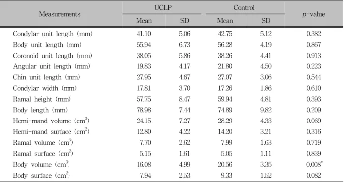
Table 5. Comparison of the linear, surface and volumetric measurements between cleft lip palate and non-cleft pa- tients
Measurements UCLP Control
p-value
Mean SD Mean SD
Condylar unit length (mm) 41.10 5.06 42.75 5.12 0.382
Body unit length (mm) 55.94 6.73 56.28 4.19 0.867
Coronoid unit length (mm) 38.05 5.86 38.26 4.41 0.913
Angular unit length (mm) 19.83 4.17 21.80 4.50 0.223
Chin unit length (mm) 27.95 4.67 27.07 3.06 0.544
Condylar width (mm) 17.81 3.70 17.26 1.86 0.610
Ramal height (mm) 57.75 8.47 59.94 4.81 0.393
Body length (mm) 78.98 7.44 74.89 9.82 0.209
Hemi-mand volume (cm3) 24.15 7.27 28.29 4.33 0.069
Hemi-mand surface (cm2) 12.80 4.22 14.20 3.21 0.316
Ramal volume (cm3) 7.70 2.62 7.99 1.63 0.719
Ramal surface (cm2) 5.15 1.61 5.05 1.11 0.839
Body volume (cm3) 16.08 4.99 20.56 3.35 0.008*
Body surface (cm2) 7.94 2.53 9.33 1.52 0.082
UCLP, Unilateral cleft lip and palate; SD, standard deviation. *p < 0.01.
were measured in cm2 or cm3, and all landmark identi- fications and measurements were made by one in- dividual to prevent interobserver variability.
To determine the errors associated with CBCT measurements, 15 of the CBCT images were randomly selected and re-measured 4 weeks after the initial measurements.
Statistical analysis
All statistical analyses were performed using the statistical package for social sciences, 13.0 (SPSS for Windows; SPSS Inc., Chicago, IL, USA). Normality of the data was tested using Shapiro-Wilks tests, and the homogeneity of variances was verified using Levene’s test. All UCLP and control patient asymmetry data was normally distributed with homogeneous variance, ex- cept for gender data. Therefore, we used parametric tests to evaluate the asymmetry data.
Wilcoxon tests were used to compare genders. To compare the measurements between the cleft and non-cleft sides in UCLP patients, and the right and left sides in control patients, we used paired-sample t-tests.
We performed independent t-tests to evaluate side- to-side differences and differences between cleft and control patients. To evaluate the differences in asym- metry between control and UCLP patients, we com- pared the right-left differences of controls with the cleft-non-cleft differences of UCLP patients. p-values less than 0.05 were considered significant. Results are reported as the means ± standard deviations.
RESULTS
A Bland and Altman plot revealed no significant dif- ferences between repeated measurements of the same radiograph (Table 3). There were also no significant differences between any of the median measurement values for male and female subjects (p > 0.05 for all). Therefore, data for both genders were pooled for further analyses.
Descriptive statistics and comparisons of the linear, surface, and volumetric measurements between the cleft and non-cleft sides of UCLP patients are pre- sented in Table 4. In UCLP patients, the coronoid unit was longer on the cleft side than on the non-cleft side
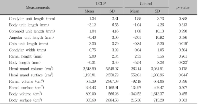
Table 6. Intergroup comparison of side-to-side differences
Measurements UCLP Control
p-value
Mean SD Mean SD
Condylar unit length (mm) 1.34 2.31 1.55 3.73 0.858
Body unit length (mm) -3.12 6.55 -1.04 4.26 0.313
Coronoid unit length (mm) 1.04 4.16 1.08 10.13 0.990
Angular unit length (mm) -0.40 3.00 -2.01 10.92 0.586
Chin unit length (mm) 3.30 3.79 -0.84 5.20 0.019*
Condylar width (mm) -0.75 3.92 -0.04 1.05 0.504
Ramal height (mm) 2.00 2.34 2.33 3.56 0.761
Body length (mm) -0.31 3.40 -5.54 8.28 0.032*
Hemi-mand volume (cm3) 2,518.59 5,545.97 262.14 3,031.91 0.178
Hemi-mand surface (cm2) 1,193.81 2,550.72 552.61 1,936.96 0.044*
Ramal volume (cm3) 563.39 2,867.08 -92.18 661.06 0.396
Ramal surface (cm2) 394.43 1,168.91 134.97 402.47 0.507
Body volume (cm3) 809.00 566.26 -342.52 1,613.37 0.455
Body surface (cm2) 305.60 2,884.58 -215.36 715.20 0.503
UCLP, Unilateral cleft lip and palate; SD, standard deviation. *p < 0.05.
(p = 0.046). This was the only significant difference between the cleft and non-cleft sides of these patients.
Only the ramal height (p = 0.024) and body length (p
= 0.021) were significantly different on the right and left sides of control patients (Table 4). Therefore, the data for both sides in each group were pooled for fur- ther statistical analysis.
Comparison of measurements between groups in- dicated that the body volume was significantly lower in UCLP patients than in controls (16.08 ± 4.99 cm3 vs. 20.56 ± 3.35 cm3; p = 0.008; Table 5). Compari- son of the differences between the cleft and non-cleft sides of UCLP patients with the differences between the left and right sides of control patients indicated that side-to-side body length differences were greater in the control group than in the UCLP group (p = 0.032), whereas side-to-side differences in chin unit length and hemi-mandibular surface area were greater in the UCLP group than in the control group (p = 0.019 and p = 0.044, respectively; Table 6).
DISCUSSION
In UCLP patients, facial and nasomaxillary skeletal
asymmetries are commonly present with the nasomaxil- lary complex being more asymmetric in affected in- dividuals than in non-cleft controls.20 Previous 2D stu- dies on facial asymmetry have reported that the man- dible appears to be the leading factor in facial asym- metry.21,22 Because quantitative measurement is a key element in the diagnosis of asymmetry, 3D structures cannot be properly analyzed with 2D radiographs.11 We therefore used 3D images to assess mandibular asymmetry in cleft lip and palate patients.
Previous studies have shown that UCLP patients reach the postpubertal growth spurt at a later age than do non-cleft patients.23 da Silva Filho et al.24 reported that cleft patients, irrespective of the type of cleft, have smaller mandibles than non-cleft patients at adulthood. Krogman et al.25 used postero-anterior ceph- alometric radiographs to assess craniofacial growth and noted a significantly larger gonial height in the UCLP group and bilateral CLP group during early and late childhood. Further, Laspos et al.26 studied postero-ante- rior radiographs of children and reported that UCLP patients had mandibles that were more asymmetric than those of controls. In contrast, Athanasiou et al.27 found that those children with cleft palates who have
undergone corrective surgery may have normal growth rates. In the current study, only post-adolescent patients were included to eliminate possible growth rate differences. We are therefore unable to discuss the cause-and-effect relationship between mandibular asym- metry and growth.
Liukkonen et al.1 reported that facial asymmetry is a natural phenomenon often due to differences in man- dibular dimensions on the right and left sides. They al- so concluded that healthy young subjects generally have some degree of mandibular asymmetry. In the present study, side-to-side comparisons of control pa- tients revealed statistically significant differences in ramal height and body length. We attribute the differ- ences in these measurements to natural asymmetry. On the other the hand, the side-to-side comparison in the UCLP group revealed that the coronoid unit length was significantly longer on the cleft side. The coronoid unit is affected by the temporalis muscle28; however, side- to-side comparison of temporalis muscle volume re- vealed no statistical difference in patients with facial asymmetry.29 It is difficult to attribute the difference in coronoid unit length directly to muscular activity be- cause the muscles and other soft tissues were not con- sidered in the current study.
Side-to-side differences in chin unit length, body unit length, and hemi-mandibular surface measurements were significantly different between the groups in this study. However, no statistically significant differences were found in any of the other measurements consi- dered. These differences may be related to genetic fac- tors or to functional activity of the skeletal muscular system, particularly in the masticatory apparatus.
According to Laspos et al.,5 UCLP patients may have cranial base/temporal region anomalies that are responsible for asymmetry of the lower facial skeleton.
Smahel and Brejcha6 studied the lateral and PA radio- graphs of 58 UCLP patients (32 complete CLP and 26 incomplete clefts of the palate) and found no signifi- cant differences between the two cleft groups, except for a shorter mandibular ramus in complete UCLP pa- tients. Smahel and Müllerová30 used lateral and poster- oanterior radiographs to study the craniofacial mor- phology in UCLP patients prior to palatoplasty and de- tected significant shortening of the mandibular body
and ramus. In contrast, Horswell and Levant inves- tigated 16 complete UCLP patients and found that the mandible was normal in every dimension.31 Kurt et al.7 compared the condylar, ramal, and condylar plus ramal height values on panoramic radiographs and found no statistically significant differences except gonial angle, and they considered that this difference might result from a compensation mechanism of the mandible on the cleft side. In the current study, only body volume was significantly different between cleft and non-cleft patients. We attribute the differences between our re- sults and those of earlier studies to the use of different research methods and landmarks used for assessment.
Additionally, differences in body volume may result from muscular activity and functional adaptation to soft tissue disharmonies. However, these were not consid- ered in the present study. Further study is needed to understand the role of soft tissues, including muscle volume and muscle activity, in the observed man- dibular asymmetry in UCLP patients.
One limitation of this study is the small sample size.
To overcome this limitation, patients’ age and gender were homogenized, and the same author carefully per- formed all measurements. The high precision of CBCT quantitative analyses contributes to the reliability of the measurements rendering small sample sizes accep- table.32 Future studies with large sample sizes are needed for further explore facial asymmetry in UCLP patients.
CONCLUSION
Mandibular asymmetry was evaluated 3-dimension- ally using the CBCT data of UCLP patients. From this evaluation, we conclude the following:
1. There is no statistically significant difference be- tween genders in mandibular asymmetry measure- ments in either group.
2. In the UCLP group, coronoid unit length was sig- nificantly longer on the cleft side than on the non-cleft side. Only ramal height and body length were significantly different between the left and right sides of non-cleft control subjects.
3. Although body volume was larger in UCLP patients
than in controls, both groups had similarly sym- metrical mandibles.
REFERENCES
1. Liukkonen M, Sillanmäki L, Peltomäki T. Mandibular asym- metry in healthy children. Acta Odontol Scand 2005;63:
168-72.
2. Pirttiniemi P, Raustia A, Kantomaa T, Pyhtinen J. Relation- ships of bicondylar position to occlusal asymmetry. Eur J Orthod 1991;13:441-5.
3. Van Elslande DC, Russett SJ, Major PW, Flores-Mir C.
Mandibular asymmetry diagnosis with panoramic imaging. Am J Orthod Dentofacial Orthop 2008;134:183-92.
4. Bishara SE, Burkey PS, Kharouf JG. Dental and facial asym- metries: a review. Angle Orthod 1994;64:89-98.
5. Laspos CP, Kyrkanides S, Tallents RH, Moss ME, Subtelny JD. Mandibular asymmetry in noncleft and unilateral cleft lip and palate individuals. Cleft Palate Craniofac J 1997;34:410-6.
6. Smahel Z, Brejcha M. Differences in craniofacial morphology between complete and incomplete unilateral cleft lip and pal- ate in adults. Cleft Palate J 1983;20:113-27.
7. Kurt G, Bayram M, Uysal T, Ozer M. Mandibular asymmetry in cleft lip and palate patients. Eur J Orthod 2010;32:19-23.
8. Horswell BB, Levant BA. Craniofacial growth in unilateral cleft lip and palate: skeletal growth from eight to eighteen years. Cleft Palate J 1988;25:114-21.
9. Persson M. Mandibular asymmetry of hereditary origin. Am J Orthod 1973;63:1-11.
10. Schmid W, Mongini F, Felisio A. A computer-based assess- ment of structural and displacement asymmetries of the mandible. Am J Orthod Dentofacial Orthop 1991;100:19-34.
11. Hwang HS, Hwang CH, Lee KH, Kang BC. Maxillofacial 3-dimensional image analysis for the diagnosis of facial asymmetry. Am J Orthod Dentofacial Orthop 2006;130:779-85.
12. Van Elslande DC, Russett SJ, Major PW, Flores-Mir C.
Mandibular asymmetry diagnosis with panoramic imaging. Am J Orthod Dentofacial Orthop 2008;134:183-92.
13. Ziegler CM, Woertche R, Brief J, Hassfeld S. Clinical in- dications for digital volume tomography in oral and max- illofacial surgery. Dentomaxillofac Radiol 2002;31:126-30.
14. Scarfe WC, Farman AG, Sukovic P. Clinical applications of cone-beam computed tomography in dental practice. J Can Dent Assoc 2006;72:75-80.
15. Kyrkanides S, Klambani M, Subtelny JD. Cranial base and fa- cial skeleton asymmetries in individuals with unilateral cleft lip and palate. Cleft Palate Craniofac J 2000;37:556-61.
16. Steiner CC. Cephalometrics in clinical practice. Angle Orthod 1959;29:8-29.
17. Grummons DC, Kappeyne van de Coppello MA. A frontal asymmetry analysis. J Clin Orthod 1987;21:448-65.
18. You KH, Lee KJ, Lee SH, Baik HS. Three-dimensional com-
puted tomography analysis of mandibular morphology in pa- tients with facial asymmetry and mandibular prognathism. Am J Orthod Dentofacial Orthop 2010;138:540.e1-8.
19. Park W, Kim BC, Yu HS, Yi CK, Lee SH. Architectural char- acteristics of the normal and deformity mandible revealed by three-dimensional functional unit analysis. Clin Oral Investig 2010;14:691-8.
20. Kyrkanides S, Richter L. Mandibular asymmetry and anti- gonial notching in individuals with unilateral cleft lip and palate. Cleft Palate Craniofac J 2002;39:30-5.
21. Severt TR, Proffit WR. The prevalence of facial asymmetry in the dentofacial deformities population at the University of North Carolina. Int J Adult Orthodon Orthognath Surg 1997;
12:171-6.
22. Maeda M, Katsumata A, Ariji Y, Muramatsu A, Yoshida K, Goto S, et al. 3D-CT evaluation of facial asymmetry in pa- tients with maxillofacial deformities. Oral Surg Oral Med Oral Pathol Oral Radiol Endod 2006;102:382-90.
23. Kyrkanides S, Bellohusen R, Subtelny JD. Skeletal asymme- tries of the nasomaxillary complex in noncleft and postsurgical unilateral cleft lip and palate individuals. Cleft Palate Cranio- fac J 1995;32:428-33.
24. da Silva Filho OG, Normando AD, Capelozza Filho L.
Mandibular growth in patients with cleft lip and/or cleft pal- ate--the influence of cleft type. Am J Orthod Dentofacial Orthop 1993;104:269-75.
25. Krogman WM, Jain RB, Oka SW. Craniofacial growth in dif- ferent cleft types from one month to ten years. Cleft Palate J 1982;19:206-11.
26. Laspos CP, Kyrkanides S, Tallents RH, Moss ME, Subtelny JD. Mandibular and maxillary asymmetry in individuals with unilateral cleft lip and palate. Cleft Palate Craniofac J 1997;34:232-9.
27. Athanasiou AE, Moyers RE, Mazaheri M, Toutountzakis N.
Frontal cephalometric evaluation of transverse dentofacial mor- phology and growth of children with isolated cleft palate. J Craniomaxillofac Surg 1991;19:249-53.
28. Moss ML, Rankow RM. The role of the functional matrix in mandibular growth. Angle Orthod 1968;38:95-103.
29. Kwon TG, Lee KH, Park HS, Ryoo HM, Kim HJ, Lee SH.
Relationship between the masticatory muscles and mandibular skeleton in mandibular prognathism with and without asym- metry. J Oral Maxillofac Surg 2007;65:1538-43.
30. Smahel Z, Müllerová Z. Craniofacial morphology in unilateral cleft lip and palate prior to palatoplasty. Cleft Palate J 1986;
23:225-32.
31. Horswell BB, Levant BA. Craniofacial growth in unilateral cleft lip and palate: skeletal growth from eight to eighteen years. Cleft Palate J 1988;25:114-21.
32. Lascala CA, Panella J, Marques MM. Analysis of the accuracy of linear measurements obtained by cone beam computed to- mography (CBCT-NewTom). Dentomaxillofac Radiol 2004;33:
291-4.