• 검색 결과가 없습니다.

Anti-osteoporotic and Antioxidant Activities by Rhizomes of Kaempferia parviflora Wall. ex Baker

N/A
N/A
Protected

Academic year: 2022

Share "Anti-osteoporotic and Antioxidant Activities by Rhizomes of Kaempferia parviflora Wall. ex Baker"

Copied!
7
0
0

로드 중.... (전체 텍스트 보기)

전체 글

(1)

http://dx.doi.org/10.20307/nps.2016.22.1.13

13

Anti-osteoporotic and Antioxidant Activities by Rhizomes of Kaempferia parviflora Wall. ex Baker

Nguyen Phuong Thao

1, 2

, Bui Thi Thuy Luyen

1

, Sang Hyun Lee

3

, Hae Dong Jang

3,

*, and Young Ho Kim

1,

*

1

College of Pharmacy, Chungnam National University, Daejeon 305-764, Republic of Korea

2

Institute of Marine Biochemistry (IMBC), Vietnam Academy of Science and Technology (VAST), 18 Hoang Quoc Viet, Cau Giay, Hanoi, Vietnam

3

Department of Food and Nutrition, Hannam University, Daejeon 305-811, Republic of Korea

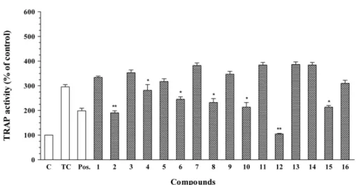
Abstract − In this report, we investigated the antioxidant (peroxyl radical-scavenging and reducing capacities) and anti-osteoporotic activities of extracts and isolated constituents (1 - 16) from the rhizomes of Kaempferia parviflora Wall. ex Baker on pre-osteoclastic RAW 264.7 cells. Compound 5 exhibited significant peroxyl radical- scavenging capacity, with TE value of 8.47 ± 0.52 µM, while compound 13 showed significant reducing capacity, with CUPRAC value of 5.66 ± 0.26 µM, at 10.0 µM. In addition, flavonoid compounds 2, 4, 6, 8, 10, 12, and terpene compound 15 showed significant inhibition of tartrate-resistant acid phosphatase (TRAP) in NF-κB ligand-induced osteoclastic RAW 264.7 cells, with values ranging from 16.97 ± 1.02 to 64.67 ± 2.76%. These results indicated that K. parviflora could be excellent sources for the antioxidant and anti-osteoporotic traditional medicinal plants.

Keywords − Kaempferia parviflora, Zingiberaceae, Antioxidant, Anti-osteoporosis, TRAP

Introduction

Natural antioxidant compounds have numerous beneficial health effects. Previous studies suggested that antioxidants reduce the risk of chronic diseases, including cancer and heart diseases, as well as cardiovascular and cerebrovas- cular events. Several antioxidants have been reported including anthocyanins, flavonoids, phenolic acids, and tanins.

1

These compounds act as radical scavengers, hydrogen donors, electron donors, and metal-chelating agents that can detoxify reactive oxygen species (ROS) and reduce ROS induced damage. Antioxidant activity is generally due to the trapping of free radicals.

2

In addition, antioxidant systems play important roles in the develop- ment of osteoporosis. Evolving evidence suggests that ROS are involved in osteogenesis, including bone formation and resorption, which are associated with the aging process and may lead to osteoporosis. Osteoporosis is a family of disorders in which systemic bone mass is reduced and the

patient is at risk of spontaneous fracture. Bone develop- ment is a normal process that involves the resorption of bone by osteoclasts and the synthesis of bone matrix by osteoblasts.

3

Kaempferia parviflora Wall ex. Baker (Black galingale), known locally in Thai as Kra-Chai-Dam, is an herbaceous plant in the Zingiberaceae. Traditionally, its black to purple rhizomes are used in local food as a flavoring agent and as a traditional medicinal plant for the treatment of a wide spectrum of illnesses. Since ancient times, it has traditionally been used as a health-promoting and vitalizing agent, and a folk medicine to lower blood glucose levels, improve blood flow, and increase vitality.

4

The effects of K. parviflora rhizomes on male sexual function have also been promoted. Several studies have examined the chemical constituents of K. parviflora and their biological activities.

Flavonoids, attached with many methoxyl groups, are demonstrated as the major constituents,

5

along with chalcone derivatives, related phenolic compounds, and kaempferiaosides.

6

Recently, extracts of this plant and compounds isolated from them, including flavonoids have been reported to exert various pharmacological activities, such as inhibition of α-glucosidase,

7

cholinesterase,

8

P- glycoprotein,

9

and NO production,

10

as well as the ability to reduce obesity,

11

inflammatory,

12

allergic,

13

and convul-

*Author for correspondence

Prof. Young Ho Kim, College of Pharmacy, Chungnam National University, Daejeon 305-764, Korea

Tel: +82-42-821-5933; E-mail: [email protected]

Prof. Hae Dong Jang, Department of Food and Nutrition, Hannam University, Daejeon 305-811, Korea

Tel: +82-42-629-8805; E-mail: [email protected]

(2)

sions,

14

and treatment of plasmodial, fungal, and myco- bacterial infections.

4

K. parviflora extracts have also been shown to modulate the function of multidrug resistance associated-proteins,

15

and induce cytotoxicity in various cancer cell lines.

16

In our screening study of medicinal plants on antioxi- dant and anti-osteoporotis activities, the methanolic extract of K. parviflora exhibited significant antioxidant and anti- osteoporosis properties. This study details anti-oxidative evaluation of extracts and constituents isolated from K.

parviflora (1 - 16, see Fig. 1) determined using the oxygen radical absorbance capacity (ORAC) and cupric ion reducing antioxidant capacity (CUPRAC) assays. In addition, the anti-osteoporosis activity of these compounds were evaluated through their inhibitory effects on the osteoclast differen- tiation from RAW 264.7 pre-osteoclast cells.

Experimental

Plant material − The rhizomes of Kaempferia parviflora Wall. Ex Baker were kindly provided from Ok Nam Kim, which was purchased from a local shop, Laos, in 2014, and identified by Prof. Young Ho Kim, College of Pharmacy, Chungnam National University. A voucher specimen (CNU-14110) was deposited at the Herbarium

of the College of Pharmacy, Chungnam National University, Republic of Korea.

Compounds − From the methanolic extracts of the rhizomes of K. parviflora sixteen compounds (1 - 16) were isolated and structurally elucidated. Stock solutions of tested compounds in DMSO were prepared, kept at −20

o

C, and diluted to the final concentration in fresh media before each experiment. For not to affect cell growth, the final DMSO concentration did not exceed 0.5% in all experiments.

Chemicals and reagents − 2,2'-Azobis (2-amidinopro-

pane) dihydrochloride (AAPH), neocuproine, Dulbecco’s

modified Eagle’s medium (DMEM), fetal bovine serum

(FBS), β-glycerophosphate, 3-(4,5-dimethylthiazol-2-yl)-

2,5-diphenyltetrazolium bromide (MTT), Triton X-100,

Hank’s balanced salt solution (HBSS), RANKL, leukocyte

acid phosphatase assay kit, sodium tartrate, p-nitrophenyl-

phosphate (PNPP), phosphate-buffered saline (PBS, pH

7.4), dihydroethidium dihydrorhodamine (DHR), and

dimethylsulfoxide (DMSO) were purchased from Sigma-

Aldrich (St. Louis, MO, USA). RAW 264.7 cells were

obtained from the American Type Culture Collection

(ATCC, Rockville, MD, USA). The natural compounds

tested in this study were isolated from Library of Natural

Products, College of Pharmacy, Chungnam National Uni-

Fig. 1. Chemical structures of compounds 1 - 16 from K. parviflora (Glc: glucosyl).

(3)

versity. Information about their isolation method, chemical structure, and purity provided in references listed in main text. All solvents were used of analytical grade and supplied by SK Chemicals (Ulsan, Korea).

General experimental procedures − Optical rotations were determined on a JASCO P-2000 polarimeter. The UV spectrum was recorded on a JASCO V-630 spectro- photometer. IR spectra were obtained on a Bruker TENSOR 37 FT-IR spectrometer. The NMR spectra were recorded on a JEOL ECA 600 MHz and JEOL JNM-AL 400 MHz spectrometer and TMS was used as an internal standard, chemical shift ( δ) was expressed in ppm with reference to the TMS signals. Column chromatography (CC) was performed on silica gel (Kieselgel 60, 70 - 230 mesh and 230 - 400 mesh, Merck), porous polymer gel (Diaion

®

HP-20, 70 × 180 mm, Mitsubishi Chemical), Sephadex

TM

LH20, octadecyl silica (ODS, Cosmosil 140 C

18

-OPN, Nacalai Tesque), and YMC RP-18 resins (30 - 50 μm, Fuji Silysia Chemical). Thin layer chromatography (TLC) used pre-coated silica gel 60 F

254

(1.05554.0001, Merck) and RP-18 F

254S

plates (1.15685.0001, Merck) and compounds were visualized by spraying with aqueous 10% H

2

SO

4

and heating for 3 - 5 minutes.

Extraction and isolation − The rhizomes of K.

parviflora Wall. ex Baker were dried at room temperature.

The dried material (0.8 kg) was extracted with MeOH using ultrasonic maceration (3 × 2.5 h) at 35

o

C, after which the solutions were filtered, the combined filtrates were concentrated under reduced pressure to give a crude extract. After concentration, the MeOH extract (18.26 g, M) was suspended in H

2

O and then partitioned successively with CH

2

Cl

2

to give CH

2

Cl

2

fraction (10.03 g, C) and water layer (8.20 g, W), respectively. Fraction C was chromatographed over silica gel, eluted with n-hexane in acetone (0 → 100%, stepwise), yielding six fractions (C-1 to C-6). Fraction C-2 (1.85 g) was chromatographed over a silica gel column eluted with n-hexane-acetone (9:1) and further purified by YMC RP-18 CC using MeOH- H

2

O (4:1) as the eluent to afford 5-hydroxy-3,7-dime- thoxyflavone (3, 56.8 mg), 5-hydroxy-3,7,3',4'-tetramethoxy- flavone (11, 52.5 mg), 2-methoxyanthraquinone (13, 4.2 mg), 2-hydroxy-9,10-anthraquinone (14, 5.1 mg), and β- sitosterol (15, 5.8 mg). Next, fraction C-3 (0.9 g) was chromatographed over a silica gel CC eluted with CH

2

Cl

2

- MeOH (17:1) to obtain 5-hydroxy-7-methoxyflavone (1, 27.5 mg), 5-hydroxy-7,4'-dimethoxyflavone (7, 25.6 mg), and 5-hydroxy-3,7,4'-trimethoxyflavone (9, 57.3 mg). Si- milarly, fraction C-4 (1.2 g) was separated by YMC RP- 18 chromatography using acetone-H

2

O (2:1) and Sephadex

TM

LH-20 using CH

2

Cl

2

-MeOH (1:1) as eluent to give 5,7-

dimethoxyflavone (2, 75.5 mg), 3,5,7-trimethoxyflavone (4, 20.9 mg), 5,7,4'-trimethoxyflavone (6, 8.6 mg), and 3,5,7,3',4'-pentamethoxyflavone (10, 55.6 mg). Next, fraction C-5 (1.32 g) was chromatographed on a column of Sephadex

TM

LH-20 and eluted with H

2

O and MeOH, successively, to give three subfractions (C-5.1 to C-5.3).

Subfraction C-5.1 (0.4 g) was separated by YMC RP-18 CC, using MeOH-H

2

O (3:1) as eluent, and further purified by over silica gel CC, eluted with CH

2

Cl

2

-MeOH (10:1), to obtain 4'-hydroxy-5,7-dimethoxyflavone (5, 52.8 mg) and daucosterol (16, 4.5 mg). Finally, subfraction C-5.2 (0.5 g) was separated by CC over silica gel, using CH

2

Cl

2

-MeOH-H

2

O (8:1:0.1) as eluents, and further puri- fied by YMC RP-18 CC, eluted with MeOH-H

2

O (3:1), to give 3,5,7,4'-tetramethoxyflavone (8, 8.6 mg), and 4',5- dihydroxy-3,7,4'-trimethoxyflavone (12, 10.5 mg).

Cupric ion reducing antioxidant capacity (CUPRAC) assay − The electron-donating capacities of each compound to reduce Cu(II) to Cu(I) ions were assessed according to the method of Aruoma et al.

17

Forty microliters of different concentrations of each compound dissolved in ethanol were mixed with 160 μL of a mixture containing 0.5 mM CuCl

2

and 0.75 mM neocuproine; a Cu(I) ions specific chelator, in 10.0 mM phosphate buffer, pH 7.4. Absorbance was measured using a microplate reader at 454 nm for 1 h. Increased absorbance of the reaction mixture indicated greater reducing power.

Oxygen radical absorbance capacity (ORAC) assay − The ORAC assay, which has been employed extensively in previous antioxidant studies,

18

was carried out using a Tecan GENios multifunctional plate reader (Salzburg, Austria) with fluorescent filters (excitation wavelength:

485 nm, emission filter: 535 nm). In the final assay

mixture, fluorescein (40.0 nM) was used as a target of

free radical attack with AAPH (20.0 mM) as a peroxyl

radical generator in the peroxyl radical-scavenging

capacity assay.

18

The analyzer was programmed to record

fluorescein fluorescence every 2 min after AAPH had

been added. All fluorescence measurements were expressed

relative to the initial reading. Final values were calculated

based on the difference in the area under the fluorescence

decay curve between the blank and test samples. All data

were expressed as net protection area (net area). Trolox

(1.0 μM) was used as the positive control to scavenge

peroxyl radicals. It was used as a control standard and

prepared fresh daily. The ORAC value was calculated by

dividing the area under the sample curve by the area

under the Trolox curve, with both areas being corrected

by subtracting the area under the blank curve. One ORAC

unit was assigned as the net area of protection provided

(4)

by Trolox at a final concentration of 1.0 μM. The area under the curve of the sample was compared to the area under the curve for Trolox, and the antioxidative value was expressed in micromoles of TE per liter.

Cell culture − RAW 264.7 [macrophages (pre-osteoclasts) from BALB/c mouse] cells were cultured in 96-well plates (1 × 10

4

cells/mL) containing DMEM supplemented with 10% (v/v) FBS for 2 days. The medium was then replaced with test samples in a differentiation medium containing 50.0 ng/mL RANKL. The differentiation medium was changed every 2 days.

Cell cytotoxicity by MTT assay − RAW 264.7 cells were cultured in 24-well plates (2 × 10

4

cells/mL) con- taining DMEM supplemented with 10% (v/v) FBS and 1% (v/v) antibiotics in a humidied atmosphere of 5% CO

2

at 37

o

C for 5 days, washed with PBS, and pretreated with different concentrations (1.0 - 20.0 μM) of samples to be tested. After 5 days’ incubation, MTT reagent was added to each well, and the plate was incubated at 37

o

C for 1 h.

The medium was removed, and the plate was washed twice with PBS. The intracellular insoluble formazan was dissolved in DMSO. The absorbance of each cell was recorded in DMSO. The absorbance of each cell was then measured at 570 nm using an ELISA (Tecan, Salzburg, Austria) reader, and the percentage proliferation was calculated.

19

TRAP staining − RAW 264.7 cells were seeded in 12- well plates (3 × 10

4

cells/well) containing DMEM medium plus 10% FBS, and the medium was replaced with test samples in differentiation medium containing 50.0 ng/mL RANKL. The differentiation medium was changed every 2 days. After 5 days, the medium was removed, and the cell monolayer was gently washed twice using PBS. The cells were fixed in 3.5% formaldehyde for 10 min and washed with distilled water. The cells were incubated at 37

o

C in a humid and light-protected incubator for 1 h in the reaction mixture of a leukocyte acid phosphatase assay kit (Sigma-Aldrich, St. Louis, MO, USA, Cat. No.

387), as directed by the manufacturer. The cells were washed three times with distilled water, and TRAP- positive multinucleated cells containing three or more nuclei were counted under a light microscope.

20

TRAP activity − After differentiating the RAW 264.7 cells into osteoclasts for 5 days, the medium was removed, and the cell monolayer was gently washed twice using ice-cold PBS. The cells were fixed in 3.5%

formaldehyde for 10 min and ethanol-acetone (1:1) for 1 min. Subsequently, the dried cells were incubated in 50.0 mM citrate buffer (pH 4.5) containing 10.0 mM sodium tartrate and 6.0 mM PNPP. After 1 h incubation,

the reaction mixtures were transferred to new well plates containing an equal volume of 0.1N NaOH. Absorbance was measured at 405 nm using an enzyme-linked immu- noassay reader, and TRAP activity was expressed as the percent of the control.

20

Statistical analysis − Data were presented as the means

± standard deviation (SD) of at least three independent experiments performed in triplicate. Statistical analysis was carried out using the SPSS statistical package (SPSS, Chicago, IL, USA) program, and the significances between the control and experimental groups were analysed using Student’s t-test. Differences were considered to be signi- ficant at

*

P < 0.05 and

**

P < 0.01.

Result and Discussion

The methanolic extract of K. parviflora was suspended in H

2

O and then partitioned sequentially with CH

2

Cl

2

to yield CH

2

Cl

2

fraction and H

2

O layer. All fractions were first evaluated for their antioxidant activity at concen- trations of 5.0 µg/mL and 10.0 µg/mL. The MeOH extract as well as CH

2

Cl

2

fraction showed antioxidant activities (see Table 1). At 10.0 µg/mL, the ORAC and CUPRAC values were 7.85 ± 0.08 and 3.35 ± 0.19 µM fold higher than the protection provide by 1.0 µM of Trolox, respec- tively. In addition, all fractions were also screened for their anti-osteoporosis activity. The MeOH extract and CH

2

Cl

2

fraction (5.0 µg/mL) significantly inhibited with TRAP activities by 51.03 ± 1.37% and 64.07 ± 3.58%, respectively. Therefore, the CH

2

Cl

2

fraction was selected for chemical constituent separation using various chroma- tographic methods, which yielded 16 compounds (1 - 16).

Extracts of the rhizomes of K. parviflora and its

constituents have shown different pharmacological acti-

vities,

4, 8-10, 13-16

although antioxidant activity have been

reported well.

21

The antioxidant activities of the isolated

compounds (1 - 16) were measured using ORAC, which

is commonly used for antioxidant assessment and provides

important information regarding the antioxidant capacity

of biological samples. The results showed that polymethoxy-

flavonoids (1 - 12) exhibited significant antioxidant activities,

with ORAC values ranging from 0.79 ± 0.13 to 8.47 ±

0.52 µM at 10.0 µM and from 0.02 ± 0.06 to 2.23 ±

0.05 µM at 1.0 µM (see Table 1). Among the compounds,

4'-hydroxy-5,7-dimethoxyflavone (5) showed the highest

activity, with an ORAC value of 8.47 ± 0.52 µM, at 10.0

µM. Anthraquinones (13 and 14) exhibited peroxyl radical

scavenging capacities with ORAC values of 3.85 ± 0.22,

7.04 ± 0.33 and 0.79 ± 0.09, 1.12 ± 0.09 at concentrations

of 10.0 and 1.0 µM, respectively. The remaining triterpene

(5)

(15) and triterpene glycoside (16) showed weak antioxi- dant activities with ORAC values of 1.26 ± 0.10, 0.01 ± 0.02 and 0.03 ± 0.09, 0.46 ± 0.02 at 10.0 and 1.0 µM, respectively. These results suggest that the peroxyl radical- scavenging capacities of compounds are dependent on the location and number of hydroxyl and methoxy groups in flavonoids and anthraquinones.

The reducing capacities of compounds (1 - 16) were evaluated by the amount of Cu(I) ions reduced with neocuproine, which interacts specically with Cu(I) ions.

Reducing capacity appears to be associated with electron transfer, which was a well-established mechanism contri- buting to antioxidant capacity. At concentration of 10.0 µM, 2-methoxyanthraquinone (13) showed significant reducing capacities, with CUPRAC value of 5.66 ± 0.26 µM. Other compounds (1 - 12 and 14 - 16) showed weak or inactive effects at the tested concentrations. The results were not sufficient for discussion regarding the structure–

activity relationship of polymethoxyflavonoid and/or other components. However, more studies may be required for the understanding of their selective potential antioxidant activities.

To date, the anti-osteoporosis effects of extracts and/or compounds isolated from K. parviflora have not been

reported. Therefore, we assayed the anti-osteoporosis activity of a methanolic extract and dichloromethane fraction on RAW 264.7 cells. The anti-osteoporotic activities of compounds 1 - 16 were evaluated based on the suppression of excessive bone breakdown by osteoclasts. The results showed that flavonoid derivatives 2, 4, 6, 8, 10, 12, and tecpene 15 suppressed osteoclast formation in a dose- dependent manner with TRAP values ranging from 16.97 ± 1.02 to 65.67 ± 2.76 (% of control) at concen- tration of 10.0 µM (see Fig. 2). Among them, compounds 2 and 12 showed the most significant when compared to daidzein used as a positive control, with values of 40.58 ± 2.28% and 65.67 ± 2.76%, respectively. However, these results could be regarded as inhibitory effects of compounds on cell viability. To address this, the com- pounds (5.0 to 20.0 µM) were tested for their cytotoxic activity on RAW 264.7 macrophage cells during a five- day differentiation period. However, no significant cytotoxic effects were observed (data not shown). Thus, these results suggest that 5,7-dimethoxyflavone (2) and 4',5- dihydroxy-3,7,4'-trimethoxyflavone (12) possess anti- osteoclastogenic activities, but do not affect cell viability.

When compared both anti-osteoporotic and antioxidant activities of 1 - 16, the significant both TRAP and peroxyl Table 1. Antioxidant activities of the extracts and isolated compounds (1 - 16) from K. parviflora

Comps. Peroxyl radical-scavenging capacity (TE, µM) Reducing capacity [Copper(I) ions, µM]

1.0 µM 10.0 µM 1.0 µM 10.0 µM

1 0.21 ± 0.06 0.94 ± 0.12 0.50 ± 0.17 0.84 ± 0.36

2 ND 1.26 ± 0.10 ND 0.08 ± 0.06

3 1.01 ± 0.04 3.59 ± 0.16 ND 0.80 ± 0.11

4 0.08 ± 0.03 1.11 ± 0.14 0.29 ± 0.30 0.00 ± 0.17

5 2.23 ± 0.05 8.47 ± 0.52 ND 0.17 ± 0.11

6 0.71 ± 0.03 1.51 ± 0.05 ND 0.17 ± 0.11

7 0.05 ± 0.03 1.01 ± 0.04 0.17 ± 0.11 0.17 ± 0.00

8 0.50 ± 0.03 1.01 ± 0.04 0.17 ± 0.11 0.34 ± 0.17

9 0.02 ± 0.06 0.95 ± 0.11 0.34 ± 0.06 0.04 ± 0.11

10 0.17 ± 0.01 0.79 ± 0.13 ND 0.00 ± 0.86

11 0.44 ± 0.14 2.66 ± 0.16 0.29 ± 0.11 1.05 ± 0.11

12 0.27 ± 0.04 1.70 ± 0.17 0.08 ± 0.06 0.00 ± 0.17

13 0.79 ± 0.09 3.85 ± 0.22 0.63 ± 0.06 5.66 ± 0.26

14 1.12 ± 0.09 7.04 ± 0.33 0.29 ± 0.11 1.05 ± 0.11

15 0.03 ± 0.09 1.26 ± 0.10 0.29 ± 0.11 0.34 ±0.06

16 0.46 ± 0.02 0.01 ± 0.02 ND ND

CH

2

Cl

2

fraction 1.43 ± 0.10 7.85 ± 0.08 0.67 ± 0.17 3.35 ± 0.19

MeOH extract 1.50 ± 0.02 9.11 ± 0.18 0.80 ± 0.13 4.53 ± 0.06

All data are expressed as the mean ± SD of three individual experiments.

Values are expressed as µM of TE. ND: Not determined.

One ORAC unit is equivalent to the net protection area provided by 1.0 µM of Trolox.

(6)

radical scavenging activities were observed in 16 isolated compounds. In addition, there is no correlation between TRAP activity and antioxidant of tested 16 compounds.

In other hand, the significant anti-osteoporotic activities of flavonoid derivatives 2, 4, 6, 8, 10, and 12 not showing strong antioxidant activities in vitro may be attributed to their indirect antioxidant activities by activating the endogenous antioxidant defense system. Therefore, more research may be needed to determine whether the signifi- cant anti-osteoporotic activites compound 2, 4, 6, 8, 10, and 12 result from indirect antioxidant activity.

As regards its previous studies, the range of the biological activities of the major flavonoid constituents of K. parviflora rhizomes has not yet been fully elucidated.

5,7-Dimethoxyflavone (2) was found to have anti-inflam- matory activity.

12

5-Hydroxy-7-methoxyflavone (1), 3,5,7- trimethoxyflavone (4), 5,7,4'-trimethoxyflavone (6), 5- hydroxy-3,7,4'-trimethoxyflavone (9), and 3,5,7,3',4'-pen- tamethoxyflavone (10) were found to protect against D- galactosamine-induced cytotoxicity in primary cultured mouse hepatocytes.

6

3,5,7,4'-Tetramethoxyflavone (8) and 3,5,7,3',4'-pentamethoxyflavone (10) have an effect similar to that of nobiletin, which strongly promotes the differen- tiation of 3T3-L1 preadipocytes into mature adipocytes via transcriptional activation of peroxisome proliferator- activated receptor γ (PPARγ), yet lacks ligand-binding activity.

22

The active constituents of the anti-allergic activity of K. parviflora have been identified as 5-hydroxy- 7-methoxyflavone (1), 5-hydroxy-7,4'-dimethoxyflavone (7), and 5-hydroxy-3,7,3',4'-tetramethoxyflavone (11).

13

In addition, 3,5,7,3',4'-pentamethoxyflavone (10) inhibits P-

glycoprotein function.

9

These recent reports concur with our findings suggesting that flavonoid derivatives are the major constituents that contribute to biological effects of K. parviflora Wall. ex Baker.

Acknowledgments

This work was supported by research fund of Chungnam National University.

References

(1) Hu, F. B.; Willett, W. C. J. Am. Med. Assoc. 2002, 288, 2569-2578.

(2) Pietta, P. G. J. Nat. Prod. 2000, 63, 1035-1042.

(3) Kong, Y. Y.; Yoshida, H.; Sarosi, I.; Tan, H. L.; Timms, E.;

Capparelli, C.; Morony, S.; Oliveira-dos-Santos, A. J.; Van, G.; Itie, A.;

Khoo, W.; Wakeham, A.; Dunstan, C. R.; Lacey, D. L.; Mak, T. W.;

Boyle, W. J.; Penninger, J. M. Nature 1999, 397, 315-323.

(4) Yenjai, C.; Prasanphen, K.; Daodeeb, S.; Wongpanich, V.; Kittakoop, P. Fitoterapia 2004, 75, 89-92.

(5) Sutthanut, K.; Sripanidkulchai, B.; Yenjai, C.; Jay, M. J. Chromatogr.

A 2007, 1143, 227-233.

(6) Chaipech, S.; Morikawa, T.; Ninomiya, K.; Yoshikawa, M.;

Pongpiriyadacha, Y.; Hayakawa, T.; Muraoka, O. Chem. Pharm. Bull.

2012, 60, 62-69.

(7) Azuma, T.; Kayano, S. I.; Matsumura, Y.; Konishi, Y.; Tanaka, Y.;

Kikuzaki, H. Food Chem. 2011, 125, 471-475.

(8) Sawasdee, P.; Sabphon, C.; Sitthiwongwanit, D.; Kokpol, U.

Phytother. Res. 2009, 23, 1792-1794.

(9) Patanasethanont, D.; Nagai, J.; Yumoto, R.; Murakami, T.;

Sutthanut, K.; Sripanidkulchai, B. O.; Yenjai, C.; Takano, M. J. Pharma.

Sci. 2007, 96, 223-233.

(10) Wattanapitayakul, S. K.; Suwatronnakorn, M.; Chularojmontri, L.;

Herunsalee, A.; Niumsakul, S.; Charuchongkolwongse, S.; Chansuvanich, N. J. Ethnopharmacol. 2007, 110, 559-562.

(11) Akase, T.; Shimada, T.; Terabayashi, S.; Ikeya, Y.; Sanada, H.;

Fig. 2. Inhibitory effects of compounds 1 - 16 on TRAP activity in RANKL-induced osteoclastic RAW 264.7 cells. TRAP activity was

measured from cultures after 5 days of treatment with RANKL and test compounds (10.0 µM). The treated control was obtained from

RAW 264.7 cells cultured with RANKL stimulation and without test compounds. Data are expressed as percentages of the treated control

(mean ± SD, n = 3,

*

P < 0.05 and

**

P < 0.01 vs TC. C: control, which was not treated; TC: treated control, which was treated with

RANKL). Daidzein (10.0 µM) was use as a positive control (Pos.).

(7)

Aburada, M. J. Nat. Med. 2011, 65, 73-80.

(12) Wong, C. S.; Tansakul, P.; Tewtrakul, S. J. Ethnopharmacol. 2009, 124, 576-580.

(13) Tewtrakul, S.; Subhadhirasakul, S.; Kummee, S. J. Ethnopharmacol.

2008, 116, 191-193.

(14) Wattanapitayakul, S. K.; Chularojmontri, L.; Herunsalee, A.;

Charuchongkolwongse, S.; Chansuvanich, N. Fitoterapia 2008, 79, 214- 216.

(15) Patanasethanont, D.; Nagai, J.; Matsuura, C.; Fukui, K.; Sutthanut, K.; Sripanidkulchai, B.O.; Yumoto, R.; Takano, M. Eur. J. Pharmacol.

2007, 566, 67-74.

(16) Yenjai, C.; Wanich, S. Bioorg. Med. Chem. Lett. 2010, 20, 2821- 2823.

(17) Aruoma, O. I.; Deiana, M.; Jenner, A.; Halliwell, B.; Kaur, H.;

Banni, S.; Corongiu, F. P.; Dessí, M. A.; Aeschbach, R. J. Agric. Food Chem. 1998, 46, 5181-5187.

(18) Kurihara, H.; Fukami, H.; Asami, S.; Toyoda, Y.; Nakai, M.;

Shibata, H.; Yao, X. S. Biol. Pharm. Bull. 2004, 27, 1093-1098.

(19) van de Loosdrecht, A. A.; Beelen, R. H. J.; Ossenkoppele, G. J.;

Broekhoven, M. G.; Langenhuijsen, M. M. A. C. J. Immunol. Methods 1994, 174, 311-320.

(20) Lee, S. H.; Ding, Y.; Yan, X. T.; Kim, Y. H.; Jang, H. D. J. Nat.

Prod. 2013, 76, 615-620.

(21) Vichitphan, S.; Vichitphan, K.; Sirikhansaeng, P. KMITL Sci. Tech.

J. 2007, 7, 97-105.

(22) Horikawa, T.; Shimada, T.; Okabe, Y.; Kinoshita, K.; Koyama, K.;

Miyamoto, K. I.; Ichinose, K.; Takahashi, K.; Aburada, M. Biol. Pharm.

Bull. 2012, 35, 686-692.

Received June 10, 2015

Revised July 20, 2015

Accepted July 22, 2015

수치

Fig. 2. Inhibitory effects of compounds 1 - 16 on TRAP activity in RANKL-induced osteoclastic RAW 264.7 cells

참조

관련 문서

To confirm the antioxidative effects of the extracts, free radical scavenging activities (1,1-diphenyl-2-picrylhydrazyl), total antioxidant capacity by

High levels of ABTS radical [12.1 mg Trolox equivalent antioxidant capacity (TEAC)/g residue] and DPPH radical (4.4 mg TEAC/g residue) scavenging activity and reducing power (A

In order to measured antioxidant activities, we measured total phenol and flavonoids contents, DPPH free radical scavenging activity and reducing

Therefore, the antioxidant activities of COSs were investigated in DPPH radical scavenging assays and superoxide anion and xanthine oxidase inhibition studies... Biological

DPPH, reducing power, ferric reducing antioxidant power (FRAP), and ABTS radical scavenging activ- ity decreased in this order: 70% methanol, 70% ethanol, EA, CM, n-butanol

After that, we determined anthocyanin, total phenol, flavonoid content, DPPH radical scavenging activity, ABTS radical scavenging activity, ferric reducing antioxidant

Correlation between total antioxidant capacities measured by the DPPH and ABTS radical scavenging abilities for Schizandra fruit extracts by microwave-assisted extraction..

In the superoxide radical scavenging activities of ginseng and cultivated wild ginseng extracts, antioxidant activities of cultivated wild ginseng extracts was showed