Ectopic Pancreas Presenting as a Duodenal Obstructing Mass
4
0
0
전체 글
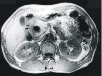
(2) 66. 대한외과학회지:제 68 권 제 1 호 2005. ꠏꠏꠏꠏꠏꠏꠏꠏꠏꠏꠏꠏꠏꠏꠏꠏꠏꠏꠏꠏꠏꠏꠏꠏꠏꠏꠏꠏꠏꠏꠏꠏꠏꠏꠏꠏꠏꠏꠏꠏꠏꠏꠏꠏꠏꠏꠏꠏꠏꠏꠏꠏꠏꠏꠏꠏꠏꠏꠏꠏꠏꠏꠏꠏꠏꠏꠏꠏꠏꠏꠏꠏꠏꠏꠏꠏꠏꠏꠏꠏꠏꠏꠏꠏꠏꠏꠏꠏꠏꠏꠏꠏꠏꠏꠏꠏꠏꠏꠏꠏꠏꠏꠏꠏꠏꠏꠏꠏꠏꠏꠏꠏꠏꠏꠏ. Fig. 1. Abdominal CT scan demonstrate a solid mass (arrows) in the medial wall of second portion of the duodenum. Fig. 3. Duodunoscopic findings shows a submucosal mass (arrows) at the second portion of the duodenum.. Fig. 2. Axial T1-weighted MRI shows a high signal intensified mass (arrows) in the medial wall of the duodenum.. 보였다(Fig. 3). 내시경적 역행성 담췌관 조영술에서는 십이 지장 종괴로 인해서 원위부 총담관이 압박을 받아 좁아진 소견을 보였으나 췌담관은 정상 소견을 보였다. 수술 소견: 십이지장의 폐쇄성 종괴 진단 하에 개복술을 시행 받았다. 수술 소견상 십이지장 하행 부위에 약 4×4× 3 cm 크기의 단단한 종괴가 만져졌으며, 췌장 두부와 원위 부 총담관에 밀접하게 유착되어 있었다. 췌-십이지장 절제 술이 시행되었다. 조직 병리학적 소견: 절제된 표본은 십이지장 하행 부위 에서 발생한 점막하층의 종괴였으며, 절단면상 고형의 흰 색 종괴 양상이었다(Fig. 4). 현미경적 검사상 십이지장의 근육층에 포상세포와 관으로 구성된 췌장 조직이 관찰되었 으며, 췌장은 만성 췌장염 소견을 보였다(Fig. 5). 술 후 경과: 환자는 수술 후 14일째 퇴원하였다.. Fig. 4. Surgical specimen shows a submucosal solid mass (arrows) at the second portion of the duodenum, with whitish color in cross-section.. 고. 찰. 이소성 췌장은 상부 위장관 내시경, 수술 또는 부검시 드 물지 않게 발견되는 질환이다. 부검시 이소성 췌장의 빈도 는 1∼2% 정도로 알려져 있으며,(1-4) Barbosa 등(5)은 개복 술을 시행 받은 환자의 0.5 %에서 발견된다고 보고하였다. 이소성 췌장이 발생하는 흔한 장소로는 위(25∼38.2%), 십 이지장(17∼36.3%), 공장(15∼21.7%)이다. 또한 이소성 췌 장은 식도, 폐, 종격동, 담낭, 총담관, 담낭관, 바터 팽대부, 비장, 장간막, 림프절 등에서 발견된다.(1) 현미경적으로 포 상세포와 관을 가진 이소성 췌장 소엽은 대부분 점막하층 에 위치하고 있고, 근육섬유 사이에 침윤하고 있으며, 흔히.
(3) 윤현철 외:십이지장의 폐쇄성 종괴로 발현된 이소성 췌장. 67. ꠏꠏꠏꠏꠏꠏꠏꠏꠏꠏꠏꠏꠏꠏꠏꠏꠏꠏꠏꠏꠏꠏꠏꠏꠏꠏꠏꠏꠏꠏꠏꠏꠏꠏꠏꠏꠏꠏꠏꠏꠏꠏꠏꠏꠏꠏꠏꠏꠏꠏꠏꠏꠏꠏꠏꠏꠏꠏꠏꠏꠏꠏꠏꠏꠏꠏꠏꠏꠏꠏꠏꠏꠏꠏꠏꠏꠏꠏꠏꠏꠏꠏꠏꠏꠏꠏꠏꠏꠏꠏꠏꠏꠏꠏꠏꠏꠏꠏꠏꠏꠏꠏꠏꠏꠏꠏꠏꠏꠏꠏꠏꠏꠏꠏꠏ. A. B. Fig. 5. (A) Microscopic findings shows pancreatic tissues(arrows) at the muscluar layer of the duodenum (H&E stain, ×20), (B) with acinar cells and ductal structure (H&E stain, ×200).. 근육층의 과도 증식과 동반된다.(6) 일반적으로, 이소성 췌 장을 가진 대부분의 환자들은 증상이 없거나, 복통, 상복부 불편감, 오심, 구토, 출혈 등 비특이적인 증상을 보인다.(7,8) 이소성 췌장의 특이 증상은 종괴 효과나 잠재된 병리에 의 해서 나타날 수 있다.(1) 종괴 효과에 의한 증상의 발현은 이소성 췌장의 위치에 따라 다른데. 소장에서 발생한 이소 성 췌장에 의한 장중첩은 가장 흔한 증상이며, 다른 특이한 임상 증상으로는 폐쇄성 황달, 유문 폐쇄 등이 있다.(9,10) 이소성 췌장의 잠재된 병리는 췌장 자체에서 생길 수 있는 모든 변화가 발생할 수 있다고 알려져 있다.(1,10) 이소성 췌장의 외분비선에서 발생한 췌장암, 낭종, 췌장염, 농양 등 도 보고되었다.(11,12) 또한 이소성 췌장의 내분비선에서 발 생한 인슐린 종양, 가스트린 종양 그리고 말단비대증을 동 반한 성장 호르몬 분비 종양 같은 소도세포종양의 발생도 보고되었다.(13-15) 방사선적, 내시경적 그리고 생검 시술의 발달에도 불구하고 이소성 췌장의 진단은 어렵다고 알려져 있다.(7) 과거에는 이소성 췌장의 술 전 진단은 위나 십이지 장의 근위부에 점막 외부의 부드러운, 중심부 함몰을 가진 단일 결함 소견 같은 방사선적 소견으로 이루어졌다. 현재 는 대부분 내시경으로 진단이 되는데, 이를 이용한 내시경 하 초음파검사나 세침 흡입검사는 점막하 병변이 발견되었 을 경우 이의 감별 진단에 도움이 된다.(16) 이소성 췌장의 치료에 대해서는 아직도 논란이 되고 있다. Barbosa 등(5)은 41예에서 61%가 이소성 췌장 자체에 의한 증상을 가졌다고 보고하였다. 그러나 Dolan 등(3)은 수술적 치료와 비슷하게 내과적 치료로 증상이 조절되기 때문에 복부 불편감의 대 부분이 이소성 췌장에서 발생하지 않았다고 결론지었다. Jochimsen 등(17)은 위궤양, 위식도 역류 또는 담도 질환 같 은 동반 질환을 배제한 후에도 지속되는 증상이 있는 이소 성 췌장은 외과적 절제를 권유하였다. Tanaka 등(18)은 수술 시 우연히 발견된 이소성 췌장은 후에 증상을 유발할 수. 있기 때문에 절제를 권유하고 있다. 증상이 없는 환자는 병 변이 양성이 확실하면 관찰하고, 악성과 구별이 명확하지 않다면 절제를 시행한다. 또한 증상이 있을 경우에 다른 원 인이 배제되면 절제가 권유되고 있다.(19) Barbosa 등(5)은 이소성 췌장의 조직이 췌장 자체보다 악성의 경향이 강하 다고 주장하였으나, 이소성 췌장은 일반적으로 양성이므로 국소절제술만으로 충분하다고 알려져있다.(17-19). REFERENCES 1) MaKoto K, Takashi Y, Toshio Y. Ectopic pancreas in the stomach presenting as an inflammatory Abdominal mass. The American Journal of Gastroenterology 1989;84:663-6. 2) Caberwal D, Kogan SJ, Levitt SB. Ectopic pancreas presenting as an umbilical mass. Journal of Pediatric Surgery 1977;12: 593-5. 3) Dolan RV, ReMine WH, Dockerty MB. The fate of heterotropic pancreatic tissue: A study of 212 cases. Arch Surgery 1974;109:762-5. 4) Thoeni RF, Gedgaudas RK. Ectopic pancreas: Usual and unusual features. Gastrointestinal Radiology 1980;5:37-42. 5) Barbosa J de C, Dockerty MB, Waugh JM. Pancreatic heterotopia: Review of the literature and report of 41 authenticated surgical case, of which 25 were clinically significant. Surg Gynecol Obstet 1946;82:527-42. 6) Hayes-Jordan A, Idowu O, Cohen R. Ectopic pancreas as the cause of gastric outlet obstruction in a newborn. Pediatric Radiology 1998;28:868-70. 7) Chung JP, Lee SI, Kim KH, Chi HS, Jeong HJ, Moon YM, et al. Duodenal ectopic pancreas complicated bychronic pancreatitis and pseudocyst formation. J Korean Med Sci 1994;9: 351-6. 8) Wang CK, Kuo YT, Yeung KW. CT appearance of ectopic.
(4) 68. 대한외과학회지:제 68 권 제 1 호 2005. ꠏꠏꠏꠏꠏꠏꠏꠏꠏꠏꠏꠏꠏꠏꠏꠏꠏꠏꠏꠏꠏꠏꠏꠏꠏꠏꠏꠏꠏꠏꠏꠏꠏꠏꠏꠏꠏꠏꠏꠏꠏꠏꠏꠏꠏꠏꠏꠏꠏꠏꠏꠏꠏꠏꠏꠏꠏꠏꠏꠏꠏꠏꠏꠏꠏꠏꠏꠏꠏꠏꠏꠏꠏꠏꠏꠏꠏꠏꠏꠏꠏꠏꠏꠏꠏꠏꠏꠏꠏꠏꠏꠏꠏꠏꠏꠏꠏꠏꠏꠏꠏꠏꠏꠏꠏꠏꠏꠏꠏꠏꠏꠏꠏꠏꠏ pancreas: a case report Abdominal imaging 1998;23:332-3. 9) Carleton CC, Ackerbaum R. Intussusception secondary to aberrant pancreas in a child. JAMA 1976;236:1047. 10) Anseline P, Grundfest S, Carey W, Weiss R :Pancreatic heterotropia:A rare cause of bowel obstruction. Surgery 1981;90: 110-3. 11) Qizilbash AH. Acute pancreatitis occurring in heterotropic pancreatic tissue in the gallbladder. Can J Surg 1976;19:413-4. 12) Dutkiewics Z, pogodski E. Tumor of the stomach due to a heterotropy of pancreatic tissue. Pol Przegl Chir 1972;44: 127-9. 13) Rose C, Kassaram RA, Lind JF. Ectopic gastric pancreas: A review and report of 4 cases. Diagn Imaging 1980;49:214-8. 14) Barrocas A, Fontenelle LJ, Williams MJ. Gastric heterotropic pancreas. A case report and review of literature. Am Surg 1973;5:361-5.. 15) Melmed S, Ezrin C, Kovasc K, Goodman RS, Frohm LA. Acromegaly due to secretion of growth hormone by an ectopic pancreatic islet cell tumor. N Engl J Med 1985;312:9-17. 16) Matsui M, Goto H, Niwa Y, Arisawa T, Hirooka Y, Hayakawa T. Preliminary results of fine needle aspiration biopsy histology in upper gastrointestinal tumors. Endoscopy 1998;30: 750-5. 17) Jochimsen PR, Shirazi SS, Lewis JW. Symptomatic ectopic pancreas relieved by surgical excision. Surg Gynecol Obstet 1981;153:49-52. 18) Tanaka K, Tsunoda T, Eto T, Yamada M, Jajima Y, Shimogama H, et al. Diagnosis and management of heterotopic pancreas. Int Surg 1998;78:32-5. 19) Chen CH, Yang CC, Yeh YH, Chou DA, Kuo CL. Ectopic pancreas located in the major duodenal papillav: case report and review. Gastrointest Endosc 2001;53:121-3..
(5)
수치
관련 문서
Acute mediastinitis secondary to delayed vascular injury by a central venous catheter and total parenteral nutrition.. Gyeong-Jo Byeon 1 , Eun-Jung Kim 2 , Ji-Young Yoon 2 ,
Here, we present a rare case of schwannoma on the scalp with a review of magnetic resonance imaging (MRI) findings, which was initially misdiagnosed as an epidermal cyst or
Basal Cell Carcinoma Presenting as a Perianal Ulcer and Treated with Radiotherapy.. Hyun Soo Lee, Sue
Oral Hairy Leukoplakia Which Occurred as a Presenting Sign of Acute Myeloid Leukemia in a Child.. Hyun-Ho Cho, M.D., Su-Han Kim, M.D., Sang-Hee Seo, M.D., Do-Sang Jung,
A Case of Acute Tubulointerstitial Nephritis Associated with Rifampin Therapy Presenting as Fanconi-like Syndrome.. Jun Tae Park, Sik Lee, Won Kim, Sung Kwang Park, and Kyung
Sunyoung Kang 1 , Seung Shin Park 1 , Jae Hyun Bae 1 , Kyu Eun Lee 2 , Jung Hee Kim 1 , Chan Soo Shin 1 Departments of 1 Internal Medicine, 2 General Surgery, Seoul
Delta Neutrophil Index as a Prognostic Marker of Early Mortality in Gram Negative Bacteremia.. Hye Won Kim * , Ji Hyun Yoon * , Sung Joon Jin, Sun Bean Kim, Nam Su Ku, Su Jin Jeong,
