154
Copyrightⓒ2013 Journal of the Korean Society of Magnetic Resonance in Medicine Ectopic pancreas is defined as pancreatic tissuelacking anatomic and vascular continuity with the main body of the gland (1). Pancreatic fatty infiltra- tion-replacement is commonly diffuse involvement, most frequently seen in elderly or obese patients, and may be associated with diseases such as chronic pancreatitis and cystic fibrosis (2). Pathogenesis of fat replacement (also termed lipomatosis, adipose atrophy, or fat infiltration) of the exocrine pancreas is not well established. In our case, peripheral steatosis was detected in ectopic pancreas, and mimicked as exogastric mass. The radiographic features of this condition have not been described, to the author’s knowledge, based on a computerized search of the literature. The clinical importance of this case is to demonstrate that this finding may not be misdiagnosed
as exogastric mass and highly suggesting feature for ectopic pancreas.
A 38-year-old man was admitted to our institute for evaluation of a known gastric mass. He was diagnosed it on endoscopy 2 years ago, and suffered from unintentional weight loss of 2 kg for 2 months. He had adult-onset diabetes mellitus for 3 years, which was controlled by medication. He had no history of hypertension, alcohol abuse, smoking and previous surgery. Physical examination was unremarkable.
Laboratory examination revealed elevations in serum glucose 350 mg/dL (normal reference: 70-100 mg/dL), cholesterol 238 (normal reference: 238 mg/dL), and alanine transaminase (ALT) 55 IU/L (normal reference: 4-43 IU/L). Blood cell count, hemoglobin, total bilirubin, serum lipase, aspartate aminotrans- ferase (AST), alkaline phosphatase (ALP), creatinine, and blood urea nitrogen (BUN) levels were within normal range.
Contrast-enhanced computed tomography (CT) demonstrated 2.5 cm sized, well-defined mass with peripheral fatty infiltration on the anterior wall of the
CASE REPORT INTRODUCTION
�Received; March 14, 2013�Revised; May 13, 2013
�Accepted; May 13, 2013
Corresponding author : Mi Young Kim, M.D.
Department of Radiology, Inha University Hospital, 7-206, 3-ga, Shinheung-dong, Jung-gu, Incheon 400-711, Korea.
Tel. 82-32-890-2769, Fax. 82-32-890-2743 E-mail : [email protected]
Perilesional Steatosis in Ectopic Pancreas Mimicking Exogastric Mass : A Case Report
Mi Yeon Nam1, Mi Young Kim1, Yeo Ju Kim1, Chang Hae Suh1, Suk Jin Choi2, Jae Sung Cho3
1Department of Radiology, Inha University Hospital, Incheon, Korea
2Department of Pathology, Inha University Hospital, Incheon, Korea
3Biology, New York University, USA
We report an unusual case of ectopic pancreas that appeared on radiologic images as a lobulated, submucosal mass enclosed by fat component in the gastric lower body. Although, ectopic pancreas including fat component is extremely rare, in the setting of gastric submucosal mass with containing perilesional fat, these findings should be considered in ectopic pancreas as part of the differential diagnosis.
Index words : Ectopic pancreas∙Submucosal tumor∙Stomach∙Magnetic resonance image
www.ksmrm.org JKSMRM 17(2) : 154-157, 2013
pISSN 1226-9751 / eISSN 2288-3800 http://dx.doi.org/10.13104/jksmrm.2013.17.2.154
Case Report
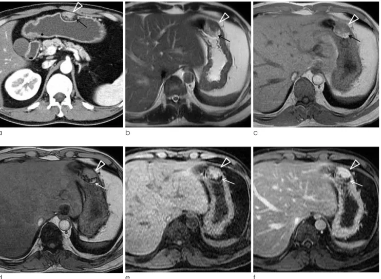
gastric lower body (Fig. 1a). The main pancreas showed normal contour and attenuation. MR images (Signa HDxt 1.5T, GE Healthcare, Milwaukee, WI) revealed a well-defined submucosal mass with smooth margins, enclosed by fat component. T2-weighted image (single shot fast spin echo sequence, TR/TE 1500/92.4 ms, flip angle 90。) demonstrated that high signal intensity margin enclose iso-signal intensity contour mass with pancreas (Fig. 1b). On T1-weighted 2D dual gradient echo (TR/TE 135/4.3 ms, flip angle 60。), inphased image (Fig. 1c) showed that hyperin- tense fatty infiltraion enclose iso intense lobulated
mass, and opposed phase (Fig. 1d) showed signal drop on margin of fatty lesion. Non-enhanced and contrast enhanced (Omniscan, GE Healthcare) fat supressed T1-weighted images (TR/TE 4.7/2.1 ms, flip angle 12。) using the liver acquisition with volume acceleration (LAVA) sequence revealed enhancing ectopic pancreas and peripheral non-enhancing suppressed fatty component (Fig. 1e, f).
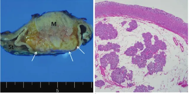
The patient underwent a gastric wedge resection including the tumor. The tumor was 5.5×3.0×3.0 cm in size, covered by gastric mucosa, arising from the muscular layer and containing pancreatic tissue with
http://dx.doi.org/10.13104/jksmrm.2013.17.2.154 http://www.ksmrm.org
Perilesional Steatosis in Ectopic Pancreas Mimicking Exogastric Mass � Mi Yeon Nam, et al.
155
a b c
d e f
Fig. 1. 38-year-old man with ectopic pancreas in gastric lower body.
a. Axial CT scan shows a well-defined ovoid endoluminal submucosal mass (arrowhead) and perilesional fat replacement (arrow) in anterior wall of gastric lower body.
b. Axial T2-weighted MR image shows isointense mass (arrowhead) and periperhal hyperintense fatty tissue (arrow).
c, d. T1-weighted 2D dual gradient echo inphased image shows that hyperintense fatty infiltration enclose iso intense (arrowhead) lobulated mass with pancreas, as well as signal drop on opposed phase (arrow).
e, f. Axial fat suppressed T1-weighted MR image and contrast-enhanced T1-weighted MR image show hyperintense mass (arrowhead) with highly enhancement and peripheral saturated hypointense fatty contents (arrow).
perilesional fat tissue (Fig. 1g). Histologic examination demonstrated ectopic pancreas (Fig. 1h). The postop- erative course was uneventful, and he was discharged on postoperative 10 days.
Ectopic pancreas is a common developmental anomaly with a reported incidence of 0.55-14% at autopsy (1, 3, 4). They consist histologically of all pancreatic elements, including acini, islet cells, and ductal structures (2, 5, 6). Fatty replacement of pancreas may be uniform or may be unevenly distrib- uted, and Langerhans are usually not affected by fatty replacement (4). No etiology has been established for fat replacement of the pancreas; however, several predisposing factors have been suggested: age, obesity, diabetes mellitus, chronic pancreatitis, hepatic disease, dietary deficiency, viral infection, steroid therapy, obstruction of the pancreatic duct, and fibrocystic disease (7, 8). Experimental and clinical studies have shown that fat replacement of the pancreas does result from ligation of the pancreatic duct or obstruction of the pancreatic duct by a tumor or a calculus.
We postulate that in our case, obesity and diabetes
mellitus were significant etiologic factors responsible (7, 8). Hepatic disease, chronic pancreatitis and adult- onset diabetes mellitus may have been contributory (7, 8). Although the pathophysiology was not well known, uniform fatty replacement of pancreas is the most frequent CT finding in adolescent and adult patients with cystic fibrosis, and pancreatic glandular tissue is significantly reduced in size (5, 7). Fatty lesion of pancreas is also detected in cases of lipomatous pseudohypertrophy, lipoma, liposarcoma, fibrolipoma, and teratoma (4). Endoscopy may be performed to differentiate an ectopic pancreas from neoplastic lesions. Ectopic pancreas can be developed into complications such as pancreatitis, pseudocyst, insuli- noma, adenoma, and malignant transformation (1, 3).
These complications cause clinical symptoms such as abdominal pain, gastrointestinal bleeding, and obstruc- tion (1, 3). Surgical resection is required for sympto- matic patients with ectopic pancreas that has atypical radiographic features.
Heterotopic pancreas in the stomach may easily be misinterpreted as other gastric submucosal tumor.
According to previous study (3), CT findings were interpreted correctly as heterotopic pancreas in only two (17%) cases, and the remaining 10 (83%) cases were misinterpreted as leiomyoma in four, as carcinoid DISCUSSION
156
JKSMRM 17(2) : 154-157, 2013http://www.ksmrm.org http://dx.doi.org/10.13104/jksmrm.2013.17.2.154
g h
Fig. 1. g. Gross cut surface of the specimen shows a well-demarcated submucosal mass (M) surrounded by adipose tissue (arrows) and deeply burried within the muscularis propria.
h. On microscopic finding, the heterotopic pancreas lobules are composed of pancreatic acini and ducts within the muscular bundles and surrounded by adipose tissue (H & E stain, original magnification ×12.5)
http://dx.doi.org/10.13104/jksmrm.2013.17.2.154 http://www.ksmrm.org Perilesional Steatosis in Ectopic Pancreas Mimicking Exogastric Mass � Mi Yeon Nam, et al.
157
tumor in three, as submucosal scirrhous carcinoma in two, and as lymphadenopathy in one (3). Although focal fatty infiltration of gastric submucosal tumor can be helpful for diagnosis of ectopic pancreas, the finding of perilesional steatosis can be detected in other tumors including lipoma, liposarcoma, angiolipoma, teratoma (9).
Prominent enhancement of the overlying mucosa, location, LD/SD ratio, growth pattern, and lesion border are statistically significant predictors in the differentiation of ectopic pancreas from GIST and leiomyoma (1). Glomus tumors appear as smooth submucosal masses with or without ulceration and may contain tiny flecks of calcification (10). These tumors frequently demonstrate strong enhancement on early-phase contrast enhanced images (10). Gastric schwannomas usually appear as discrete submucosal masses that are indistinguishable from other mesenchymal tumors (10). As they outgrow their blood supply, these lesions may undergo central necrosis and ulceration (11).
In conclusion, we report an unusual case of ectopic pancreas that appeared on radiologic images as a lobulated, submucosal mass enclosed by fat component in the gastric lower body. Although ectopic pancreas including fat component is extremely rare, in the setting of gastric submucosal mass with containing focal fat, these findings should be considered in
ectopic pancreas as part of the differential diagnosis.
References
1. Kim JY, Lee JM, Kim KW, et al. Ectopic pancreas: CT findings with emphasis on differentiation from small gastrointestinal stromal tumor and leiomyoma. Radiology 2009;252:292 2. Cho JS, Shin KS, Kwon ST, et al. Heterotopic pancreas in the
stomach: CT findings. Radiology 2000;217:139-144
3. Katz DS, Hines J, Math KR, Nardi PM, Mindelzun RE, Lane MJ.
Using CT to reveal fat-containing abnormalities of the pancreas.
AJR Am J Roentgenol 1999;172:393-396
4. Low G, Panu A, Millo N, Leen E. Multimodality imaging of neoplastic and nonneoplastic solid lesions of the pancreas.
Radiographics 2011;31:993-1015
5. Soyer P, Spelle L, Pelage JP, et al. Cystic fibrosis in adolescents and adults: fatty replacement of the pancreas--CT evaluation and functional correlation. Radiology 1999;210:611-615 6. Matsumoto S, Mori H, Miyake H, et al. Uneven fatty replace-
ment of the pancreas: evaluation with CT. Radiology 1995;194:453-458
7. Kawamoto K, Yamada Y, Utsunomiya T, et al. Gastrointestinal submucosal tumors: evaluation with endoscopic US. Radiology 1997;205:733
8. Kilman WJ, Berk RN. The spectrum of radiographic features of aberrant pancreatic rests involving the stomach. Radiology 1977;123:291-296
9. Ferrozzi F, Tognini G, Marchesi G, Spaggiari E, Pavone P.
Gastric tumors with fat components. CT findings and differen- tial diagnosis. Radiol Med 2000;100:343-347
10. Park SH, Han JK, Kim TK, et al. Unusual gastric tumors:
radiologic-pathologic correlation. Radiographics 1999;19:1435- 1446
11. Patel S, Bellon EM, Haaga J, Park CH. Fat replacement of the exocrine pancreas. AJR Am J Roentgenol 1980;135:843-845
통신저자 : 김미영, (400-711) 인천시 중구 신흥동 3가 7-206, 인하대학교병원 영상의학과
Tel. (032) 890-2769 Fax. (032) 890-2743 E-mail: [email protected]
주변부 지방침윤으로 인해 외위장 종괴로 보이는 이소성 췌장: 증례 보고
1인하대학교병원 영상의학과
2인하대학교병원 병리과
3뉴욕대학교 생물학과
남미연1∙김미영1∙김여주1∙서창해1∙최석진2∙조재성3
저자들은 위의 하부 체부에 지방 성분으로 둘러싸여 있는 분엽성 형태의 점막하 종괴로 나타난 이소성 췌장의 1예 를 경험하였다. 지방 성분을 포함한 이소성 췌장은 매우 드물지만, 이 증례를 바탕으로 병변 주위 지방 침윤을 보이는 위 점막하 종괴가 있을 때 이소성 췌장의 가능성을 감별해야 하므로 이를 보고하는 바이다.
대한자기공명의과학회지 17:154-157(2013)