© 2016 The Korean Ophthalmological Society
This is an Open Access article distributed under the terms of the Creative Commons Attribution Non-Commercial License (http://creativecommons.org/licenses /by-nc/3.0/) which permits unrestricted non-commercial use, distribution, and reproduction in any medium, provided the original work is properly cited.
Original Article
Patients complained of repeated epiphora need to evalu- ate nasolacrimal drainage system because the common-
est cause of epiphora is nasolacrimal duct obstruction (NLDO). The location of obstruction or stenosis can be precisely located if there are obvious causes of obstruction like previous trauma, infection, inflammatory diseases such as sarcoidosis and Wegener’s granulomatosis, or ma- lignancy [1,2]. However, most cases of NLDO are associat- ed with unknown origins, so many recent studies used computed tomography (CT) imaging to evaluate periocular pathology and nasolacrimal drainage system [3-5]. They recommend CT as a diagnostic tool for NLDO when cause of obstruction is uncertain [6-8].
Preoperative Computed Tomography Findings for Patients with Nasolacrimal Duct Obstruction or Stenosis
Seong Chan Choi, Saem Lee, Hye Sun Choi, Jae Woo Jang, Sung Joo Kim, Jung Hye Lee
Myunggok Eye Research Institute, Department of Ophthalmology, Kim’s Eye Hospital, Konyang University College of Medicine, Seoul, Korea
Purpose: To identify and analyze the role of preoperative computed tomography (CT) in patients with tearing symptoms with nasolacrimal duct obstruction (NLDO).
Methods: We retrospectively reviewed the medical records and CT results on 218 patients who complained of tearing symptoms with NLDO between January 2014 and December 2014. All patients were recruited from Kim’s Eye Hospital's outpatient clinic and assessed by clinical history, examination, and CT to evaluate perio- cular pathology and nasolacrimal drainage system. Patients with abnormal findings assessed by preoperative CT were further reviewed.
Results: CT was performed on 218 patients (average age, 58.2 ± 11.9 years). Of these, 196 (89.9%) had endo- nasal dacryocystorhinostomy, 14 (6.4%) declined surgery, and 8 (3.7%) were inoperable due to abnormal CT findings. Soft tissue opacity was the most common finding which 243 cases (85.9%) of 283 obstructed naso- lacrimal duct and 89 cases (81.7%) of 109 non-obstructed nasolacrimal duct showed it. Thirty-nine (17.8%) of 218 patients showed either maxillary sinusitis or ethmoidal sinusitis and 32 (14.7%) of 218 patients presented with periocular inflammation. Other abnormal CT findings included septal deviations, previous fractures, mass- es, and structural abnormalities of nasal cavity.
Conclusions: Preoperative CT imaging is useful in the assessment of both nasolacrimal drainage and nearby anatomical structures. This information will be helpful in planning surgical interventions and management of NLDO.
Key Words: Endonasal dacryocystorhinostomy, Nasolacrimal duct obstruction, Preoperative computed tomography
Received: July 31, 2015 Accepted: November 3, 2015
Corresponding Author: Jung Hye Lee, MD. Myunggok Eye Research Institute, Department of Ophthalmology, Kim’s Eye Hospital, Konyang University College of Medicine, #136 Yeongsin-ro, Yeongdeungpo-gu, Seoul 07301, Korea. Tel: 82-2-1577-2639, Fax: 82-2-2671-6359, E-mail:
This study was presented at the 113th annual meeting of Korean Oph- thalmological Society on April 12, 2014 in Gwangju, Korea.
Treatment of NLDO is mainly dependent on surgical managements which include external or endonasal dacryo- cystorhinostomy (DCR), balloon catheter dilatation, and silicone intubation. Endonasal DCR became a widely used standard procedure performed for NLDO because of ad- vancements in local anesthesia, and the vastly improved visibility in the operative field provided by endoscopy. A facial incision which can hamper the pumping action of orbicularis oculi muscle is not required in endonasal DCR and it has shorter recovery period than external DCR [9].
We believe that thorough clinical assessments, including taking history of any nasal or sinus disorders, doing regur- gitation test, syringing of lacrimal sac, and contrast da- cryocystography (DCG), NLDO can be evaluated enough.
Also, additional preoperative nasal endoscopy may ensure the appropriate lacrimal operation. So, CT is usually not recommended if there is certainty of patient’s diagnosis.
Although CT is not routine evaluation for watery eyes, drainage-limiting factors such as bony abnormalities, mu- cosal edema, retention of secretions, and obstructive mass- es are readily identified on CT which makes it a good diag- nostic tool. For these reason, otolaryngologists who undertake functional endoscopic sinus surgery almost rou- tinely obtain a CT scan before undertaking surgery in or- der to evaluate the exact bony structures and the mucous membranes of patients. There are many published reports of abnormalities in lacrimal sacs, orbital walls, and sinus- es, which could affect the final results of endonasal DCR [7,10-13]. Even though the incidence of abnormal anatomic findings is rare, knowing the exact state of nearby struc- tures of the lacrimal drainage system can reduce surprises during the surgery and it is useful to ensure proper treat- ments to the patients.
Still there has been no recent studies about usefulness of preoperative CT on patients with NLDO since Francis et al. [6] reported on the value of CT imaging in patients with symptoms of lacrimal drainage obstruction. Therefore, we evaluated the role of preoperational CT in patients with NLDO.
Materials and Methods
We retrospectively reviewed the medical records and CT results on 218 patients who complained of tearing symp- toms with NLDO who were planned to undergo DCR sur-
gery between January 2014 and December 2014. All pa- tients were recruited from Kim’s Eye Hospital’s outpatient clinic, and diagnosis of NLDO was made clinically by do- ing conventional probing, syringing and confirmed by DCG with contrast. DCG was done in all patients and they were examined of puncta, ocular surface, tear film and CT to rule out any eyelid malposition, disease of the eyelid margin, bony or soft tissue abnormalities. A history of sin- onasal disease, previous sinus or facial surgery, allergic rhinitis and trauma were obtained. Patients were routinely followed up at 1 week, 1 month, 3 months, 6 months, and 12 months postoperatively. The patency of nasolacrimal duct (NLD) was assessed by doing syringing of lacrimal passage on every follow ups. No additional procedures were performed during this period.
A total of 218 patients underwent supine axial imaging using a Siemens Somatom Spirit dual-slice CT scanner (Siemens, Munich, Germany) with images obtained at 1.0- mm intervals. All patients’ CT images were interpreted by radiologist and reviewed by three masked observers for presence of air within the nasolacrimal drainage system and other abnormalities around orbit. The NLD was classi- fied as full opacity, partial opacity, and no opacity. The term “no opacity” was used if air in the lacrimal sac and NLD was seen in continuity on contiguous slices.
We analyzed patient demographics and radiologic char- acteristics on CT scans, including the abnormalities of or- bit, sinus and other facial structures. Patients with abnor- mal findings assessed by preoperative CT were prescribed management and treatment according to each abnormality.
All patients with abnormal findings assessed by preopera- tive CT were further reviewed.
We also reviewed the differences of CT imaging be- tween eyes which found to have obstruction (which DCR were performed) and eyes which showed no obstruction (which are the contralateral eyes of patients who undergo unilateral DCR). Statistical analyses were performed with the commercially available software package SPSS ver.
12.0 (SPSS Inc., Chicago, IL, USA). Differences in values between eyes with NLDO and eyes without NLDO were analyzed using linear by linear association. A p-value less than 0.05 was considered significant.
Results
CT was performed on 218 patients (average age, 58.2 ± 11.9 years; range, 14 to 83 years). There were 52 men (23.9%) and 166 women (76.1%). Of these 218 patients, 196 (89.9%) had endonasal DCR, 14 (6.4%) declined surgery, and 8 (3.7%) were inoperable due to abnormal CT findings.
For the 196 patients who received endonasal DCR, 54 (27.6%) had right side DCR, 55 (28.1%) had left side DCR, and 87 (44.4%) had bilateral DCR. Patient demographics are summarized in Table 1.
Endonasal DCR was performed on 283 NLDs of 196 pa- tients. Postoperative success was evaluated by syringing and examination of rhinostomy site which was good pas- sage with patent opening. A total 184 (93.9%) of 196 pa- tients were fully satisfied. In 9 cases (4.6%), minimal block was seen with clear fluid regurgitation and to some extent their symptom was relieved. However, three patients (1.5%) with mucoid regurgitation or complete block were not re- lieved of their symptoms and needed further intervention.
The most common abnormal CT finding was soft tissue opacity, which showed no air and it reflects mucosal thick- ening whining the NLD and retention of secretions (Fig.
1A-1D). CT scans of 196 individuals who underwent DCR were reviewed in the coronal and axial plane by three in- dependent reviewers for the soft tissue opacity. Because not all the patients underwent bilateral DCR, 392 NLDs include 283 obstructed NLDs (87 bilateral : 54 left : 55 right) which underwent DCR and 109 non-obstructed NLDs (54 left : 55 right) which didn’t undergo DCR. Two hundred forty-three (85.9%) of 283 obstructed NLDs showed full opacity, 19 (6.7%) NLDs showed partial opaci- ty, and 21 (7.4%) NLDs showed no opacity. Eighty-nine
(81.7%) of 109 non-obstructed NLDs showed full opacity, 10 (9.2%) NLDs showed partial opacity, and 10 (9.2%) NLDs showed no opacity (Table 2). Patients with tearing problems showed higher incidence of soft tissue opacity in NLDs but there were no statistical differences between two groups (p = 0.362).
The second most common abnormal CT finding was chronic rhinosinusitis (CRS), which included maxillary si- nusitis and ethmoidal sinusitis. There were 39 (17.8%) of 218 patients whose CT results showed either maxillary si- nusitis or ethmoidal sinusitis. Of these, 31 (14.2%) showed maxillary sinusitis, 18 (8.3%) showed ethmoidal sinusitis, and 10 (4.6%) showed both. When patients with rhinosi- nusitis underwent surgery, they routinely received preop- erative and postoperative antibiotics. They were checked more often at our clinic after endonasal DCR.
We observed 32 cases (14.7%) of periocular inflamma- tion, which included dacryocystitis and periorbital celluli- tis without lacrimal sac inflammation; 23 patients (10.6%) had dacryocystitis, and 9 (4.1%) had periorbital cellulitis without lacrimal sac inflammation. Other abnormal CT findings were septal deviations, previous fractures, mass- es, and structural abnormalities of nasal cavity. Twenty-six cases (11.9%) had septal deviation and were referred to otolaryngologists before undertaking surgery. Of these 26 patients, 18 had severe deviation which required septoplas- ty before DCR and eight had mild deviation which under- went DCR without septoplasty. In 16 cases (7.3%), we Table 1. Clinical characteristics of patients who complained
of tearing symptoms and received computed tomography
Characteristics Value
No. of patients 218
Mean age (yr) 58.2 ± 11.9
Male : female 52 : 166
No. of patients who underwent DCR 196
Side (OD : OS : OU) 54 : 55 : 87
No. of patients who declined surgery 22 DCR = dacryocystorhinostomy; OD = right eye; OS = left eye;
OU = both eyes.
Fig. 1. (A) Computed tomography (CT) of a patient with a nor- mal nasolacrimal duct. In the axial plane CT image, air fills the nasolacrimal duct, which is the passage for tears (arrows). (B) The CT image in the coronal plane also shows empty nasolacrimal ducts (arrows). (C,D) Axial plane and coronal plane of the CT im- ages show the nasolacrimal duct obstructed by soft tissue on both sides (arrows).
A B
C D
found previous fractures; 15 patients had orbital wall frac- tures and one patient had a nasal bone fracture. All of the fractures were old events and had no effect on proceeding to surgical intervention for these patients. In seven pa- tients, masses were found on CT; five (2.3%) had paranasal sinus masses, one had a lid mass, and one had a zygoma mass.
Nasal abnormalities were found in four cases. They were two cases of mucocele, one of nasal polyp, and one of trau- ma-induced nasal deformity. Details of the abnormal CT findings and number of patients are summarized in Table 3.
Two cases of 196 DCRs developed acute rhinosinusitis within the first 5 days following DCR and both had a past history of CRS that had been asymptomatic at the time of surgery. They were treated with a 1 to 3 week course of oxymetazoline hydrochloride nasal spray and oral ofloxa- cin three times daily.
Case 1
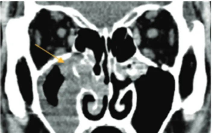
One patient came to our clinic complaining of epiphora and had a history of nasal trauma at young age. When ex- amining his nose, his right nostril was completely ob- structed by mucosa, and thus could not be examined fur- ther. His CT showed membranous obstruction of his right nostril (Fig. 2), which required ear, nose, and throat sur- gery to open the obstructed nasal cavity before performing DCR.
Case 2
One patient with a tearing problem for 2 to 3 months came to our clinic. Syringing revealed no abnormalities.
Since his DCG showed irregular passages of dye, we rec- ommended a CT examination. His CT results revealed that his right maxillary sinus and ethmoid soft tissue was filled with irregular bony disruptions, and he was referred to a
tertiary hospital for assessment of possible inverted papil- loma (Fig. 3A and 3B).
Case 3
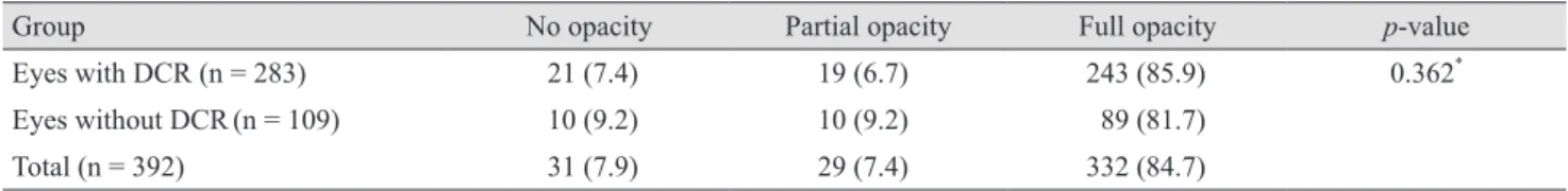
We assessed a 58-year-old male patient who presented with epiphora, anosmia, eyelid swelling, and pain in both Table 2. Overall presence of soft tissue opacity in the nasolacrimal drainage system
Group No opacity Partial opacity Full opacity p-value
Eyes with DCR (n = 283) 21 (7.4) 19 (6.7) 243 (85.9) 0.362*
Eyes without DCR(n = 109) 10 (9.2) 10 (9.2) 89 (81.7)
Total (n = 392) 31 (7.9) 29 (7.4) 332 (84.7)
Values are presented as number (%).
DCR = dacryocystorhinostomy.
*Linear by linear association.
Table 3. Summary of major computed tomography findings Computed tomography findings No. of patients (%) Asymptomatic chronic rhinosinusitis* 39 (17.8)
Maxillary sinus opacity 31 (14.2)
Ethmoid sinus opacity 18 (8.3)
Periocular inflammation 32 (14.7)
Dacryocystitis 23 (10.6)
Cellulitis without lacrimal sac
inflammation 9 (4.1)
Septal deviation 26 (11.9)
Severe† 18 (8.3)
Mild 8 (3.7)
Previous fracture 16 (7.3)
Orbital wall 15 (6.9)
Nasal bone 1 (0.5)
Mass 8 (3.7)
Paranasal sinus 6 (2.8)
Lid 1 (0.5)
Zygoma 1 (0.5)
Nasal abnormality 4 (1.8)
Mucocele 2 (1.0)
Nasal deformity 1 (0.5)
Nasal polyp 1 (0.5)
Total no. of patient with abnormal findings 125 (57.3) Total no. of patient with computed
tomography images 218 (100)
*Ten patients have both maxillary and ethmoidal sinus opacity;
†Severe septal deviationindicated a need for septoplasty.
eyes. He had no relevant medical history, and syringing re- vealed no abnormalities. However, an irregularly margin- ated soft tissue mass was found in his ethmoid. This mass extended to the medial walls of the orbit, the sphenoid si- nus, and the intra-cranial area on both sides. The radiolo- gist suspected neuroblastoma of the olfactory nerves.
Thus, if we had not performed preoperative CT, we may have missed the mass until we encountered it during endo- nasal DCR (Fig. 4A and 4B).
Case 4
A 47-year-old male patient with epiphora, swelling, and pain in right eye. The CT image in the coronal plane shows severe right ethmoiditis and maxillary sinusitis (Fig. 5).
Discussion
Many studies have used CT to evaluate nasolacrimal drainage system anatomy and it’s relation with drainage, because CT imaging is useful in evaluation of periocular pathology with detailed imaging in short scan time [3-6].
We performed CT on the 218 patients who complained of tearing symptoms; 125 abnormal findings (57.3%) were de- tected some of which led to changes in the management and treatment plans. Considering the studies with more than 50 cases, success rate varies from 77% to 99% [14]. In our study, success rate of endonasal DCR was 93.9%
which is higher than many previous studies. Even though advancement of surgical methods and surgical instruments has increased success rate of endonasal DCR, there can be many intraoperative and postoperative complications. We suppose that preoperative imaging examination can pro- vide information in assessing and preventing such compli- Fig. 2. A 70-year-old female patient with epiphora in both eyes.
She had a previous history of nasal trauma. When examining her nose, we found it was totally obstructed by nasal mucosa, and we could not examine the inner structure of nose. The coronal plane of the computed tomography image shows nasal mucosal adhe- sions in her right nose (arrow).
Fig. 3. (A) A 61-year-old male patient with epiphora in his right eye, and swelling and pain in his right medial canthal area for 2 months. The axial plane computed tomography image shows that the right maxillary sinus and the ethmoid are filled with soft tis
sue, with a bulging contour (arrow). (B) The computed tomogra- phy image in the coronal plane shows irregular filling of the right maxillary sinus and the ethmoid with irregular bony disruption (arrow).
Fig. 4. (A) A 58-year-old male patient with epiphora in both eyes, and swelling, anosmia, and pain in his right eye. The axial plane com puted tomography image shows irregular marginated evi- dence of the soft tissue mass ethmoids (arrow). (B) The coronal plane comput ed tomography image shows a soft tissue mass ex- tending to the medial wall, the sphenoid sinus, and the intracrani- al area (arrow).
Fig. 5. A 47-year-old male patient with epiphora, swelling, and pain in right eye. The computed tomography image in the coronal plane shows severe right ethmoditis and maxillary sinusitis (ar- row).
A
A
B
B
cations.
To the best of our knowledge, no previous study has evaluated the detailed characteristics of nasolacrimal drainage system within a population of individuals previ- ously diagnosed with NLDO. In this study, many eyes with or without epiphora showed opacity in NLD and there were no difference in the incidence of opacity be- tween patient with or without NLDO (p = 0.362). Previous studies reported that opaque NLDs were detected in large population which suggested that opaque NLD could be normal finding [5,15]. Loftus et al. [5] also mentioned that statistically there was no relationship between NLD opaci- fication and ipsilateral sinus disease. Loftus et al. [5] re- ported 72% of NLD opacity and Czyz et al. [4] reported 70% of NLD opacity in normal population. In our study, 81.8% of non-obstructed NLD patients showed NLD opac- ity and 85.8% of obstructed NLD patients showed NLD opacity which was slightly higher than previous studies.
Czyz et al. [4] reported that air was found to be present more fully in the upright-position group as compared with the supine-position group. High rates of opacity in this study may be due to the position of patients in performing CT. Further research is needed on this matter.
In our study, seven patients (3.2%) showed masses in the CT images and had to change treatments. Previously, there were many reports of masses found around orbit and nasal sinuses [1,7,10-13]. Lee et al. [7] reported a case of lacrimal sac tumor discovered in patients with persistent epiphora following DCR and mentioned usefulness of using CT which made it possible to diagnosis relatively early, and were able to obtain rapid, satisfactory treatment. In addi- tion, Jung et al. [11] reported oncocytic carcinoma in pa- tients with NLDO. A tumor originating from the lacrimal sac is rare, but half of the cases of lacrimal sac tumors are malignant, with a high mortality rate, indicating a need for precaution [16]. The most common presenting signs and symptoms of lacrimal sac neoplasms in adults are epiphora (53%), recurrent dacryocystitis (38%), and/or lacrimal sac mass (36%), which are similar to the symptoms seen in pa- tients with NLDO [6]. Further, tumors originating from around the orbits and paranasal sinuses can cause epipho- ra. Because recent DCRs are performed using sinus endo- scopes, it is more difficult to find tumors in these cases than when conventional DCRs are performed. Thus, knowing the exact state of nearby structures of the lacri- mal drainage system is important and CT is useful to rule
out secondary cause of NLDO and planning for surgical intervention.
Since endonasal DCR is widely used, it is important to examine the nasal cavity in order to inspect the turbinates, and to exclude nasal pathologic characteristics that could adversely affect the surgical outcomes. In addition, having enough space for surgery is important to obtain good sur- gical outcomes. For that reason, the 26 patients who showed septal deviation were referred to ear, nose, and throat specialists, and 18 patients who showed severe sep- tal deviation underwent septoplasty before DCR. The eight patients with mild septal deviation underwent DCR with- out septoplasty.
Preoperative inflammation is an important factor affect- ing surgical outcomes and complication rates. Shams and Selva [17] mentioned that CRS may be a risk factor for the development of postoperative acute rhinosinusitis. Our study had 71 patients with presurgical inflammation, 39 with CRS and 32 with periocular inflammation, including periocular cellulitis and dacryocystitis. Three patients were referred to a tertiary hospital because of severe inflamma- tion that required urgent systemic antibiotics, or because they had concurrent systemic disease (Fig. 5). Other pa- tients, with only mild inflammation, did undergo surgery, but received preoperative and postoperative antibiotics.
Patients with pus in lacrimal sac were further examined with bacterial and fungus culture. Also patients with in- flammation were checked more often at outpatient clinic after endonasal DCR. In our study, acute postoperative rhinosinusitis was found in two patients (6.9%) who had a history of CRS but no symptom at the time of surgery; this occurrence rate was slightly higher than that reported by Shams and Selva [17]. Considering all cases of DCR, oc- currence rate was 2.0% and it is similar to the previous re- ports [18,19]. CT of the paranasal sinuses should be ob- tained in evaluating patients with CRS or recurrent acute rhinosinusitis because it offers an objective method for monitoring recurrent or chronic disease [20,21].
By detecting the aforementioned abnormalities using CT, we could provide proper care and management for our patients. The patients with abnormal CT finding received treatments tailored to their needs. In addition, 37 patients needed consultation for other departments, and subse- quently had radical changes in both management and treatment.
The cause of acquired lacrimal drainage obstruction
may be primary or secondary. Primary acquired NLDO results from inflammation of unknown origin, but second- ary acquired NLDO may results from a wide variety of causes like infection, trauma, inflammation, and neoplasm.
CT as a diagnostic tool offers many advantages. It allows surgeons to more effectively and safely plan surgical treat- ment, especially in the presence of fractures, tumors, nasal sinus abnormalities, or other inflammations near the lacri- mal drainage system. Although more proximal obstruc- tions may be difficult to view, CT is regarded as a safe, ef- ficient means to assist in the diagnosis of paranasal sinus pathology. Moreover, it provides patients with detailed di- agnoses and prognoses, and helps them make informed treatment decisions. Finally, it can help determine if there are other, treatable causes of patients’ NLDOs. Due to pre- operative CT, we could prevent surprises and complica- tions which could happen during or after DCR. Also by using prophylactic antibiotics and frequent follow-up of patients with inflammation which were detected by CT, there were only two of 218 outbreaks of acute postopera- tive rhinosinusitis.
Thus, preoperative CT imaging is a useful assessment tool of nasolacrimal drainage and nearby anatomical struc- tures especially if patients have previous history of CRS or suspicious of secondary NLDO. This information will be helpful in creating plans of NLDO treatment and manage- ment.
Conflict of Interest
No potential conflict of interest relevant to this article was reported.
References
1. Dalgleish R. Idiopathic acquired lacrimal drainage obstruc- tion. Br J Ophthalmol 1967;51:463-8.
2. Linberg JV, McCormick SA. Primary acquired nasolacri- mal duct obstruction: a clinicopathologic report and biopsy technique. Ophthalmology 1986;93:1055-63.
3. Czyz CN, Bacon TS, Stacey AW, et al. Nasolacrimal sys- tem aeration on computed tomographic imaging: sex and age variation. Ophthal Plast Reconstr Surg 2016;32:11-6.
4. Czyz CN, Bacon TS, Stacey AW, et al. Nasolacrimal sys-
tem aeration on computed tomographic imaging: effects of patient positioning and scan orientation. Clin Ophthalmol 2015;9:469-73.
5. Loftus WK, Kew J, Metreweli C. Nasolacrimal duct opaci- ty on CT. Br J Radiol 1996;69:630-1.
6. Francis IC, Kappagoda MB, Cole IE, et al. Computed to- mography of the lacrimal drainage system: retrospective study of 107 cases of dacryostenosis. Ophthal Plast Recon- str Surg 1999;15:217-26.
7. Lee KH, Han SH, Yoon JS. Case reports of lacrimal sac tu- mors discovered in patients with persistent epiphora fol- lowing dacryocystorhinostomy. Korean J Ophthalmol 2015;29:66-7.
8. Nagi KS, Meyer DR. Utilization patterns for diagnostic imaging in the evaluation of epiphora due to lacrimal ob- struction: a national survey. Ophthal Plast Reconstr Surg 2010;26:168-71.
9. Ibrahim HA, Batterbury M, Banhegyi G, McGalliard J.
Endonasal laser dacryocystorhinostomy and external da- cryocystorhinostomy outcome profile in a general ophthal- mic service unit: a comparative retrospective study. Oph- thalmic Surg Lasers 2001;32:220-7.
10. Elner VM, Burnstine MA, Goodman ML, Dortzbach RK.
Inverted papillomas that invade the orbit. Arch Ophthalmol 1995;113:1178-83.
11. Jung JH, Shin DH, Cho KS, Choi HY. Nasolacrimal duct obstruction caused by oncocytic carcinoma. Korean J Ophthalmol 2013;27:126-9.
12. Roth SI, August CZ, Lissner GS, O’Grady RB. Hemangio- pericytoma of the lacrimal sac. Ophthalmology 1991;98:
925-7.
13. Stefanyszyn MA, Hidayat AA, Pe’er JJ, Flanagan JC. Lac- rimal sac tumors. Ophthal Plast Reconstr Surg 1994;10:169- 84.
14. Woog JJ, Kennedy RH, Custer PL, et al. Endonasal dacryo- cystorhinostomy: a report by the American Academy of Ophthalmology. Ophthalmology 2001;108:2369-77.
15. Russell EJ, Czervionke L, Huckman M, et al. CT of the in- feromedial orbit and the lacrimal drainage apparatus: nor- mal and pathologic anatomy. AJR Am J Roentgenol 1985;
145:1147-54.
16. Merkonidis C, Brewis C, Yung M, Nussbaumer M. Is rou- tine biopsy of the lacrimal sac wall indicated at dacryo- cystorhinostomy? A prospective study and literature re- view. Br J Ophthalmol 2005;89:1589-91.
17. Shams PN, Selva D. Acute post-operative rhinosinusitis
following endonasal dacryocystorhinostomy. Eye (Lond) 2013;27:1130-6.
18. Fayet B, Racy E, Assouline M. Complications of standard- ized endonasal dacryocystorhinostomy with unciformecto- my. Ophthalmology 2004;111:837-45.
19. Leong SC, Macewen CJ, White PS. A systematic review of outcomes after dacryocystorhinostomy in adults. Am J Rhinol Allergy 2010;24:81-90.
20. Kenny TJ, Duncavage J, Bracikowski J, et al. Prospective analysis of sinus symptoms and correlation with paranasal computed tomography scan. Otolaryngol Head Neck Surg 2001;125:40-3.
21. Bhattacharyya T, Piccirillo J, Wippold FJ 2nd. Relationship between patient-based descriptions of sinusitis and parana- sal sinus computed tomographic findings. Arch Otolaryn- gol Head Neck Surg 1997;123:1189-92.