Penile leiomyosarcoma is a very rare disease and clini- cally and pathologically classified into the superficial and deep type (1). To our knowledge, the imaging find- ings of penile leiomyosarcoma are rarely reported in the existing radiological English literature. We report the ul- trasonographic and CT findings of a deep type of a pe- nile leiomyosarcoma that helped characterize a penile mass and review the published literature.
Case Report
A 47-year-old male presented at our hospital with a palpable penile mass, which was present for the last 10 years. The patient’s medical history was not remarkable with symptoms limited to pain during palpation, which
developed one week prior. Upon physical examination a non-mobile hard mass was palpated on the distal shaft of the penis (Fig. 1).
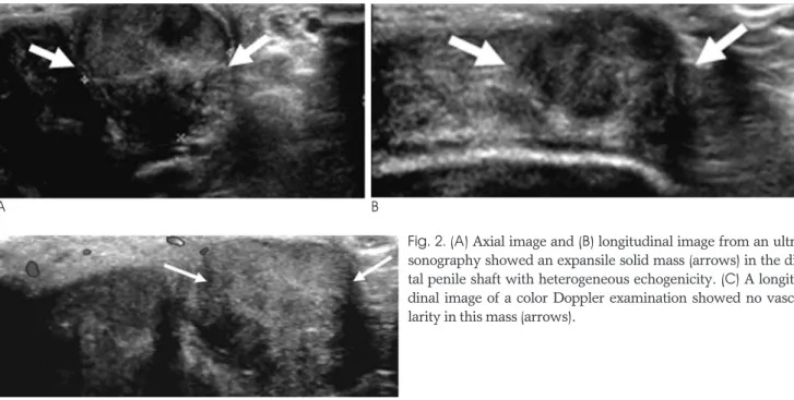
An ultrasonographic examination showed an expan- sile and heterogeneous hypoechoic solid mass measur- ing 1.9 1.9 2.5 cm involving the distal shaft of the left side of the penis (Figs. 2A, B). The corpus spongio- sum and urethra were not involved, and the overlying skin appeared normal. A color Doppler study examina- tion revealed no vascularity in this mass (Fig. 2C).
Subsequent CT imaging showed an expansile, soft tissue mass on the left side of distal penile shaft. This mass showed slightly thick, peripheral rim enhancement on the contrast-enhanced CT images, with an internal ho- mogeneous low density, compared to the adjacent nor- mal shaft of the penis (Fig. 3). The enlarged lymph node was not visualized in either the inguinal areas or pelvic cavity on contrast-enhanced pelvic CT images without distant metastasis.
According to the physical examination and radiologic findings, the penile mass was determined to be a benign lesion, in which the patient underwent a local excision
J Korean Soc Radiol 2009;61:405-408
─ 405 ─
Imaging Findings of Sonography and Computed Tomography for a Penile Leiomyosarcoma: A Case Report1
Jin Chung, M.D., Jae-Joon Chung, M.D., Jeong-Sik Yu, M.D., Joo Hee Kim, M.D.
1Department of Radiology, Gangnam Severance Hospital, Yonsei University College of Medicine, Seoul, Korea
Received January 28, 2009 ; Accepted July 25, 2009
Address reprint requests to : Jae-Joon Chung, M.D., Department of Radiology and Research Institute of Radiological Science, Yonsei University College of Medicine, Gangnam Severance Hospital, 146- 92 Dogok-dong, Gangnam-gu, Seoul 135-720, Korea.
Tel. 82-2-2019-3512 Fax. 82-2-3462-5472 E-mail: [email protected]
We report the ultrasonographic and computed tomography (CT) findings of a deep type of penile leiomyosarcoma that helped characterize a penile mass along with a re- view of the published literature. Leiomyosarcoma of the penis is a very rare disease characterized by a lobulated, expansile, soft tissue mass in CT images, with peripheral rim enhancement and internal homogeneous low density. The ultrasonographic find- ings revealed a lobulated and heterogeneously hypoechoic solid mass at the distal tip of the penis.
Index words :Leiomyosarcoma Penis
Ultrasonography
Tomography, X-Ray Computed
for. A gross pathologic examination of the specimen showed a lobulated, rubbery, solid mass involving the left corpora cavernosa (Fig. 4). The overlying skin was grossly normal. However, a resection revealed a homo- geneously white and rubbery to firm irregular mass.
Histologically, this mass was identified as a grade 2 leiomyosarcoma with a 2.7 cm maximum diameter, 15 mitoses /10 high power fields, and about 20-30% tumor necrosis. The immunohistochemical stain was found to be positive for smooth muscle actin (SMA), focal posi- tive for desmin, and negative for S-100. The excision was not completed due to the presence of tumor cells in the resection margins.
The patient was referred to the department of radia- tion oncology for radiotherapy. However, he wanted to
change hospital for his radiotherapy treatment. As a re- sult, the postoperative course was not available.
Discussion
Penile cancer is a rare neoplasm and accounts for ap- proximately 0.4% of all male malignancies in the United States (2, 3). Penile neoplasms generally fall into 2 cate- gories: epithelial and soft tissue tumors. Skin-based su- perficial lesions constitute the vast majority and repre- sent various types of benign and malignant epithelial neoplasms such as condylomata acuminate and squa-
Jin Chung, et al: Imaging Findings of Sonography and Computed Tomography for a Penile Leiomyosarcoma
─ 406 ─
A B
C
Fig. 2. (A) Axial image and (B) longitudinal image from an ultra- sonography showed an expansile solid mass (arrows) in the dis- tal penile shaft with heterogeneous echogenicity. (C) A longitu- dinal image of a color Doppler examination showed no vascu- larity in this mass (arrows).
Fig. 3. Contrast-enhanced axial CT image reveals an expansile contour and a relatively thick, peripheral rim enhancing lesion (arrow) with an internal, homogeneous, low density, com- pared with the adjacent normal shaft of the penis in the distal shaft of left side of penis.
Fig. 1. Appearance of penis at presentation. A non-mobile hard mass (arrows) was palpated on the distal shaft of left penis.
mous cell carcinoma. Soft tissue penile tumors tend to be deeply seated and centered in the shaft. These repre- sent the benign and malignant counterparts of tumors with a vascular, neural, and smooth muscle derivation (4).
The most common primary malignant neoplasm of the penis is squamous cell carcinoma, constituting more than 95% of cases, followed by metastatic neoplasms of the prostate, bladder, rectum, kidney, and testis, as well as those spreading by direct extension from the adjacent structures (5). The remaining types are sarcoma, melanoma, basal cell carcinoma, and lymphoma (6).
Sarcomas are uncommon penile neoplasms, which in- clude epithelioid sarcoma, Kaposi sarcoma, leiomyosar- coma, and rhabdomyosarcoma (3).
As per the Armed Forces Institute of Pathology files (7), penile leiomyosarcoma is the second most common primary sarcoma of the penile mesenchymal tissue after Kaposi sarcoma. The differential diagnoses for leiomyosarcoma of the penis includes squamous cell carcinoma, leiomyoma, nodular Kaposi sarcoma, and malignant fibrous histiocytoma (7). However, it is very difficult to differentially diagnose those tumors by imag- ing findings only.
There are a number of potential sources within the pe- nis from which leiomyosarcoma can arise: 1) the dartos muscle layer of the prepuce and shaft, 2) the arrector pili muscle associated with lanugo hairs on the penile shaft, 3) the muscular walls of the superficial vessels situated outside of the tunica albuginea, and 4) the muscular
walls of the deep vascular complex that make up the corpus cavernosum and corpus spongiosum (7). The first three are considered to be the source of the superfi- cial type, and the last, the deep type.
Penile leiomyosarcomas have been classified into the superficial and deep types (1, 8). The deep type tumors involve the glans penis and the proximal portions of the corpora cavernosa or corpus spongiosum, and are likely to grow rapidly. Patients with deep type tumors are gen- erally more elderly and are often symptomatic with ure- thral obstruction. Despite intense treatment, most pa- tients die with distant metastases within 1 or 2 years af- ter surgery (5, 7). The superficial tumor type, which most often occurs in middle-aged men, likely originates from smooth muscle cells of the superficial layers of the distal shaft or the penile prepuce, and has a high salvage rate and good prognosis (4). It seems that the leiomyosarcomas of mesenchymal penile tumors are more prone to recur and become more undifferentiated with each recurrence (9). The recurrence rate is relative- ly similar in both groups, but the metastatic potential is higher in the deep lesions (8).
Our case was a deep type involving the left corpora cavernosa without distant metastasis at diagnosis, even though the tumor mass showed an expansile and bulging contour. The presented case was found to be positive for an immunohistochemical stain identifying smooth muscle actin, focal positive for desmin, and neg- ative for S-100. In general, penile leiomyosarcomas show an immunohistochemical stain for muscle mark- ers such as vimentin, actin, smooth muscle myosin, and desmin as well as basal lamina elements such as lamina and collagen type IV (7).
The CT findings of penile leiomyosarcoma are rarely reported in the existing English literature (5). An ab- domen-pelvic CT scan is usually performed to find a metastatic lesion in abdomen and pelvis. For contrast- enhanced CT images, our case showed a relatively thick, peripheral rim enhancement with internal homo- geneous low density, compared to the adjacent normal shaft of the penis. Another penile leiomyosarcoma was depicted as a soft mass lesion on CT (5). CT imaging is useful for differential diagnosis and for finding distant metastases in the abdomen and pelvis. Imaging by ultra- sonography and MRI is useful for a correct diagnosis and to show subclinical lesions, which may affect treat- ment management.
The ultrasonographic findings of penile leiomyosarco- ma were reported in a previous case report with a hy-
J Korean Soc Radiol 2009;61:405-408
─ 407 ─ Fig. 4. Gross pathology of the surgical specimen shows a lobu- lated, rubbery solid mass.
poechoic mass and two satellite metastatic nodules at the proximal penile shaft (4). Our case showed a lobulat- ed and heterogeneously hypoechoic solid mass without an evident satellite nodule from the ultrasonographic re- sults. Another case report showed vascularity on a color Doppler image; however, our case showed no such vas- cularity (4).
In conclusion, leiomyosarcoma of the penis is a very rare disease, which is depicted as a lobulated, expansile, soft tissue mass on CT images, with peripheral rim en- hancement and an internal homogeneous low density.
The ultrasonographic findings revealed a lobulated and heterogeneously hypoechoic solid mass at the distal tip of the penis. These imaging findings could be helpful to characterize the penile mass.
References
1. Pratt RM, Ross RT. Leiomyosarcoma of the penis: a report of a case. Br J Surg 1969;56:870-872
2. Singh AK, Saokar A, Hahn PF, Harisinghani MG. Imaging of pe- nile neoplasm. Radiographics 2005;25:1629-1638
3. Pow-Sang MR, Benavente V, Pow-Sang JE, Morante C, Meza L, Baker M, et al. Cancer of the penis. Cancer Control 2002;9:305-314 4. Dobos N, Nisenbaum HL, Axel L, Van Arsdalen K, Tomaszewski
JE. Penile leiomyosarcoma: sonographic and magnetic resonance imaging findings. J Ultrasound Med 2001;20:553-557
5. Katsikas VS, Kalyvas KD, Ioannidis SS, Panagiotopoulou KP, Hitiroglou PM, Yannakoyorgos K. Leiomyosarcoma of the penis.
Sarcoma 2002;6:75-77
6. Lucia MS, Miller GJ. Histopathology of malignant lesions of the penis. Urol Clin North Am 1992;19:227-246
7. Fetsch JF, Davis Jr CJ, Miettinen M, Sesterhenn IA.
Leiomyosarcoma of the penis: a clinicopathologic study of 14 cases with review of the literature and discussion of the differential diag- nosis. Am J Surg Pathol 2004;28:115-125
8. Nanri M, Kondo T, Okuda H, Tanabe K, Toma H. A case of leiomyosarcoma of the penis. Int J Urol 2006;13:655-658
9. Valadez RA, Waters WB. Leiomyosarcoma of penis. Urology 1986;27:265-267
Jin Chung, et al: Imaging Findings of Sonography and Computed Tomography for a Penile Leiomyosarcoma
─ 408 ─
대한영상의학회지 2009;61:405-408
음경 평활근육종의 초음파와 CT 소견: 증례 보고1
1연세의대 강남세브란스병원 영상의학과 정 진∙정재준∙유정식∙김주희
심부 음경 평활근육종의 초음파 검사와 CT 소견을 보고함으로 음경의 종괴를 구분하는 데 도움을 주고 기존의 논 문들을 정리하였다. 음경 평활근육종은 매우 드문 질환이며 CT 상 여러 소엽 모양으로 팽창성의 연조직 종괴로 보 이면서 주변부에 조영 증강을 보이며 내부는 균질한 저음영으로 보인다. 초음파 검사에서는 음경의 말단 끝 부분에 소엽 모양을 보이며 비균질한 저에코의 고형 종괴로 보였다.