저작자표시-비영리-변경금지 2.0 대한민국 이용자는 아래의 조건을 따르는 경우에 한하여 자유롭게 l 이 저작물을 복제, 배포, 전송, 전시, 공연 및 방송할 수 있습니다. 다음과 같은 조건을 따라야 합니다: l 귀하는, 이 저작물의 재이용이나 배포의 경우, 이 저작물에 적용된 이용허락조건 을 명확하게 나타내어야 합니다. l 저작권자로부터 별도의 허가를 받으면 이러한 조건들은 적용되지 않습니다. 저작권법에 따른 이용자의 권리는 위의 내용에 의하여 영향을 받지 않습니다. 이것은 이용허락규약(Legal Code)을 이해하기 쉽게 요약한 것입니다. Disclaimer 저작자표시. 귀하는 원저작자를 표시하여야 합니다. 비영리. 귀하는 이 저작물을 영리 목적으로 이용할 수 없습니다. 변경금지. 귀하는 이 저작물을 개작, 변형 또는 가공할 수 없습니다.
Doctoral Thesis in Medicine
The Prevalence of Cam Deformity of the
Hip in Asymptomatic Adults
Using Computed Tomography.
Ajou University Graduate School
Major in Medicine
The Prevalence of Cam Deformity of the
Hip in Asymptomatic Adults
Using Computed Tomography.
Ye-Yeon Won, M.D., Ph.D., Advisor
I submit this thesis as doctoral thesis in medicine.
August, 2017
Ajou University Graduate School
Major in Medicine
This doctoral thesis of JUN-HAN
in Medicine is hereby approved.
Thesis Defense Committee President
Jae-Ho Cho seal
Member Ye-Yeon Won seal
Member Chang-Hoon Jeon seal
Member Myung-Hoon Hahn seal
Member Young-Uk Park seal
The Graduate School, Ajou University
i
- ABSTRACT -
The prevalence of cam deformity of the hip in asymptomatic adults
using computed tomography.
Purpose: Femoroacetabular impingement (FAI) which restricts range of hip motion is
regarded as an important cause leading to early development of degenerative arthritis. Although three-dimensional imaging such as CT and MRI are regarded as precise imaging modalities for three dimensional morphology of FAI, the modalities have several limitations for screening tool in out-patient clinic. The paucity of morphologic data of FAI in Korean makes it difficult to choose which method of radiograph is the most useful to screen out general orthopedic problems. We postulated the distribution of cam deformity have individual variation in Korean population. Thus we investigated the distribution of cam deformity in asymptomatic Korean population.
Materials and Methods: From Jan 2011 to Dec 2015, hip CT images of 120 subjects
without any history around hip joint were evaluated. A computer program which reconstructs three dimensional model from CT scan was used to provide sectional images which cross the central axis of femoral head and neck. Alpha angle was measured in each sectional images. Alpha angle above 55 degrees were regarded as cam deformity.
Results: Mean alpha angle was 43.5°(34.7-56.1°) in 3 o’clock, 51.24°(39.5-58.8°) in 2
o’clock, 52.45° (43.3-65.5°) in 1 o’clock, 44.09°(36.8-49.8°) in 12 o’clock, 40.71°(33.5-45.8°) in 11 o’clock, 39.21°(34.1-44.6°) in 10 o’clock. Alpha angle in 1 and 2 o’clock was
ii
significantly larger than any locations (P<0.01). The prevalence of cam deformity was 15% and 15.8% in 1 and 2 o’clock, respectively.
Conclusion: Cam deformity of FAI was observed on 25.8% of asymptomatic hip. The
most common region of cam deformity was antero-superior area of femoral head-neck junction (1 and 2 o’clock). The present study first reported the prevalence and the distribution of cam deformity in asymptomatic Korean population. The results would provide anatomical comprehension of cam deformity for diagnosis and surgical treatments.
Key words: Femoroacetabular impingement. Cam deformity. reconstructs three dimensional. Alpha angle. antero-superior area
iii
TABLE OF CONTENTS
ABSTRACT ··· ⅰ TABLE OF CONTENTS ··· ⅲ LIST OF FIGURES ··· ⅳ LIST OF TABLES ··· ⅴ . Ⅰ INTRODUCTION ··· 1 . Ⅱ MATERIALS AND METHODS ··· 3A. MATERIALS ··· 3 B. METHODS ··· 3 C. STATISTICAL ANALYSIS ··· 4 . Ⅲ RESULTS ··· 8 . Ⅳ DISCUSSION ··· 11 . Ⅴ CONCLUSION ··· 16 REFERENCES ··· 17 국문요약 ··· 21
iv
LIST OF FIGURES
Fig. 1.
The radial CT image
. ··· 6Fig. 2. Measurement of
the alpha angle
. ··· 7v
LIST OF TABLES
Table 1. The Subjects’ Demographic Data ··· 5
Table 2. Mean alpha angle at 10:00h to 3:00h locations by sex and total ··· 9
Table 3.
Mean alpha angle at 1:00 and 2:00 compare each area
··· 9Table 4. Number and distribution of cam abnormalities according to sex ··· 10
1
-I. INTRODUCTION
The design concept of femoroacetabular impingement (FAI) was first described by Ganz et al. in 1991(Ganz et al., 1991). FAI is when the hip joint is due to abnormal of the hip, when the hip joint movement it refers to the impingement between the acetabulum rim and head-neck junction. Three types of FAI are recognized. The first involves an excess of bone along the upper surface of the femoral head, known as a Cam deformity. The second is due to an excess of growth of the upper lip of the acetabular cup and is known as a 'Pincer' deformity. The third is a combination of the two. However, the diagnosis is often difficult, and radiographic evaluation is controversial. Studies have suggested that 'Cam' deformities are more common in the male, while 'Pincer' deformities are more common in females. However, the most common situation, approximately 70%, is a combination of both (Beck et al., 2005; Bardakos and Villar, 2009). It is known to cause functional artefacts such as reducing the range of motion of the hip joint as a cause of premature arthritis of the hip joint (Ganz et al., 2003; Kubiak-Langer et al., 2007; Bardakos and Villar, 2009). Especially, it is known to occur in professional athletes who move the hip joint excessively (Anderson et al., 2001) (Siebenrock et al., 2011). It is thought that it will occur in the general people who do yoga and pilates that flexion and internal rotation the hips excessively.
FAI has been pointed out as an important cause of hip joint pain in young adults and it has been pointed out as a possible cause of degenerative arthritis of the hip joint (Ganz et al., 2003; Beck et al., 2005), and the prevalence rate is 10-15% (Leunig and Ganz, 2005). In recent years, FAI has emerged as a cause of hip joint pain and degenerative arthritis in people who are relatively young and excessively sports person in Korea, so early diagnosis and
2
-proper treatment of this disease can prevent the development of degenerative osteoarthritis (Clohisy et al., 2010).
The alpha angle is one of the widely used index for evaluating deformity of the femoral head-neck junction, is determined as Cam type FAI if the alpha angle value is greater than 55 ° (Notzli et al., 2002). Recently, it has been reported that alpha angle was measured by dividing the femoral head-neck junction into several sections in order to know the morphological abnormality of the femur three-dimensionally using proximal femoral CT or MRI images, but this is a study of Westerners (Dudda et al., 2009; Rakhra et al., 2009; Van Houcke et al., 2015). In Korea, there have been studies to investigate the prevalence of FAI by radiographs, and the results of the prevalence have been reported, but the morphological abnormality of the femoral head-neck junction has not been analyzed in three dimensions (Kim et al., 2015; Ahn et al., 2016).In conclusion, there is a lack of data on FAI of normal adults in Korea and lack of data related to the findings of plain radiography.
The authors used computed tomography (CT) images of patients without symptoms: (1) frequency of radiologic findings of hip joint Cam type in asymptomatic adults, (2) when the femoral head is observed in the three-dimensional to investigate the distribution of the Cam type, (3) to investigate the frequency difference between male and female of radiological findings of Cam type deformity.
3
-II. Materials and Methods
A. Materials
From Jan. 2011 to Dec. 2015, we performed CT using a patient who visited the orthopedic department of Ajou University Hospital, Suwon, Korea for trauma. The subjects were 60 men and 60 woment under 55 years old. The purpose of this retrospective study was to investigate the clinical characteristics of the patients who were diagnosed as degenerative osteoarthritis and osteonecrosis of the femoral head, LCP (Legg-Calve-Perthes), DDH (Developmental Dislocation of the Hip) patients were excluded from the study. The average age of the patients was 38.5 years (20-54 years), the height was 166.4cm (147-186cm), the weight was 62.5kg (38-95kg) and the BMI was 22.4 (15.2-30.5)(Table1).
B. Methods
CTs were scanned use of a 64-slice CT scanner (Brilliance 64, Philips, Israel;
Somatom definision flash and edge, Siemens, Germany). The scanning parameters
were as follows: 120 kV, 300 mAs, 64 * 0.625mm, rotation time: 0.75 ; A slice
thickness was 3mm.)
The CT images selected by the PACS (Picture Archiving Communications System) were transferred to the INFINITT Xelis (VERSION: Xelis 1.0.6.2BN13) 3D program. The proximal femur was reconstructed at intervals of 30 degrees around the line connecting the center of the head and the center of the neck. The proximal femur, including the head, was
4
-seen at 12 o'clock when standing sideways. The superior part of the femur was 12 o'clock, the anterior part was 3 o'clock, and the posterior part was 9 o'clock (Fig 1). The alpha angle in each section was measured. The alpha angle is formed by the axis of the femoral neck (AB) and a line (AC) drawn from the femoral head center to the point where the head extends beyond the margin of the best-fit circle (Fig2) (Notzli et al., 2002; Kang et al., 2010; Schmitz et al., 2013). Alpha angle above 55 degrees were regarded as cam deformity.
C. Statistical analysis
Each data was statistically analyzed using SPSS (version 22 SPSS Inc, Chicago, Illinois) statistical program. The mean and standard deviation of each analysis were expressed in terms of type and range (95%). The difference between the sexes was determined using the independent sample T test. P values of 0.05 or less were considered statistically significant.
5
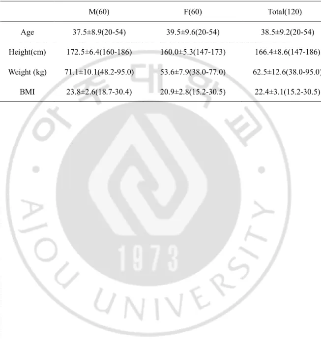
-Table 1: The Subjects’ Demographic Data
M(60) F(60) Total(120)
Age 37.5±8.9(20-54) 39.5±9.6(20-54) 38.5±9.2(20-54) Height(cm) 172.5±6.4(160-186) 160.0±5.3(147-173) 166.4±8.6(147-186) Weight (kg) 71.1±10.1(48.2-95.0) 53.6±7.9(38.0-77.0) 62.5±12.6(38.0-95.0)
6
-Fig 1A-B: (A) The radial CT image, which are perpendicular to the femoral
head-neck axis, are defined on a sagittal oblique localizer. (B) The radial cuts rotate
clockwise in 30°-intervals around the femoral head-neck axis. The alpha angle
measurements were performed throughout the cranial hemisphere form 10 o’clock to
3 o’clock.
7
-Fig 2: The alpha angle is formed by the axis of the femoral neck (AB) and a line
(AC) drawn from the femoral head center to the point where the head extends
beyond the margin of the best-fit circle.
8
-III. RESULTS
Of the 120 patients, 60 were males and the mean age was 37.45 ± 9.03 (19-54 years), 60 were females and the mean age was 39.51 ± 9.61 (20-54 years), smaller than the males, but there was no statistically significant difference.
When we look at the results of the clockwise system used by the author’s methods, the mean value of the alpha angle from 3 to 10 o'clock is greater for men than women for each region.There was no statistically significant difference in the remaining parts except 3 and 10 o'clock (Table2).
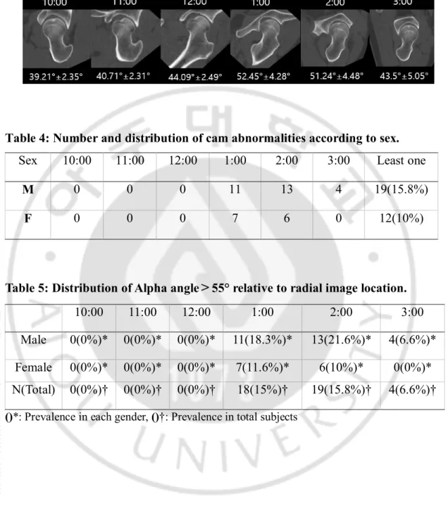
Mean alpha angle was 43.5°(34.7-56.1°) in 3 o’clock, 51.24°(39.5-58.8°) in 2 o’clock, 52.45° (43.3-65.5°) in 1 o’clock, 44.09°(36.8-49.8°) in 12 o’clock, 40.71°(33.5-45.8°) in 11 o’clock, 39.21°(34.1-44.6°) in 10 o’clock (Fig 3). Alpha angle in 1 and 2 o’clock was significantly larger than any locations (P<0.01) (Table 3).
The prevalence of cam deformity was 15%, 15.8% and 3.3% in 1, 2 and 3 o’clock, respectively. Among them, 18.3% (11 cases), 21.6% (13 cases) and 6.6% (4 cases) were males respectively, and 11.6% (7 cases), 10% (6 cases) and 0% were females, the prevalence of males was higher than females (Table 4). The prevalence of atypical abnormalities was 25.8% (31 cases), male 15.8% (19 cases) and female 10% (12 cases) (Table 5).In addition, 8 cases were reported in which two or more region cam type deformations were observed.
9
-Table2: Mean alpha angle at 10:00h to 3:00h locations by sex and total.
Table3: Mean alpha angle at 1:00 and 2:00 compare each area.
M F Total P Value (M vs. F) N 60 60 120 10:00 (°) 39.71±2.36(34.8-44.6) 38.69±2.26(34.1-42.6) 39.21±2.35(34.1-44.6) 0.029 11:00 (°) 41.05±2.24(36.5-45.6) 40.35±2.36(33.5-45.8) 40.71±2.31(33.5-45.8) 0.129 12:00 (°) 44.34±2.32(39.4-49.8) 43.83±2.65(36.8-49.4) 44.09±2.49(36.8-49.8) 0.310 1:00 (°) 53.17±4.27(47-65.5) 51.71±4.21(43.3±65.1) 52.45±4.28(43.3-65.5) 0.090 2:00 (°) 51.7±4.86(39.6-58.8) 50.77±4.04(39.5-58.3) 51.24±4.48(39.5-58.8) 0.304 3:00 (°) 44.52±5.71(34.7-56.1) 42.45±4.04(36.1-52.7) 43.5±5.05(34.7-56.1) 0.040
Radial image P Value Radial image P Value
1:00(52.45°) 10:00(39.21°) 0.000 2:00(51.24°) 10:00(39.21°) 0.000 11:00(40.71°) 0.000 11:00(40.71°) 0.000 12:00(44.09°) 0.000 12:00(44.09°) 0.000 2:00(51.24°) 0.664 1:00(52.45°) 0.664 3:00(43.50°) 0.000 3:00(43.50°) 0.000
- 10 -
Fig 3: Mean alpha angle at 10:00h to 3:00h.
Table 4: Number and distribution of cam abnormalities according to sex.
Sex
10:00
11:00
12:00
1:00
2:00
3:00
Least one
M
0
0
0
11
13
4
19(15.8%)
F
0
0
0
7
6
0
12(10%)
Table 5: Distribution of Alpha angle>55° relative to radial image location.
10:00
11:00
12:00
1:00
2:00
3:00
Male
0(0%)* 0(0%)* 0(0%)*
11(18.3%)*
13(21.6%)*
4(6.6%)*
Female
0(0%)* 0(0%)* 0(0%)*
7(11.6%)*
6(10%)*
0(0%)*
N(Total) 0(0%)† 0(0%)† 0(0%)†
18(15%)†
19(15.8%)†
4(6.6%)†
- 11 -
Ⅳ
. DISCUSSION
Plane radiography is the first screening test for suspected femoracetabular-impingement. Through this, it is determined whether there is a structural abnormality of the bone in the femoral head-neck junction. Physical examination revealed pain in the examination of the hip joint femoral acetabulum, loss of spherical head due to bony prominence of the femoral head, or morphological abnormality that could cause collision because the acetabulum overly covers the femoral head, the disease can be diagnosed clinically.However, there are cases in which asymptomatic patients are observed among radiologically abnormal findings of the hip joint. Therefore, clinical evaluation, physical findings and comprehensive judgment are required for the diagnosis of femoracetabular-impingement.
MRI and MRA (arthrography) are known to help identify acetabular rupture or other lesions within the hip joint. CT scans are more accurate than MRI in assessing morphological anomalies because they can observe bone abnormalities three-dimensionally. The use of 3D-CT reconstruction provides the operator with the additional advantage of a visual assessment of a cam lesion. Unlike plain radiographs, CT allows more consistent positioning of the patients, better image quality and more reliable readings (Biswas et al., 2009; Dolan et al., 2011). The main disadvantages in using CT as a supplementary investigation are the additional costs and the increased radiation exposure to the patient(Biswas et al., 2009). This study was carried out with CT in order to observe the morphological abnormality of bone three dimensionally.
Surgical treatment of FAI is aimed at making the abnormal shape of the hip normal, and it is the cause of arthritis when not treated surgically. Therefore, it is suggested that surgical correction is needed(Ganz et al., 2003).
- 12 -
the affected part. Such surgery has problems such as the risk associated with exposure of the hip during surgery, nonunion of the electronic part, and late recovery period.
The purpose of arthroscopic treatment is to proceed through a process similar to open surgery.It is difficult to accurately identify the anatomical structure of the surgical site and to confirm the position of the surgical tool within the hip. In contrast,since the incision site is small, there is less damage to the lesion and less bleeding, Arthroscopic treatment is continuously expanding because of its short duration and low complication rate(Sampson, 2006). This is the first comprehensive study using reformatted CT which would provide valuable data for surgeons performing open or arthroscopic bumpectomy.
In order to know the morphological abnormality of the femoral head-neck junction in three dimensions, previous study comparing angles according to positions by rotating the cross section measuring the alpha angle. It is known as an anterior-superior region, and to occur mainly when the hip joint is flexion or internal rotational (Ito et al., 2001; Lavigne et al., 2004).
We identified the mean value of alpha angle in Korea patients at the anterior (3:00), anterior-superior (2:00 and 1:00) and superior (12:00) region. According to previous reports that the alpha angle at the anterior-superior region using radial images is relatively large and shows the best differential between normal and abnormal hips (Hack et al., 2010; Kang et al., 2010; Chakraverty et al., 2013; Khan and Witt, 2014; Mimura et al., 2015).Khan and Witt evaluated the size and location of measurements of the alpha angle using CT scans in 42 patients with cam-type FAI and showed that the greatest alpha angle was found at the 2 o’clock region, followed by the 1 o’clock region(Khan and Witt, 2014). Our results also revealed similar propensity: greater values of alpha angle and higher abnormal rates in the one and two o’clock region than in the three o’clock and twelve o’clock region.
- 13 -
MRI images. It was confirmed that the region where the maximum alpha angle is distributed is between the anterior part and the superior part (1 and 2 o’clock). Mean alpha angle was 64.7° (44.2-99.7°) in 1 o’clock, 65.9° (44.2-99.3°) in 2 o’clock, respectively (Rakhra et al., 2009). Hack et al. Reported 400 cases of both hip joints in 200 normal adults who had no hip disease or pain and had no surgical treatment. Alpha angles were measured at the anterior (3 o'clock) and anterior-superior (1 o'clock). The average alpha angle was 40.78°(27 ° -69.9 °) in the anterior region and 50.15 ° (32.6 ° -76.3 °) in the anterior-superior region (1:30)(Hack et al., 2010). Sutter et al. reported that the alpha angle of the anterior-superior region of the femur was the largest at 53.3 ° after five-point rotation of the head-neck axis in normal adults without hip joint pain (Sutter et al., 2012). The authors' study also compared the angles of the six regions measured by rotating the head-neck axis. The results showed that the alpha angle of the anterior-superior region was (1:00: 52.45 °, 2:00: 51.24 °), this is the largest value, which is consistent with previous studies.However, the authors' study showed that the angle of the head was rotated in six regions by rotating the head-neck axis, which is the first reported study of asymptomatic adults in Korea.
According to Siebenrock et al. and Agricola et al., the prevalence of Cam-type FAI was reported to be more common in men than in women, which is reported to be related to differences in femoral anatomical features of males and females and excessive exercise in adults (Siebenrock et al., 2011; Siebenrock et al., 2013; Agricola et al., 2014). The authors also found that the frequency of Cam-type deformity was 15.8% for males and 10% for females, which is consistent with previous studies.
Van Houcke et al. in a study of normal Asian adults and normal European adults with no hip pain reported that the mean alpha angle of the anterior-superior (1:30) was 52 ° for Asian men and 49 ° for women (P <0.001), and 56 ° for European men and 56° for women (P = 0.01), respectively. The prevalence of Cam type FAI (alpha angle> 55 °) was reported to be
- 14 -
22% in Asian males and 15% in females and 35% in European males and 32% in females. There was a statistically significant difference in alpha angle between Asian males and females, while there was no statistically significant difference in alpha angle between males and females in Europe (Van Houcke et al., 2015). In the present study, the average alpha angle of males was higher than that of females at 1 o'clock, 2 o'clock, but there was no statistical difference.The frequency of Cam-type deformity was 18.3% for males and 11.6% for females at 1 o’clock and frequency of Cam-type deformity was 21.6% for males and 10% for females at 2 o'clock.These results are similar to the prevalence of FAI of Cam deformity in Asian adults. However, the prevalence of FAI in Cam variants in European adults is smaller than that in adults. This is thought to be related to race differences.
Nepple et al. [26] compared the CT images with the radiographs (pelvic AP view, 45 ° Dunn, frog leg lateral view) by dividing the superior part of the femur and the anterior part of the femur in 30 ° intervals using a clock system. The pelvic AP view, Dunn view and the frog leg lateral view were consistent with the angle of the superior region of the femur, the anterior-superior region, and the anterior region, respectively, and we could diagnose the Cam type FAI using three simple radiographs rather than CT with severe radiation exposure (Nepple et al., 2013).We did not compare the radiographs with the radiographs of the thighs. However, we used CT of the normal adult in Korea to view the morphological anomalies of the thighs in three dimensions and report the normal range at each site.
Most studies on FAI have focused on symptomatic patients (Takeyama et al., 2009; Fukushima et al., 2014).However, recent data based on CT images in asymptomatic Asian patients show high prevalence of FAI. Kim et al. reported that 18% of patients with anterior part alpha angle of greater than 55 ° and 27% of patients with a CE-angle of greater than 40 ° had a 473 hips on asymptomatic CT images. However, they studied CT images, but the alpha angle was measured only in one area and the angle in the other area was not measured (Kim
- 15 -
et al., 2015). Therefore, it may be possible to miss the cam-type deformity that may occur at other sites.
The limitations of this study were that 120 specimens of the study were few, and the medical records were reviewed, and it was not confirmed that they were asymptomatic through physical examination and follow-up, we could not evaluate the correlation and the presence of secondary deformation by radiometric measurement of the Pincer type deformity. The alpha angle was measured by one person.
- 16 -
. CONCLUSION
Ⅴ
Cam deformity of FAI was observed on 25.8% of asymptomatic hip. The most common region of cam deformity was antero-superior area of femoral head-neck junction (1 and 2 o’clock). The present study first reported the prevalence and the distribution of cam deformity in asymptomatic Korean population. The results would provide anatomical comprehension of cam deformity for diagnosis and surgical treatments.
- 17 -
REFERENCES
1. Agricola R, Heijboer MP, Ginai AZ, Roels P, Zadpoor AA, Verhaar JA, Weinans H, Waarsing JH: A cam deformity is gradually acquired during skeletal maturation in adolescent and young male soccer players: a prospective study with minimum 2-year follow-up. Am J Sports Med 42: 798-806, 2014
2. Ahn T, Kim CH, Kim TH, Chang JS, Jeong MY, Aditya K, Yoon PW: What is the Prevalence of Radiographic Hip Findings Associated With Femoroacetabular Impingement in Asymptomatic Asian Volunteers? Clin Orthop Relat Res 474: 2655-2661, 2016
3. Anderson K, Strickland SM, Warren R: Hip and groin injuries in athletes. Am J
Sports Med 29: 521-533, 2001
4. Bardakos NV, Villar RN: Predictors of progression of osteoarthritis in femoroacetabular impingement: a radiological study with a minimum of ten years follow-up. J Bone Joint Surg Br 91: 162-169, 2009
5. Beck M, Kalhor M, Leunig M, Ganz R: Hip morphology influences the pattern of damage to the acetabular cartilage: femoroacetabular impingement as a cause of early osteoarthritis of the hip. J Bone Joint Surg Br 87: 1012-1018, 2005
6. Biswas D, Bible JE, Bohan M, Simpson AK, Whang PG, Grauer JN: Radiation exposure from musculoskeletal computerized tomographic scans. J Bone Joint Surg
Am 91: 1882-1889, 2009
7. Chakraverty JK, Sullivan C, Gan C, Narayanaswamy S, Kamath S: Cam and pincer femoroacetabular impingement: CT findings of features resembling femoroacetabular impingement in a young population without symptoms. AJR Am J
Roentgenol 200: 389-395, 2013
8. Clohisy JC, St John LC, Schutz AL: Surgical treatment of femoroacetabular impingement: a systematic review of the literature. Clin Orthop Relat Res 468: 555-564, 2010
9. Dolan MM, Heyworth BE, Bedi A, Duke G, Kelly BT: CT reveals a high incidence of osseous abnormalities in hips with labral tears. Clin Orthop Relat Res 469: 831-838, 2011
- 18 -
10. Dudda M, Albers C, Mamisch TC, Werlen S, Beck M: Do normal radiographs exclude asphericity of the femoral head-neck junction? Clin Orthop Relat Res 467: 651-659, 2009
11. Fukushima K, Uchiyama K, Takahira N, Moriya M, Yamamoto T, Itoman M, Takaso M: Prevalence of radiographic findings of femoroacetabular impingement in the Japanese population. J Orthop Surg Res 9: 25, 2014
12. Ganz R, Bamert P, Hausner P, Isler B, Vrevc F: [Cervico-acetabular impingement after femoral neck fracture]. Unfallchirurg 94: 172-175, 1991
13. Ganz R, Parvizi J, Beck M, Leunig M, Notzli H, Siebenrock KA: Femoroacetabular impingement: a cause for osteoarthritis of the hip. Clin Orthop Relat Res: 112-120, 2003
14. Hack K, Di Primio G, Rakhra K, Beaule PE: Prevalence of cam-type femoroacetabular impingement morphology in asymptomatic volunteers. J Bone
Joint Surg Am 92: 2436-2444, 2010
15. Ito K, Minka MA, 2nd, Leunig M, Werlen S, Ganz R: Femoroacetabular impingement and the cam-effect. A MRI-based quantitative anatomical study of the femoral head-neck offset. J Bone Joint Surg Br 83: 171-176, 2001
16. Kang AC, Gooding AJ, Coates MH, Goh TD, Armour P, Rietveld J: Computed tomography assessment of hip joints in asymptomatic individuals in relation to femoroacetabular impingement. Am J Sports Med 38: 1160-1165, 2010
17. Khan O, Witt J: Evaluation of the magnitude and location of Cam deformity using three dimensional CT analysis. Bone Joint J 96-B: 1167-1171, 2014
18. Kim J, Choi JA, Lee E, Lee KR: Prevalence of Imaging Features on CT Thought to Be Associated With Femoroacetabular Impingement: A Retrospective Analysis of 473 Asymptomatic Adult Hip Joints. AJR Am J Roentgenol 205: W100-105, 2015 19. Kubiak-Langer M, Tannast M, Murphy SB, Siebenrock KA, Langlotz F: Range of
motion in anterior femoroacetabular impingement. Clin Orthop Relat Res 458: 117-124, 2007
20. Lavigne M, Parvizi J, Beck M, Siebenrock KA, Ganz R, Leunig M: Anterior femoroacetabular impingement: part I. Techniques of joint preserving surgery. Clin
Orthop Relat Res: 61-66, 2004
21. Leunig M, Ganz R: [Femoroacetabular impingement. A common cause of hip complaints leading to arthrosis]. Unfallchirurg 108: 9-10, 12-17, 2005
- 19 -
22. Mimura T, Kawasaki T, Itakura S, Hirata T, Fuzikawa H, Mori K, Imai S: Prevalence of radiological femoroacetabular impingement in Japanese hip joints: detailed investigation with computed tomography. J Orthop Sci 20: 649-656, 2015
23. Nepple JJ, Prather H, Trousdale RT, Clohisy JC, Beaule PE, Glyn-Jones S, Kim YJ: Clinical diagnosis of femoroacetabular impingement. J Am Acad Orthop Surg 21 Suppl 1: S16-19, 2013
24. Notzli HP, Wyss TF, Stoecklin CH, Schmid MR, Treiber K, Hodler J: The contour of the femoral head-neck junction as a predictor for the risk of anterior impingement. J
Bone Joint Surg Br 84: 556-560, 2002
25. Rakhra KS, Sheikh AM, Allen D, Beaule PE: Comparison of MRI alpha angle measurement planes in femoroacetabular impingement. Clin Orthop Relat Res 467: 660-665, 2009
26. Sampson TG: Arthroscopic treatment of femoroacetabular impingement: a proposed technique with clinical experience. Instr Course Lect 55: 337-346, 2006
27. Schmitz MR, Bittersohl B, Zaps D, Bomar JD, Pennock AT, Hosalkar HS: Spectrum of radiographic femoroacetabular impingement morphology in adolescents and young adults: an EOS-based double-cohort study. J Bone Joint Surg Am 95: e90, 2013
28. Siebenrock KA, Ferner F, Noble PC, Santore RF, Werlen S, Mamisch TC: The cam-type deformity of the proximal femur arises in childhood in response to vigorous sporting activity. Clin Orthop Relat Res 469: 3229-3240, 2011
29. Siebenrock KA, Kaschka I, Frauchiger L, Werlen S, Schwab JM: Prevalence of cam-type deformity and hip pain in elite ice hockey players before and after the end of growth. Am J Sports Med 41: 2308-2313, 2013
30. Sutter R, Dietrich TJ, Zingg PO, Pfirrmann CW: How useful is the alpha angle for discriminating between symptomatic patients with cam-type femoroacetabular impingement and asymptomatic volunteers? Radiology 264: 514-521, 2012
31. Takeyama A, Naito M, Shiramizu K, Kiyama T: Prevalence of femoroacetabular impingement in Asian patients with osteoarthritis of the hip. Int Orthop 33: 1229-1232, 2009
32. Van Houcke J, Yau WP, Yan CH, Huysse W, Dechamps H, Lau WH, Wong CS, Pattyn C, Audenaert EA: Prevalence of radiographic parameters predisposing to femoroacetabular impingement in young asymptomatic Chinese and white subjects.
- 20 -
- 21 - - 국문요약 -
무증상성인에서
컴퓨터단층촬영을 이용한 고관절
cam 변형의 빈도
아주대학교 대학원 의학과 한 준 (지도교수: 원 예 연)목적: 대퇴비구 충돌(Femoroacetabular impingement: FAI)은 고관절의 운동 범위를 감소시키는 것은 물론 초기 관절염의 초래하는 중요한 원인으로 추정되고 있다. 단순 방사선 촬영 단독보다 CT 나 MRI 와 같은 단층 촬영이 더욱 정확한 진단적 방법으로 간주되지만 외래에서 매번 적용하기에는 제한이 따른다. 우리나라 성인의 FAI에 대한 단층 촬영의 데이터가 부족하여 저자들은 한국인에서 cam 형 충돌의 위치가 개인마다 다를 것으로 가설을 수립하였다. 이에 컴퓨터 단층촬영을 이용하여 무증상성인의 cam 형 변형의 빈도를 측정하고자 한다. 대상 및 방법: 2011년 1월부터 2015년 12월까지 본원에서 120개 고관절에 특이병력 없는 무증상 성인 120명의 고관절 CT영상을 이용하였다. CT영상을 INFINITT Xelis 프로그램을 이용하여 골 두-경부 중심점을 잇는 선을 중심으로 근위 대퇴부를 시계 시스템을 이용하여 3차원적으로 분할하여 각 구간에서 알파각도를 측정하였다. 알파각도는 55도 보다 큰 값이면 고관절 Cam형 충돌로 판정하였다. 결과: 3시부터 10시(대퇴골 전방에서 후방)방향으로의 평균 알파각은 3시에서 43.5°(34.7-56.1°), 2시에서 51.24°(39.5-58.8°), 1시에서 52.45° (43.3-65.5°), 12시에서 44.09°(36.8-49.8°), 11시에서 40.71°(33.5-45.8°)였고 10시에서는 39.21°(34.1-44.6°)이었다. 전상방부(1시 및 2시)의 평균 알파각이 제일 컸고(P<0.01) 고관절Cam형 충돌과 관련된 영상의학적 소견의 빈도는 1시 및 2시에서 각각
- 22 - 15% 및 15.8%였다. 결론: 무증상 성인에서 cam형 충돌은 25.8%에서 발견 되었고 가장 호발하는 위치는 전상방부(1시 및 2시)였다. 한국에서 처음 시계 시스템으로 알파값을 측정한 결과를 보여주어 FAI에대한 관절경적 치료나 돌출부 제거술(bumpectomy)을 하는 수술의사들에게 도움이 될 것으로 생각된다. 색인단어: 대퇴비구 충돌, Cam 형충돌, 3차원적분할, 알파각, 전상방부