• 검색 결과가 없습니다.

Prevalences of Incidental Findings in Trauma Patients by Abdominal and Pelvic Computed Tomography

N/A
N/A
Protected

Academic year: 2021

Share "Prevalences of Incidental Findings in Trauma Patients by Abdominal and Pelvic Computed Tomography"

Copied!
7
0
0

로드 중.... (전체 텍스트 보기)

전체 글

(1)

Vol. 29, No. 3, September, 2016 http://dx.doi.org/10.20408/jti.2016.29.3.61

� Address for Correspondence : Seung Hwan Lee, M.D.

Department of Surgery, Yonsei University College of Medicine, 50-1 Yonsei-ro, Seodaemun-gu, Seoul 03722, Korea

Tel : 82-2-2228-2108, Fax : 82-2-313-8289, E-mail : seunghwan@yuhs.ac

Submitted : July 7, 2016 Revised : July 13, 2016 Accepted : September 12, 2016 This study was not funded and the authors have no potential conflict of interest to declare.

Prevalences of Incidental Findings in Trauma Patients by Abdominal and Pelvic Computed Tomography

Jin Young Lee, M.D., Myung Jae Jung, M.D., Jae Gil Lee, M.D., Ph.D., Seung Hwan Lee, M.D.

Department of Surgery, Yonsei University College of Medicine, Seoul, Korea

Purpose: Abdominal and pelvic computed tomography (APCT) is frequently used as a diagnostic tool in trauma patients. However, trauma unrelated, incidental findings are frequently encountered. The aim of this study was to determine the prevalences of incidental findings on APCT scans in trauma patients.

Methods: The archived records of 801 trauma patients treated from January 2013 to December 2015 were reviewed retrospectively. Six hundred and forty of these patients underwent contrast enhanced APCT in an emergency depart- ment and were included in this study, and 205 (32.1%) of these patients had incidental findings. These findings were divided into two categories: category I, meaning a radiological benign finding not requiring further evaluation or fol- low-up, and category II, requiring further evaluation and follow-up.

Results: One hundred and sixty (24.8%) patients were allocated to category I and 45 (7.2%) to category II. The most frequent incidental findings were discovered in kidneys (34.6%), followed by liver (28.8%), and gallbladder (15.6%).

The most frequent finding in category I was a benign cyst (60.1%), followed by a simple stone (15.6%), and heman- gioma (11.9%). Adenomyomatosis of the gallbladder (17.8%) was the most common lesion in category II, followed by atypical mass (15.6%), complicated stone (15.6%) and cystic neoplasm (15.6%).

Conclusion: The prevalence of an incidental finding on APCT scans was 32.1%. Although category II lesions were not common in trauma patients, these findings should be communicated to patients, and when necessary referred to a pri- mary care physician. Systems are required for producing appropriate discharge summaries and informing patients about the implications of incidental findings. [ J Trauma Inj 2016; 29: 61-67 ]

Key Words: Incidental findings, Trauma, Abdominal and pelvic computed tomography (APCT)

I. Introduction

The first commercially viable computed tomogra- phy (CT) scanner was installed in Atkinson Morley Hospital in Wimbledon, England in 1971,(1) and since CT technology has vastly improved in speed, slice count, and image quality. Multidetector CT (MDCT) is able to decrease scan times to less than a minute, and obtain high quality images that of less than 1 mm slices.(2) Due to these benefits, CT has become the

preferred means for the initial evaluation of trauma patients.(3-5)

The increased use of CT has not only improved the

immediate diagnosis of injury, but it has also

increased detection of incidental findings (IFs).(6-9)

These IFs may be beneficial to patients, for example,

they may result in the early detection of a significant

pathology, but they also increase anxiety and health-

care costs because of additional investigations under-

taken.(10) Many reports have been issued regarding

(2)

the clinical implications of IFs in non-trauma patients,(11-13) and the presence of incidental CT findings in trauma patients has been documented on several occasions.(6-9,14-16) Furthermore, the need for further diagnostic work-up, referral, and treat- ment is difficult for trauma surgeons to deter- mine,(15) and may have serious consequences.

Currently, the work-up of IFs varies between clini- cians and regions, and no well-established classifica- tion exists.(6,7,17)

We performed this study to document the fre- quency of IF detection by abdominal and pelvic computed tomography (APCT) and to determine how many follow-ups were performed in these patients.

II. Materials and Methods

1. Study design and population

This retrospective observational study was con- ducted at a single center from January 2013 to December 2015. During this period 801 trauma patients were admitted to emergency department. Six hun- dred and forty of these patients that underwent

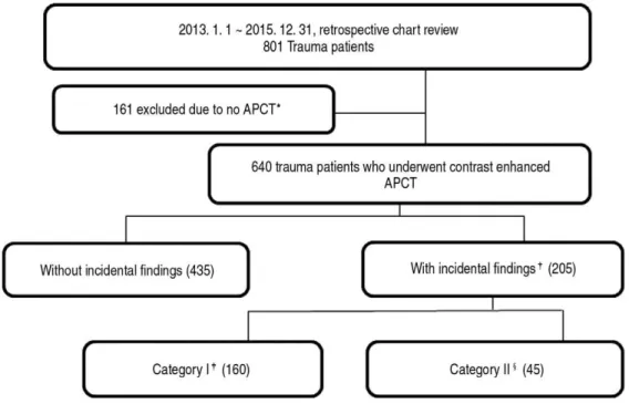
APCT were enrolled in the present study, and divid- ed into two groups: the group with IFs (the IF group; n=205) and the group without IFs (the non- IF group; n=435). The IF group was subdivided into category I (finding not followed; n=160) and category II (finding followed; n=45) (Fig. 1).

A trauma surgeon determined whether a CT scan was required after the patient had undergone an initial primary survey. CT images were immediately reviewed by a senior resident radiologist, and a final reading was subsequently issued by the staff radiol- ogist several days later.

The study was approved by our Institutional Review Board (IRB No. 4-2016-0293), which waived the requirement for informed consent because of the retrospective nature of the study.

2. Variables and definitions

The baseline characteristics included were; age, sex, hospital stay, intensive care unit (ICU) stay, trauma related variables, such as, injury mechanism, injury region, injury severity score (ISS), revised trauma score (RTS), trauma and injury severity score (TRISS),

Fig. 1. Study protocol.

Fig. 1. * APCT: Abdominal and pelvic computed tomography

Fig. 1.

Incidental findings: findings on APCT unrelated to the injury identified by formal radiologic reading Fig. 1.

Category I: radiologically benign - no need for further evaluation or follow-up

Fig. 1.

§

Category II: requiring further evaluation and follow-up

(3)

and mortality.

IFs were defined as findings on APCT, confirmed by formal reading, are unrelated to traumatic injury.

Using previously described classifications,(6,7,15,18) IFs were classified into two categories. Category I was defined as radiological benign requiring no further evaluation or follow-up, e.g., a simple cyst or tiny simple stone, and category II defined a pathology con- cern, such as, adrenal adenoma or a large (>2 cm) or impacted stones, requiring further evaluation, follow- up, and treatment. One investigator independently categorized incidental findings according to these def- initions, and then another investigator confirmed these assignments. Disagreements were discussed and resolved by consensus with other investigators.

3. Statistical analysis

Statistical analysis was performed using IBM SPSS Statistics ver. 20.0 (IBM Co. Armonk, NY). The

baseline and clinical characteristics of patients in the IF and non-IF groups were compared by uni- variate analysis. The anatomical distributions and subgroups of IFs were compared using numbers of IFs and not patient numbers. Categorical data are presented as numbers (%) and were compared using the Chi-square or Fisher’ s exact test. Continuous variables are expressed as means and standard deviations or medians and inter-quantile ranges (IQR), and intergroup comparisons were conducted using the Student’ s t-test or the Mann-Whiney U test. Statistical significance was accepted for p value<0.05.

III. Results

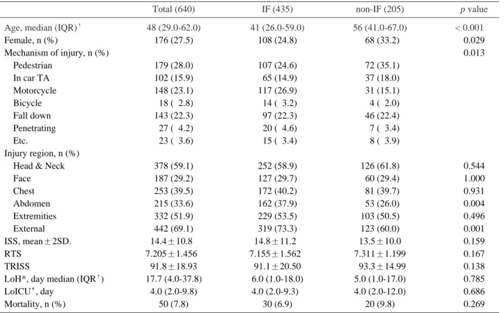
Median age of the 640 study subjects was 48 years, and 27% were women. Median age in the IF group was significantly greater than in the non-IF group (56 vs. 41 years), and the proportion of women

Table 1. Baseline characteristics of all study subjects and of patient groups

Total (640) IF (435) non-IF (205) p value

Age, median (IQR)

48 (29.0-62.0) 41 (26.0-59.0) 56 (41.0-67.0) < 0.00100

Female, n (%) 176 (27.5) 108 (24.8) 068 (33.2) 0.029

Mechanism of injury, n (%) 0.013

Pedestrian 179 (28.0) 107 (24.6) 072 (35.1)

In car TA 102 (15.9) 065 (14.9) 037 (18.0)

Motorcycle 148 (23.1) 117 (26.9) 031 (15.1)

Bicycle 018 (02.8) 014 (03.2) 004 (02.0)

Fall down 143 (22.3) 097 (22.3) 046 (22.4)

Penetrating 027 (04.2) 020 (04.6) 007 (03.4)

Etc. 023 (03.6) 015 (03.4) 008 (03.9)

Injury region, n (%)

Head & Neck 378 (59.1) 252 (58.9) 126 (61.8) 0.544

Face 187 (29.2) 127 (29.7) 060 (29.4) 1.000

Chest 253 (39.5) 172 (40.2) 081 (39.7) 0.931

Abdomen 215 (33.6) 162 (37.9) 053 (26.0) 0.004

Extremities 332 (51.9) 229 (53.5) 103 (50.5) 0.496

External 442 (69.1) 319 (73.3) 123 (60.0) 0.001

ISS, mean±2SD. 14.4±10.80 14.8±11.20 13.5±10.0 0.159

RTS 7.205±1.4560 7.155±1.5620 7.311±1.199 0.167

TRISS 91.8±18.93 91.1±20.50 093.3±14.99 0.138

LoH*, day median (IQR

) 17.7 (4.0-37.8) 6.0 (1.0-18.0) 5.0 (1.0-17.0) 0.785

LoICU

, day 4.0 (2.0-9.8) 4.0 (2.0-9.3)0 4.0 (2.0-12.0) 0.686

Mortality, n (%) 50 (7.8) 30 (6.9) 20 (9.8) 0.269

* LoH: Length of hospital stay

IQR: Inter-quartile range

LoICU: Length of intensive care unit stay

(4)

was significant higher in the IF group (33.2% vs.

24.8%, respectively). Trauma related variables, length of hospital stays and mortalities were not significantly different between these two groups (Table 1). No significant differences were observed

between the baseline characteristics of categories I and II (Table 2).

IFs were most frequently discovered in kidneys (34.6%), followed by liver (28.8%), and gallbladder (15.6%). Similarly, kidneys (41.9%) were most fre-

Table 2. Comparison of baseline characteristics of patients with category I and II

Category I (160) Category II (45) p value

Age, median (IQR)

56 (42-67) 55 (41-68) 0.951

Female, n (%) 54 (34.0) 14 (30.4) 0.724

Mechanism of injury, n (%) 0.621

Pedestrian 56 (35.2) 16 (34.8)

In car TA 26 (16.4) 11 (23.9)

Motorcycle 26 (16.4) 05 (10.9)

Bicycle 04 (02.5) 00 (00.0)

Fall down 37 (23.3) 09 (19.6)

Penetrating 05 (03.1) 02 (04.3)

Etc. 05 (03.1) 03 (06.5)

Injury region, n (%)

Head & Neck 98 (61.6) 28 (62.2) 1.000

Face 45 (28.3) 15 (33.3) 0.579

Chest 61 (38.4) 20 (44.4) 0.493

Abdomen 41 (25.8) 12 (26.7) 1.000

Extremities 80 (50.3) 23 (51.1) 1.000

External 94 (59.1) 29 (63.0) 0.733

ISS, mean±2SD 13.3±9.50 14.5±11.60 0.480

RTS 7.379±0.977 7.076±1.7600 0.269

TRISS 094.7±11.00 88.4±23.70 0.086

LoH, median (IQR) 5.3 (1.0-16.8) 5.0 (1.8-18.5) 0.858

LoICU 4.0 (2.0-14.0) 2.5 (2.0-7.5)0 0.202

Mortality, n (%) 13 (08.2) 7 (15.2). 0.165

Follow-up, n (%) 9 (23.0)

§

IQR: Inter-quartile range

§

9 (23.0): excluding patients that expired or transferred.

Table 3. Anatomical distributions and frequencies of incidental findings in categories I and II Total

Category I (184) Category II (52) p value (IF group; 238)

Kidney 71 (34.6) 67 (41.9) 04 (08.7) < 0.0010.

Liver 59 (28.8) 47 (29.4) 12 (26.7) 0.853

Gallbladder 32 (15.6) 18 (11.2) 14 (31.1) 0.002

Uterus with adnexa 17 (08.3) 15 (09.4) 02 (04.4) 0.373

Intestine 13 (06.3) 08 (05.0) 05 (11.1) 0.165

Urinary tract with bladder 13 (06.3) 09 (05.6) 02 (08.9) 0.488

Pancreas 10 (04.9) 06 (03.8) 04 (08.9) 0.231

Adrenal gland 09 (04.4) 04 (02.5) 05 (11.1) 0.026

Bone 04 (02.0) 03 (01.9) 01 (02.2) 1.000

Vascular 04 (02.0) 01 (00.6) 03 (06.7) 0.034

Others 06 (06.0) 06 (03.8) 00 (00.0) 0.343

Data are presented in number (%).

(5)

quent in category I also followed by liver (29.4%), and gallbladder (11.2%). However, in category II, IFs were commonly discovered in gallbladder (31.1%) fol- lowed by liver (26.7%) and the gastro-intestinal tract or the adrenal gland (Table 3).

The most frequent finding in category I was a benign cyst (60.1%), followed by simple stone (15.6%), and hemangioma (11.9%). Other findings in category I were diverticulum (5.0%), hyperplasia (4.4%), and hemangioma (11.9%). Adenomyomatosis of gallblad- der (17.8%) was most common in category II, followed atypical mass (15.6%), complicated stone (15.6%), and cystic neoplasm (15.6%) (Table 4). Malignancy was found in 2 patients, namely, hepatocellular carcino- ma and prostate cancer.

IV. Discussion

In this study, 32% of trauma patients (n=205) evaluated by APCT had at least one IF, which con- curs with previously reported rates.(6-9) One hun- dred and sixty patients (24.8%) were classified as category I, and 45 (7.2%) patients as category II.

Only 9 (23%) patients in category II were followed- up or further evaluated.

Previous studies,(6,16) have shown that CT IF rates increase with age, and the present study adds weight to this association, and median age was greater in the IF group than in the non-IF group.

However, median ages in category I and II were not significantly different. Elderly patients becoming more active, and thus, are experiencing severe injuries more often, which explains, at least in part, the increasing detection of IFs.(19) Furthermore, given similar injury severities, mortality appears to

be higher among older patients,(20) and hence, these patients need more careful examinations and longer observations.

Adenomyomatosis may be seen in as many as 9%

of cholecystectomy specimens, and accounts for - 25% of all polypoid lesions in the gallbladder.(21,22) Furthermore, detection rates increase with age and are higher for women.(23) The current view is that adenomyomatosis is not a premalignant condi- tion,(21,23,24) but it is difficult to differentiate focal adenomyomatosis and malignancy, and to make matters worse adenocarcinoma of the gallbladder can occur in localized adenomyomatosis.(25) For this reason, we classified adenomyomatosis as category II in the present study. Furthermore, it was the most frequently detected IF in this category.

IFs increase the workload of trauma surgeons, because they are not primary physicians. The treat- ment of trauma patients is complex, and the man- agement of trauma injuries is prioritized, and during initial trauma care, many IFs might be unimportant.

Trauma care is based on rapid diagnosis and treat- ment, and most injuries do not lead to permanent disability or require long-term follow-up. On the other hand, non-traumatic diagnoses often require a less aggressive approach, and the early identifica- tion and treatment of some IFs not only increases patient survival but also decreases morbidity.

(18,26,27) Although the optimal management of patients with many types of IFs remains a topic of discussion, system is required to include incidental CT findings on discharge summaries and to inform patients of these findings and their implications. In a previous study, it was reported that only 48% of patients with serious findings had any documenta-

Table 4. Description of subgroups in Categories I and II

Category I (177) Category II (45)

Benign cyst 96 (60.1) Adenomyomatosis 8 (17.8)

Simple stone 25 (15.6) Stone (complicated) 7 (15.6)

Hemangioma 19 (11.9) Cystic Neoplasm 7 (15.6)

Hyperplasia 07 (04.4) Atypical mass 7 (15.6)

Myoma 07 (04.4) Adenoma 6 (13.3)

Lipoma 03 (01.9) Aneurysm 3 (06.7)

Diverticulum 08 (05.0) Malignancy 2 (04.4)

Others 12 (07.5) Others 5 (11.1)

Data are presented in number (%).

(6)

tion of treatment or scheduled follow-up.(28) In the present study, we found that only 23% (n=9) of patients in category II were followed up or further evaluated, although it is possible patients with IFs were given verbal follow-up instructions, and that these were not recorded in discharge summaries.

The present study has several limitations that require consideration. First, the study involved a single-center, retrospective chart review, and thus, its results may not be applicable to all hospitals.

Furthermore, it is possible our regional patient pop- ulation had different health risk factors that increased the risks of cancer, and cardiovascular, and cerebrovascular disease. Second, APCT was per- formed because of trauma, and thus, the radiolo- gists focused on traumatic injuries and not on other anomalies. Third, the clinical significances of IFs were assessed retrospectively based on the presumed need for follow-up and thus, the classification used was highly subjective. Fourth, we did not investigate the management or outcomes of IFs and thus were unable to access the risks or benefits of our find- ings. We recommended that follow-up study be con- ducted to investigate these issues.

V. Conclusion

The use of APCT for the evaluating trauma patients has generated a large number of IFs (32%).

In the present study, category II (requiring further evaluation and follow-up) constituted 7.2% (n=45) of the 640 study subjects. We believe such findings should be communicated to patients and that when appropriate some patients be referred to primary care physicians. Furthermore, according to our records, follow-up of these patients was poor, which demonstrates a system that adds this information to discharge summaries and ensures patients are ade- quately informed is urgently required.

VI. Acknowledgements

Authors’contributions: JYL and SHL designed the study, JYL collected the data. JYL did the statistical analyses with SHL, JGL and MJJ. JYL wrote the article.

All authors read and approved the submitted version.

REFERENCES

01) Beckmann EC. CT scanning the early days. Br J Radiol 2006;

79: 5-8.

02) Prokop M. General principles of MDCT. Eur J Radiol 2003;

45: S4-10.

03) Salim A, Sangthong B, Martin M, Brown C, Plurad D, Demetriades D. Whole body imaging in blunt multisystem trauma patients without obvious signs of injury: results of a prospective study. Arch Surg 2006; 141: 468-73; discussion 73-5.

04) Sampson MA, Colquhoun KB, Hennessy NL. Computed tomography whole body imaging in multi-trauma: 7 years experience. Clin Radiol 2006; 61: 365-9.

05) Wisbach GG, Sise MJ, Sack DI, Swanson SM, Sundquist SM, Paci GM, et al. What is the role of chest X-ray in the initial assessment of stable trauma patients? J Trauma 2007; 62: 74- 8; discussion 8-9.

06) Paluska TR, Sise MJ, Sack DI, Sise CB, Egan MC, Biondi M.

Incidental CT findings in trauma patients: incidence and impli- cations for care of the injured. J Trauma 2007; 62: 157-61.

07) Barrett TW, Schierling M, Zhou C, Colfax JD, Russ S, Conatser P, et al. Prevalence of incidental findings in trauma patients detected by computed tomography imaging. Am J Emerg Med 2009; 27: 428-35.

08) Ekeh AP, Walusimbi M, Brigham E, Woods RJ, McCarthy MC. The prevalence of incidental findings on abdominal com- puted tomography scans of trauma patients. J Emerg Med 2010; 38: 484-9.

09) Munk MD, Peitzman AB, Hostler DP, Wolfson AB.

Frequency and follow-up of incidental findings on trauma computed tomography scans: experience at a level one trauma center. J Emerg Med 2010; 38: 346-50.

10) Berlin L. Potential legal ramifications of whole-body CT screening: taking a peek into Pandora’s box. AJR Am J Roentgenol 2003; 180: 317-22.

11) Elgin EE, O’Malley PG, Feuerstein I, Taylor AJ. Frequency and severity of “incidentalomas” encountered during electron beam computed tomography for coronary calcium in middle- aged army personnel. Am J Cardiol 2002; 90: 543-5.

12) Hunold P, Schmermund A, Seibel RM, Gronemeyer DH, Erbel R. Prevalence and clinical significance of accidental findings in electron-beam tomographic scans for coronary artery calcification. Eur Heart J 2001; 22: 1748-58.

13) Messersmith WA, Brown DF, Barry MJ. The prevalence and implications of incidental findings on ED abdominal CT scans. Am J Emerg Med 2001; 19: 479-81.

14) van Vugt R, Dekker HM, Deunk J, van der Vijver RJ, van Vugt AB, Kool DR, et al. Incidental Findings on Routine Thoracoabdominal Computed Tomography in Blunt Trauma Patients. J Trauma 2011.

15) Ruesseler M, Schill A, Lehnert T, Wyen H, Wutzler S, Marzi I, et al. Incidental findings in patients with multiple injuries:

how to proceed? J Trauma Acute Care Surg 2013; 75: 848-53.

(7)

16) Seah MK, Murphy CG, McDonald S, Carrothers A. Incidental findings on whole-body trauma computed tomography:

Experience at a major trauma centre. Injury 2016; 47: 691-4.

17) Berland LL, Silverman SG, Gore RM, Mayo-Smith WW, Megibow AJ, Yee J, et al. Managing incidental findings on abdominal CT: white paper of the ACR incidental findings committee. J Am Coll Radiol 2010; 7: 754-73.

18) Iezzi R, Cotroneo AR, Filippone A, Di Fabio F, Merlino B, Bonomo L. Extravascular incidental findings at multislice CT angiography of the abdominal aorta and lower extremity arter- ies: a retrospective review study. Abdom Imaging 2007; 32:

489-94.

19) Wutzler S, Lefering R, Laurer HL, Walcher F, Wyen H, Marzi I. [Changes in geriatric traumatology. An analysis of 14,869 patients from the German Trauma Registry].

Unfallchirurg 2008; 111: 592-8.

20) Tornetta P, 3rd, Mostafavi H, Riina J, Turen C, Reimer B, Levine R, et al. Morbidity and mortality in elderly trauma patients. J Trauma 1999; 46: 702-6.

21) Gallahan WC, Conway JD. Diagnosis and management of gallbladder polyps. Gastroenterol Clin North Am 2010; 39:

359-67.

22) Ootani T, Shirai Y, Tsukada K, Muto T. Relationship between gallbladder carcinoma and the segmental type of adenomy-

omatosis of the gallbladder. Cancer 1992; 69: 2647-52.

23) Mellnick VM, Menias CO, Sandrasegaran K, Hara AK, Kielar AZ, Brunt EM, et al. Polypoid Lesions of the Gallbladder:

Disease Spectrum with Pathologic Correlation-Erratum.

Radiographics 2015; 35: 1316.

24) Kim JH, Jeong IH, Han JH, Kim JH, Hwang JC, Yoo BM, et al. Clinical/pathological analysis of gallbladder adenomy- omatosis; type and pathogenesis. Hepatogastroenterology 2010; 57: 420-5.

25) Aldridge MC, Gruffaz F, Castaing D, Bismuth H.

Adenomyomatosis of the gallbladder. A premalignant lesion?

Surgery 1991; 109: 107-10.

26) Eskandary H, Sabba M, Khajehpour F, Eskandari M.

Incidental findings in brain computed tomography scans of 3000 head trauma patients. Surg Neurol 2005; 63: 550-3; dis- cussion 3.

27) Shetty SK, Maher MM, Hahn PF, Halpern EF, Aquino SL.

Significance of incidental thyroid lesions detected on CT: cor- relation among CT, sonography, and pathology. AJR Am J Roentgenol 2006; 187: 1349-56.

28) Semelka RC, Armao DM, Elias J, Jr., Huda W. Imaging

strategies to reduce the risk of radiation in CT studies, includ-

ing selective substitution with MRI. J Magn Reson Imaging

2007; 25: 900-9.

수치

Fig. 1. Study protocol.
Table 1. Baseline characteristics of all study subjects and of patient groups
Table 3. Anatomical distributions and frequencies of incidental findings in categories I and II Total
Table 4. Description of subgroups in Categories I and II

참조

관련 문서

CT (Computed Tomography) – TDLAS (Tunable Diode Laser Absorption Spectroscopy) is a non-intrusive diagnostic technique that allows for spatially resolved measurements

Over the following 3 months, the patient developed elevated alpha-fetoprotein, and positron emission tomography showed a cystic mass in the right side of the pelvic cavity

Kim K, Lee SJ, Seo J, Suh YJ, Cho I, Hong G-R, Ha J-W, Kim YJ and Shim CY (2022) Assessment of aortic valve area on cardiac computed tomography in symptomatic bicuspid

Forces exerted by a flowing fluid on a pipe bend, enlargement, or contraction in a pipeline may be computed by a application of the impulse-momentum principle...

KEY WORDS : Computed tomography, Dual energy, Renal stone, Uric acid stone, Non-uric acid stone,

 In the present study, as a preliminary study, a LACC-based Compton CT was developed to estimate the activity of the spot inside the radioactive waste drum. To

We evaluated the general characteristics, mechanism of injury, Revised Trauma Score(RTS), Injury Severity Score(ISS), Trauma Injury Severity Score(TRISS). More men

Purpose: We investigated the impact of treatment on electroencephalogram (EEG) findings, and determined efficacy of antiepileptic drugs according to EEG findings