© 2018 The Korean Ophthalmological Society
This is an Open Access article distributed under the terms of the Creative Commons Attribution Non-Commercial License (http://creativecommons.org/licenses /by-nc/3.0/) which permits unrestricted non-commercial use, distribution, and reproduction in any medium, provided the original work is properly cited.
Original Article
A tear film consists of three layers: lipid, aqueous, and mucin. The lipid layer, which is the outer layer of tear film, is an oily secretion produced in the Meibomian glands [1].
The lipid layer is integral in the maintenance of tear film through stabilization of the tear film’s surface in the spreading and redistribution of aqueous tear fluid after blinking and suppression of the evaporation of tear fluid [2-6]. In recent years, many studies have shown that lipid layer thickness (LLT) represents the condition and func- tion of the Meibomian gland in Meibomian gland dysfunc- tion or dry eye.
Some research has investigated the components of tears
Received: January 22, 2018 Accepted: March 16, 2018
Corresponding Author: Helen Lew, MD, PhD. Department of Ophthalmol- ogy, CHA Bundang Medical Center, CHA University, #59 Yatap-ro, Bun- dang-gu, Seongnam 13496, Korea. Tel: 82-31-780-5330, Fax: 82-31-780-5333, E-mail: [email protected]
Evaluation of Tear Film Lipid Layer Thickness Measurements Obtained Using an Ocular Surface Interferometer in Nasolacrimal
Duct Obstruction Patients
Sang Min Lee, Sok Joong Chung, Helen Lew
Department of Ophthalmology, CHA Bundang Medical Center, CHA University, Seongnam, Korea
Purpose: To compare the tear film lipid layer thickness (LLT) between patients with incomplete nasolacrimal duct obstruction (NLDO) and normal controls and to analyze the changes in tear film LLT and blinking pat- tern after silicone tube intubation in NLDO patients.
Methods: We reviewed the medical records of 68 eyes in 52 incomplete NLDO patients who underwent sili- cone tube intubation from January 2017 to July 2017. The LLT, blinking pattern, and Meibomian gland image were measured with the LipiView II ocular surface interferometer. The Meibomian gland drop-out ratio was measured using the polygon selection tool in the Image J program. Tear meniscus height, which is the other lacrimal indicator, was assessed with spectral-domain optical coherence tomography.
Results: Tear meniscus height was significantly decreased after silicone tube intubation (p < 0.01). Preopera- tive minimum, maximum, and average LLT values were 62.4 ± 24.0, 86.7 ± 17.9, and 71.7 ± 23.3 nm, respec- tively. Significant changes in the minimum, maximum, and average LLT (74.8 ± 23.6, 98.8 ± 11.0, and 91.6 ± 16.1 nm, respectively) were observed after silicone tube intubation (p < 0.001, p = 0.001, and p < 0.001). The partial blinking/total blinking ratio in 20 seconds and the Meibomian gland drop-out ratio showed no signifi- cant change after silicone tube intubation.
Conclusions: Overall, the LLT was increased after silicone tube intubation. Silicone tube intubation may be helpful in maintaining LLT with a normalized of amount of tears.
Key Words: Intubation, Lacrimal duct obstruction, Lipids, Nasolacrimal duct, Tears
Korean J Ophthalmol Vol.32, No.6, 2018
in patients with nasolacrimal duct obstruction (NLDO) [7,8]. According to these previous studies, these tears were more alkaline, had a higher calcium concentration, con- tained inflammatory cytokines, and were unstable in the proportion of tear proteins compared with normal patients [7,8]. In 2001, there was a report that tear LLT after da- cryocystorhinostomy [9]. Kubo et al. [9] reported that the tear lipid layer was thicker after dacryocystorhinostomy in four patients (five eyes).
After development of an ocular surface interferometer, many studies were published regarding the tear lipid layer in dry eye or Meibomian gland dysfunction patients. How- ever, there are few studies on the lipid layer or tear film in epiphora or NLDO patients. There have also been no stud- ies on whether the lipid layer is increased with aqueous as the amount of tears increases in epiphora patients or whether there is no difference between these patients and normal controls.
In this study, we evaluated the LLT and blinking pattern of incomplete NLDO patients who underwent silicone tube intubation.
Materials and Methods
Clinical evaluation
We retrospectively reviewed the medical records of NLDO patients who underwent silicone tube intubation at CHA Bundang Medical Center in Seongnam, South Ko- rea, from January 2017 to July 2017. This study was ap- proved by the institutional review board of CHA Bundang Medical Center in Seongnam (CHAMC IRB 2018-01-027), South Korea, and was performed in accordance with the tenets of the Declaration of Helsinki. Informed consent was obtained from all study participants.
All patients had been diagnosed with incomplete NLDO through examinations, including Munk’s scale, syringing, and dacryocystography (DCG). The severity of epiphora was graded using Munk’s scale, and patients classified as grades 2 to 4 were included [10] in the study. All partici- pants diagnosed with incomplete NLDO had shown partial passage with regurgitation during syringing, which was confirmed by dacryocystography. Patients who passed the syringing test and were classified as having functional NLDO were excluded from our sample. Orbital computed
tomography was performed to rule out secondary NLDO resulting from causes such as orbital tumor, nasal cavity tumor, or anatomical abnormality in the bony structures.
Patients with other ocular diseases, such as dry eye syn- drome, corneal disease, or an eyelid disorder, were exclud- ed. Silicone tube intubation was performed under local an- esthesia with lidocaine mixed with a 1 : 100,000 dilution of epinephrine by the same surgeon in all patients. A 0.94-mm (outer diameter) silicone tube (Yuwon Meditech, Wonju, Korea) was used in all procedures. Patients with a history of previous lacrimal surgery and who were followed up for a period of less than three months were excluded.
Measurement of tear meniscus height, LLT, blinking pattern, and Meibomian gland drop-out ratio
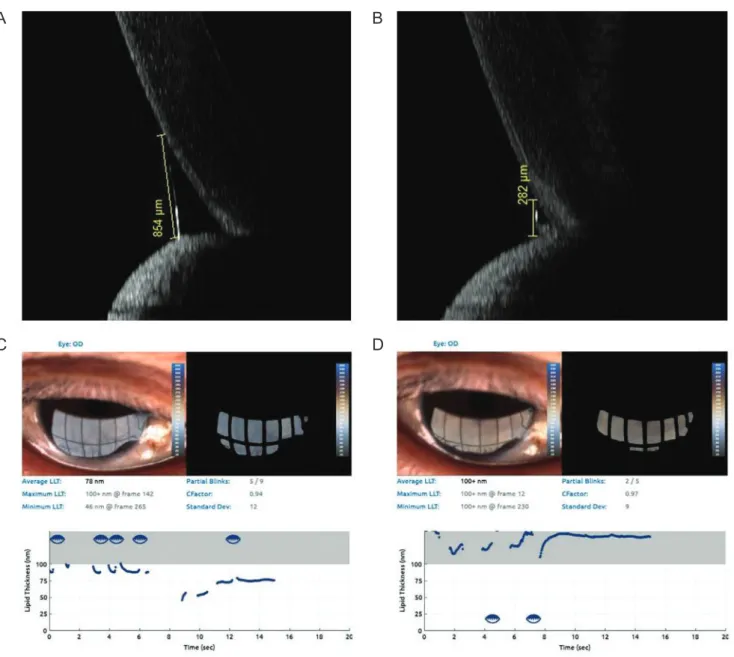
Tear meniscus height (TMH) was assessed by one op- tometrist using spectral-domain optical coherence tomog- raphy (Spectralis; Heidelberg Engineering, Heidelberg, Germany). TMH was measured from the cornea-meniscus junction to the lower lid-meniscus junction with a scan line perpendicular to the mucocutaneous junction. The exam- ination was performed twice for each patient. TMH was used for confirming the amount of tears both before and after silicone tube intubation (Fig. 1A, 1B) [11].
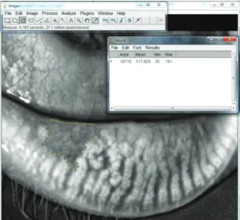
The LLT and blinking pattern were assessed using the LipiView II (TearScience, Morrisville, NC, USA) ocular surface interferometer by one optometrist. The LLT infor- mation included the minimum, maximum, and average for 20 seconds (Fig. 1C, 1D). The blinking pattern was ob- tained by determining the total blinking and partial blink- ing rates in 20 seconds and then calculating the partial blinking/total blinking ratio. The Meibomian gland drop- out ratio, the ratio of the Meibomian gland drop-out area to the total area, was measured using the polygon selection tool in the Image J program (National Institutes of Health, Bethesda, MD, USA) (Fig. 2) [12].
Statistical analyses
Statistical analyses were performed using IBM SPSS Statistics ver. 20.0 (IBM Corp., Armonk, NY, USA). Inde- pendent t-test was used to compare the overall LLT, blink- ing pattern, Meibomian gland drop-out ratio, and TMH of control and nasolacrimal duct patients. Paired t-test was used to compare the overall LLT, blinking pattern, Meibo-
mian gland drop-out ratio, and TMH both before and after silicone tube intubation. A p-value <0.05 was considered to indicate significance.
Results
This study evaluated 52 incomplete NLDO patients (68 eyes), including 16 male (30.8%) and 36 female (69.2%) pa- tients, and 21 controls (42 eyes), including 6 male (28.6%)
and 15 female (71.4%) patients. The average age and dura- tion of incomplete NLDO patients were 60.5 ± 12.9 years and 4.82 ± 3.95 months, respectively. The mean age of the controls was 54.1 ± 10.6 years, and there were no significant differences (p > 0.05). The average age and disease dura- tion were 64.5 ± 14.7 years and 5.05 ± 5.16 months, respec- tively, in male incomplete NLDO patients and 58.7 ± 11.8 years and 3.35 ± 4.57 months, respectively, in female in- complete NLDO patients. The mean Munk score was 4.2 ± 1.2, 4.0 ± 1.3 in male patients and 4.3 ± 1.2 in female patients.
Fig. 1. Tear meniscus height, lipid layer thickness and blinking patterns before and after silicone tube intubation. (A,B) Preoperative and postoperative tear meniscus height obtained using spectral-domain optical coherence tomography (Spectralis, Heidelberg Engineering, Heidelberg, Germany). (C,D) Preoperative and postoperative lipid layer thickness and blinking patterns as measured with LipiView II (TearScience, Morrisville, NC, USA)
B A
D C
Korean J Ophthalmol Vol.32, No.6, 2018
There were no significant differences in male and female patients (p > 0.05).
TMH is significantly increased in incomplete NLDO pa- tients; it decreased to show improvement after silicone tube intubation (p < 0.001) (Table 1 and Fig. 3A). There was no significant difference in LLT between control and incom- plete NLDO patients. Before performing silicone tube in- tubation, the minimum, maximum, and average LLT rates were 62.4 ± 24.0, 86.7 ± 17.9, and 71.7 ± 23.3 nm, respec- tively. Significant changes in the minimum, maximum, and average LLT (74.8 ± 23.6, 98.8 ± 11.0, and 91.6 ± 16.1 nm, respectively) were observed after silicone tube intubation
(p < 0.001, p = 0.001, and p < 0.001) (Table 1 and Fig. 3B).
The numbers of total and partial blinking events in 20 seconds were 8.2 ± 3.6 and 3.9 ± 1.9, respectively, in the control group. Preoperative total blinking and partial blinking for 20 seconds were 8.5 ± 5.3 and 4.1 ± 3.8, re- spectively, while the corresponding postoperative events totaled 7.9 ± 4.9 and 4.1 ± 3.8. There was no significant dif- ference in partial blinking or partial blinking/total blinking ratio between the controls and the incomplete NLDO pa- tients (p = 0.422, p = 0.078) (Table 1). In addition, there was no significant difference between the preoperative and postoperative partial blinking/total blinking ratios p = 0.830 (Table 1 and Fig. 3C). Finally, no significant differences in Meibomian gland drop-out ratio in control, pre, and post- operative groups (p = 0.293, p = 0.628) (Table 1 and Fig.
3D) were noted.
Discussion
According to previous studies, tear protein components in patients are more alkaline, higher in calcium concentra- tions and inflammatory cytokines, and unstable compared with normal persons [7,8]. Kubo et al. [9] reported that the tear lipid layer was thicker after dacryocystorhinostomy in four patients (five eyes). In their study, researchers deter- mined the LLT by observing the specular reflected light from the tear’s surface, a similar method to the ocular sur- face interferometer used in this study.
Tear lipids are secreted from the Meibomian glands and play an important role in maintaining the stability of the tear film. The importance of the tear lipid layer in myalgia dysfunction and dry eye has been emphasized. The LipiV- Fig. 2. Measurement of Meibomian gland drop-out ratio using the
Image J program (National Institutes of Health, Bethesda, MD, USA). Meibomian gland drop-out ratio, ratio of Meibomian gland drop-out area to total area, was measured using a polygonal selec- tion tool of the Image J program.
Table 1. Comparison of LLT, blinking pattern, and TMH between control and epiphora patients
Control Nasolacrimal duct obstruction
p-value† Preoperative Postoperative p-value*
TMH (μm) 207.1 ± 116.9 453.9 ± 309.9 264.5 ± 169.1 <0.001 <0.001
Average LLT (nm) 70.6 ± 25.3 71.7 ± 23.3 91.6 ± 16.1 <0.001 0.897
Total blinking (times/20 sec) 8.2 ± 3.6 8.5 ± 5.3 7.9 ± 4.9 0.506 0.536
Partial blinking (times/20 sec) 3.9 ± 1.9 4.2 ± 3.8 4.1 ± 3.8 0.960 0.422
Partial blinking/total blinking ratio 41.05 ± 26.30 52.28 ± 35.33 53.34 ± 35.78 0.830 0.078
Meibomian gland drop-out ratio 8.57 ± 3.65 9.48 ± 4.76 9.56 ± 4.87 0.628 0.293
LLT = lipid layer thickness; TMH = tear meniscus height.
*Paired t-test; †Independent t-test.
iew II ocular surface interferometer is a new measurement tool that can measure tear film thickness, eyelid blinking, and Meibomian glands structure [11,13]. More recent stud- ies have been conducted due to its development. Most of these studies have focused on the relationship between the tear film lipid layer and Meibomian gland dysfunction or dry eye. Eom et al. [12] reported that the LLT in South Ko- reans is 65.0 ± 19.1 interferometery color units, which is the same value obtained with a nanometer and had a similar numeric value to our study.
In this study, we investigated the tear lipid layer and pre- and postoperative changes in patients with incomplete NLDO, a typical condition of excessive tear production. In this study, the overall tear film LLT was not different be- tween the control and incomplete NLDO patients and in-
creased following silicone tube insertion. This finding is similar to the results of a previous study by Kubo et al. [9], who reported that the lipid layer could not be extended to the corneal surface by decreased tear volume and leads to a thicker and irregular lipid layer. However, in our study, the LLT in the postoperative group was significantly higher than that of the control group, but there was no significant difference in TMH. Therefore, we considered the following possibility. The lipid layer is increased with tearing to maintain homeostasis of the tear composition because the balance of tear film components is important for tear film stability, and a compensatory system is considered neces- sary in response to changes in these components [14,15].
An increased postoperative LLT might be due to the main- tained high lipid layer as the amount of tears decreases and Fig. 3. Box plot of the measured values between control and epiphora patients (before and after silicone tube intubation). (A) Tear meniscus height, (B) lipid layer thickness, (C) partial blinking/total blinking ratio, and (D) Meibomian gland drop-out ratio (*p < 0.05).
2,000
1,500
1,000
500
0
Control Preoperative Postoperative Postoperative
Postoperative Postoperative
100
80
60
40
20
Control Preoperative
1.0 0.8 0.6 0.4 0.2
0.0
Control
25 20 15 10 5
0
Control
Preoperative Preoperative
B A
D C
Korean J Ophthalmol Vol.32, No.6, 2018
lipid secretion does not change immediately. The other possibility is that the blinking dynamics produce a differ- ence of LLT after silicone tube intubation. In our study, we could only obtain the blinking pattern by ratio of partial and total blinking, and there were no significant differenc- es in the preoperative, postoperative, and control groups.
We must also consider that any blinking-related Meibo- mian gland squeezing pressure may be affected by the sili- cone tube intubation. Further studies will be needed to in- vestigate the lid blinking power along the lid margin using new instruments like cinematography to prove these ideas.
A limitation of this study is that it was carried out only by simple silicone tube insertion in incomplete NLDO.
Other techniques, such as DCR, will also be needed in the future. In addition, studies on diseases causing tearing oth- er than incomplete NLDO may be necessary. In this study, it was meaningful to measure the tear lipid layer in patients with epiphora as this has not been performed in previous tear lipid layer studies. Further study is needed to deter- mine whether the increase of tear lipid layer after silicone tube implantation is due to normalization of aqueous tear volume or to the increase of tear lipid secretion in Meibo- mian glands. In addition, the correlation between the tear lipid layer and tearing symptoms must be determined.
Conflict of Interest
No potential conflict of interest relevant to this article was reported.
References
1. Mishima S, Maurice DM. The oily layer of the tear film and evaporation from the corneal surface. Exp Eye Res 1961;1:39-45.
2. Rosenfeld L, Fuller GG. Consequences of interfacial visco- elasticity on thin film stability. Langmuir 2012;28:14238-44.
3. Goto E, Tseng SC. Differentiation of lipid tear deficiency
dry eye by kinetic analysis of tear interference images. Arch Ophthalmol 2003;121:173-80.
4. Yokoi N, Yamada H, Mizukusa Y, et al. Rheology of tear film lipid layer spread in normal and aqueous tear-deficient dry eyes. Invest Ophthalmol Vis Sci 2008;49:5319-24.
5. Tomlinson A, Doane MG, McFadyen A. Inputs and outputs of the lacrimal system: review of production and evapora- tive loss. Ocul Surf 2009;7:186-98.
6. Bron AJ, Tiffany JM, Gouveia SM, et al. Functional aspects of the tear film lipid layer. Exp Eye Res 2004;78:347-60.
7. Lee JK, Kim TH. Changes in cytokines in tears after endo- scopic endonasal dacryocystorhinostomy for primary acquired nasolacrimal duct obstruction. Eye (Lond) 2014;28:600-7.
8. Lew H, Yun YS, Lee SY. Electrolytes and electrophoretic studies of tear proteins in tears of patients with nasolacrimal duct obstruction. Ophthalmologica 2005;219:142-6.
9. Kubo M, Sakuraba T, Arai Y, Nakazawa M. Tear lipid layer interference changes after dacryocystorhinostomy. Jpn J Ophthalmol 2001;45:653-6.
10. Munk PL, Lin DT, Morris DC. Epiphora: treatment by means of dacryocystoplasty with balloon dilation of the na- solacrimal drainage apparatus. Radiology 1990;177:687-90.
11. Sung Y, Park JS, Lew H. Measurement of lacrimal punctum using spectralis domain anterior optical coherence tomogra- phy. Acta Ophthalmol 2017;95:e619-24.
12. Eom Y, Lee JS, Kang SY, et al. Correlation between quan- titative measurements of tear film lipid layer thickness and meibomian gland loss in patients with obstructive meibo- mian gland dysfunction and normal controls. Am J Oph- thalmol 2013;155:1104-10.
13. Finis D, Pischel N, Schrader S, Geerling G. Evaluation of lipid layer thickness measurement of the tear film as a diagnostic tool for Meibomian gland dysfunction. Cornea 2013;32:1549-53.
14. Arita R, Fukuoka S, Morishige N. new insights into the lip- id layer of the tear film and meibomian glands. Eye Contact Lens 2017;43:335-9.
15. Yokoi N, Takehisa Y, Kinoshita S. Correlation of tear lipid layer interference patterns with the diagnosis and severity of dry eye. Am J Ophthalmol 1996;122:818-24.