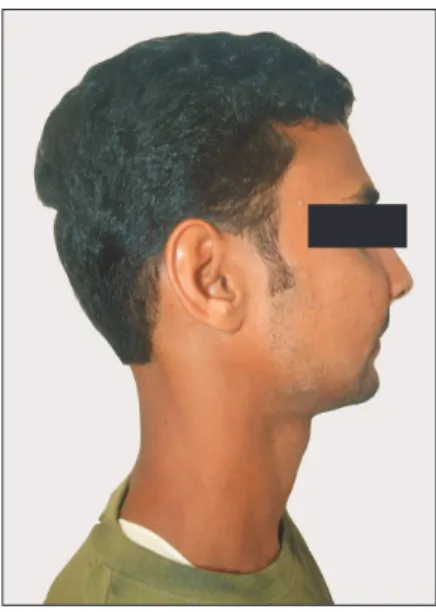
Kunhitharuvai Memorial Charitable Trust Dental College and Hospitals with the chief complaint of swelling in the upper right face region for the previous six months.(Fig. 1) His- tory revealed altered sensation on the right mid-face region for six months, noticed during his daily chores. No history of pain was present. The swelling was present for 4 months, fi rst noticed as a small hardness that progressively developed into the current size. Swelling was diffuse in nature and was
I. Introduction
Sinonasal sarcoidosis (SNS) is a rare condition that was fi rst described by Boeck in 1905
1. Occurrence is either iso- lated or part of a multisystem
2. In general, sarcoidosis is a chronic non-caseating granulomatous disease of unknown origin. It is usually associated with a lung lesion, and occur- rence without lung involvement is very rare
3. Sarcoidosis is characterized by a non-caseating granuloma found during bi- opsy. The alleged prevalence rate varies from 0.7% to 10%
4, and appears to occur in individuals living in a moist habitat.
II. Case Report
A 27-year-old male patient reported to the Department of Oral and Maxillofacial Surgery, outpatient department of
Shermil Sayd
Department of Oral and Maxillofacial Surgery, Kunhitharuvai Memorial Charitable Trust (KMCT) Dental College and Hospitals, Manassery, Mukkam, Kozhikkode, Kerala 673602, India
TEL: +91-9446230425 E-mail: [email protected]
ORCID: http://orcid.org/0000-0002-1765-8955
This is an open-access article distributed under the terms of the Creative Commons Attribution Non-Commercial License (http://creativecommons.org/licenses/by-nc/4.0/), which permits unrestricted non-commercial use, distribution, and reproduction in any medium, provided the original work is properly cited.
CC
Kunhitharuvai Memorial Charitable Trust (KMCT) Dental College and Hospitals, Kozhikode, India
Abstract
(J Korean Assoc Oral Maxillofac Surg 2015;41:217-221)
Sinonasal sarcoidosis in the head and neck region is infrequent. Its occurrence can be either isolated in combination with other systems. The literature reveals that the occurrence of sinonasal sarcoidosis without lung involvement is rare. In general, sarcoidosis is a chronic non-caseating granulomatous disease of unknown origin, often identifi ed after biopsy. In this article, we report on a benign tumor of the face that produced a diagnostic dilemma, necessitating refi nement of the surgical access and
in toto
removal of the benign tumor.Key words: Paranasal sinus, Maxillary sinus, Sarcoidosis, Granulomatous lesion, Facial paresthesia
[paper submitted 2015. 1. 15 / revised 1st 2015. 3. 8, 2nd 2015. 4. 7 / accepted 2015. 4. 15]
Copyright Ⓒ 2015 The Korean Association of Oral and Maxillofacial Surgeons. All rights reserved.
Fig. 1. Preoperative swelling on the right midface region.
Benny Joseph et al: Sinonasal sarcoidosis of the maxillary sinus and infraorbital nerve: a case report. J Korean Assoc Oral Maxillofac Surg 2015
sional biopsy and was posted for excision in toto, using blunt dissection with vestibular incision for the superficial lesion and the Caldwell-Luc approach for the sinus lesion, under general anesthesia.
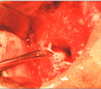
A vestibular incision was made extending from the right central incisor region to the right molar region.(Fig. 3) The tumor mass was encapsulated (5×3 cm), and the dissection proceeded with the aim of in toto removal of the lesion.(Fig. 4) During removal of the superfi cial lesion, an attachment of the mass to the infraorbital nerve (ION) with minimal erosion of the infraorbital foramen was found, which was not previously appreciated in the CT scan. A modified Caldwell Luc pro- cedure was performed by creating a window inferior and in- volving the foramen in order to further access the lesion.(Fig.
5) The whole nerve and lesion was excised in toto. Involve- only noticeable on close examination. Intraoral examination
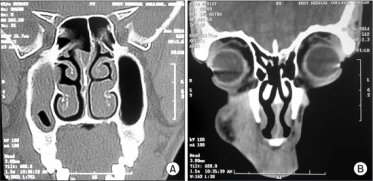
revealed no specific findings. There was no tooth mobility, or tenderness on gingiva. There was no presence of any cari- ous tooth or tenderness on percussion. All periodontal health parameters were within normal limits. There was no previous history of any sarcoidosis. On inspection, the swelling was ovoid in shape, and on palpation, a soft-swelling, non-tender, smooth tumor with well-defined borders was found. Com- puted tomography (CT) scan revealed a well-defi ned dense soft tissue tumor with homogenous enhancement extending from the right infra orbital foramen to the buccal vestibule along with sinus involvement.(Fig. 2) Chest X-ray revealed no lesions with cross specialty consultations. The case was provisionally diagnosed as a granulomatous tumor after inci-
A B
Fig. 2. Computed tomographies show sinus lesion (A) and soft tissue lesion (B).
Benny Joseph et al: Sinonasal sarcoidosis of the maxillary sinus and infraorbital nerve: a case report.
J Korean Assoc Oral Maxillofac Surg 2015
Fig. 3. Placement of intraoral vestibular incision to gain access to the lesion.
Benny Joseph et al: Sinonasal sarcoidosis of the maxillary sinus and infraorbital nerve: a case report. J Korean Assoc Oral Maxillofac Surg 2015
Fig. 4. Excised mass
in toto
.Benny Joseph et al: Sinonasal sarcoidosis of the maxillary sinus and infraorbital nerve: a case report. J Korean Assoc Oral Maxillofac Surg 2015
tion of the nasolabial fold.(Fig. 7) At the one-month review, the patient was referred to another center for further investi- gation to rule out any other organ involvement with sarcoid- osis.
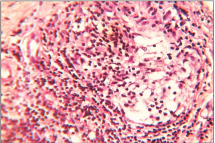
Sarcoidosis is a classic epithelioid granuloma producing disease. The most signifi cant difference between sarcoidosis and tuberculosis is that sarcoidosis lacks caseous necrosis and acid fast organisms. Formation of compact granulomas con- sists of epithelioid cells with scattered multinucleated giant cells, usually of the Langhans type. Peripheral lymphocytes, plasma cells, and fibroblasts are usually present, and there may be formation of dense collagen. Within the granulomas, ment of the ION suggested a possible inclusion of the termi-
nal branches of the facial nerve as well. Incisions were closed primarily after antral pack placement.(Fig. 6) The specimen was sent for histopathological studies. Final diagnosis of SNS was made upon clinical, radiographical and histopathological studies.
The postoperative recovery period was uneventful and sup- portive glucocorticoid therapy was later given. One-month recall showed paresthesia over the right buccal, lateral nasal, superior labial, infraorbital regions and diminished motor function over the right maxillary region along with diminu-
Fig. 5. Nerve relation with the tumor.
Benny Joseph et al: Sinonasal sarcoidosis of the maxillary sinus and infraorbital nerve: a case report. J Korean Assoc Oral Maxillofac Surg 2015
Fig. 6. Antral pack and closure.
Benny Joseph et al: Sinonasal sarcoidosis of the maxillary sinus and infraorbital nerve: a case report. J Korean Assoc Oral Maxillofac Surg 2015
Fig. 7. One-month postoperative.
Benny Joseph et al: Sinonasal sarcoidosis of the maxillary sinus and infraorbital nerve: a case report. J Korean Assoc Oral Maxillofac Surg 2015
Fig. 8. H&E stained low power (×10) section shows abundant chronic infl ammatory cells interspersed with different type of multi- nucleated giant cells.
Benny Joseph et al: Sinonasal sarcoidosis of the maxillary sinus and infraorbital nerve: a case report. J Korean Assoc Oral Maxillofac Surg 2015
be confirmed. The Kviem-Siltzbach procedure is a specific diagnostic test for sarcoidosis in which specially prepared tissue derived from the spleen of a known sarcoidosis patient is intradermally injected, and the injected site is biopsied 4 to 6 weeks later. If non-caseating granulomas are seen, this is highly specifi c for the diagnosis of sarcoidosis. The diagnosis of sarcoidosis can never be certain, and over time, other fea- tures may arise that lead to an alternative diagnosis. Evidence for new organ involvement may eventually occur, confi rming the diagnosis of sarcoidosis
7.
Krespi et al.
8proposed local treatment as the mainstay treatment. Endoscopic sinus surgery is the preferred modal- ity of surgical treatment to alleviate chronic local symptoms
9. The literature describes CT fi ndings of mucosal thickening, sinus opacifi cation and signs of chronic sinus infl ammation like destruction, erosions or bone lesions
1. Treatment of SNS includes topical or systemic steroids and sinonasal surgery.
Response to treatment varies from complete remission to no improvement at all. Repeated sinus surgeries, despite sys- temic and topical steroid treatment, have been reported in the literature, implying that SNS is a rather serious variant of sarcoidosis. Another complication is the high relapse rate in spite of the long-term therapy
10. In this case there was neural involvement, which is a rare complication of SNS
11.
SNS should be included in the differential diagnosis for swelling occurring in the maxillofacial region, despite its rare occurrence. A more skeptical diagnostic approach is recom- mended to survey the extent of nerve involvement and pos- sible intracranial extension. In toto surgical removal followed by supportive glucocorticoid therapy and follow up should be considered. More future perspective studies are suggested to inclusion bodies may be seen.(Fig. 8-10)
The required permissions have already been obtained from the patient and can be submitted on request.
III. Discussion
Isolated SNS is a rare entity that clinically presents with signs of chronic infl ammatory rhinitis and responds poorly to conventional treatment
1. Complete ear, nose and throat (ENT) examination and guided biopsy is required for diagnostic confi rmation
5. Differential diagnosis includes Wegener gran- ulomatosis, Churg-Strauss syndrome, aspergillosis, rare ENT localizations of Crohn’s disease and actinomycosis
6. Most frequent sites include the nasal septum and inferior turbi- nates, followed by paranasal sinuses, nasal bone and cartilage and sub-cutaneous tissues in that region
2.
Diagnosis of sarcoidosis requires consideration of all clini- cal and pathological features. The etiology of sarcoidosis is still unknown, so diagnosis can never be made with 100%
certainty. Patients are evaluated for sarcoidosis in two scenar- ios. In the fi rst scenario, the patient may undergo biopsy of the asymptomatic tumor to reveal a non-caseating granuloma.
And if the features are consistent with granuloma and there are no other alternative causes for non-caseating granuloma, the patient can be diagnosed with granuloma. This was the scenario in this case study. The second scenario is a patient presenting with signs or symptoms suggesting sarcoidosis, such as the presence of bilateral adenopathy in an otherwise asymptomatic patient or a patient with uveitis or a rash con- sistent with sarcoidosis. At this point, a diagnostic procedure should be performed and the presence of sarcoidosis should
Fig. 9. H&E stained high power (×40) section shows chronic in- fl ammatory cells.
Benny Joseph et al: Sinonasal sarcoidosis of the maxillary sinus and infraorbital nerve: a case report. J Korean Assoc Oral Maxillofac Surg 2015
Fig. 10. H&E stained low power (×10) section shows chronic in- fl ammatory cells, multinucleated giant cells and connective tissue.
Benny Joseph et al: Sinonasal sarcoidosis of the maxillary sinus and infraorbital nerve: a case report. J Korean Assoc Oral Maxillofac Surg 2015
ciation with lupus pernio. Thorax 1976;31:660-4.
7. Crystal RG. Sarcoidosis. In: Wonsiewicz M, Englis MR, eds.
Harrison's principles of internal medicine. 16th ed. New York:
McGraw-Hill; 2005:2022-3.
8. Krespi YP, Kuriloff DB, Aner M. Sarcoidosis of the sinonasal tract:
a new staging system. Otolaryngol Head Neck Surg 1995;112:221- 7.
9. Long CM, Smith TL, Loehrl TA, Komorowski RA, Toohill RJ. Si- nonasal disease in patients with sarcoidosis. Am J Rhinol 2001;15:
211-5.
10. Kirsten AM, Watz H, Kirsten D. Sarcoidosis with involvement of the paranasal sinuses: a retrospective analysis of 12 biopsy-proven cases. BMC Pulm Med 2013;13:59.
11. Mazziotti S, Gaeta M, Blandino A, Vinci S, Pandolfo I. Perineural spread in a case of sinonasal sarcoidosis: case report. AJNR Am J Neuroradiol 2001;22:1207-8.
ORCID
Benny Joseph, http://orcid.org/0000-0002-5659-3218 Suresh Vyloppilli, http://orcid.org/0000-0002-9423-3774 Shermil Sayd, http://orcid.org/0000-0002-1765-8955 Niyas Ummer, http://orcid.org/0000-0002-9063-0211
References
1. Scadding JG. Sarcoidosis. London: Eyre & Spottiswoode; 1967.
2. Braun JJ, Gentine A, Pauli G. Sinonasal sarcoidosis: review and report of fi fteen cases. Laryngoscope 2004;114:1960-3.