427 ciduous canines were found on the right side.(Fig. 1, 2) There was previous history of extraction of maxillary and mandibu- lar deciduous canines on the left side 2 months prior. Radio- graphic investigation using orthopantomogram was carried out and showed bilateral maxillary and mandibular impacted permanent canines.(Fig. 3) There were no associated odon- Dear Editor,
Detailed radiographic study using orthopantomograms to determine the frequency of impacted teeth and categorization of impacted canines is present in the literature1. This letter calls attention to a case of asymptomatic bilateral maxillary and mandibular impacted permanent canines in a 10-year- old Indian boy who visited our dental department for removal of corner milk teeth on the right upper and lower arches. On clinical examination, retained maxillary and mandibular de-
LETTER TO THE EDITOR
Thorakkal Shamim
Department of Dentistry, Government Taluk Head Quarters Hospital, Malappuram 676519, India
TEL: +91-989-5447351 E-mail: [email protected]
ORCID: http://orcid.org/0000-0002-6389-5417
This is an open-access article distributed under the terms of the Creative Commons Attribution Non-Commercial License (http://creativecommons.org/
licenses/by-nc/4.0/), which permits unrestricted non-commercial use, distribution, and reproduction in any medium, provided the original work is properly cited.
CC
Asymptomatic bilateral maxillary and mandibular impacted permanent canines: serendipity in dental outpatient department
Thorakkal Shamim, Prabha Surendran Renjini
Department of Dentistry, Government Taluk Head Quarters Hospital, Malappuram, India
Copyright © 2017 The Korean Association of Oral and Maxillofacial Surgeons. All rights reserved.
https://doi.org/10.5125/jkaoms.2017.43.6.427 pISSN 2234-7550·eISSN 2234-5930
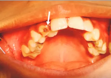
Fig. 1. Retained maxillary deciduous canine with palatal eruption of a maxillary permanent lateral incisor on the right side (arrow).
Thorakkal Shamim et al: Asymptomatic bilateral maxillary and mandibular impacted permanent canines: serendipity in dental outpatient department. J Korean Assoc Oral Maxillofac Surg 2017
Fig. 3. Panoramic radiograph showing bilateral maxillary and mandibular impacted permanent canines (arrows).
Thorakkal Shamim et al: Asymptomatic bilateral maxillary and mandibular impacted permanent canines: serendipity in dental outpatient department. J Korean Assoc Oral Maxillofac Surg 2017
Fig. 2. Retained mandibular deciduous canine on the right side (arrow).
Thorakkal Shamim et al: Asymptomatic bilateral maxillary and mandibular impacted permanent canines: serendipity in dental outpatient department. J Korean Assoc Oral Maxillofac Surg 2017
J Korean Assoc Oral Maxillofac Surg 2017;43:427-428
428
togenic neoplasms, and the patient was asymptomatic. Since there is no facility for orthodontic and surgical correction in our center, the patient was referred to a higher center for fur- ther treatment.
Conflict of Interest
No potential conflict of interest relevant to this article was reported.
ORCID
Thorakkal Shamim, http://orcid.org/0000-0002-6389-5417 Prabha Surendran Renjini, http://orcid.org/0000-0003-4143-0392
Reference
1. Al-Zoubi H, Alharbi AA, Ferguson DJ, Zafar MS. Frequency of impacted teeth and categorization of impacted canines: a retro- spective radiographic study using orthopantomograms. Eur J Dent 2017;11:117-21.