278
of implant migration into the maxillary sinus includes the classic Caldwell-Luc operation and the endoscopic approach via the nose5. In this case report, we will discuss a dental implant that disappeared following its migration into the left maxillary sinus.
II. Case Report
A 53-year-old male patient was referred to us for retrieval of a displaced dental implant that had migrated into his left maxillary sinus. Three months before his referral, the patient underwent a surgical procedure where eight dental implants were inserted to rehabilitate his fully edentulous maxilla.
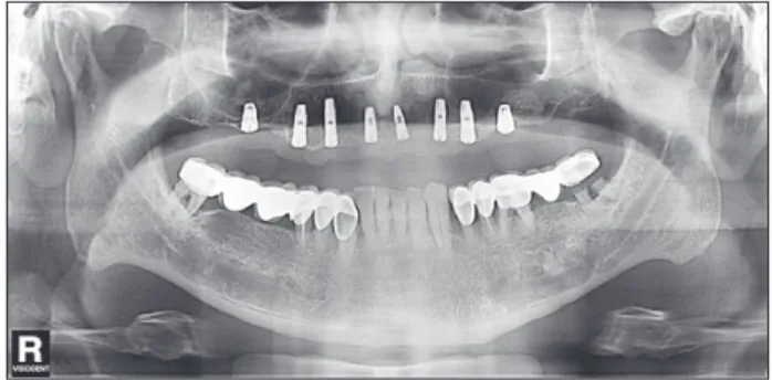
(Fig. 1) Using sinus floor elevation, implants placed in the
I. Introduction
Using dental implants for prosthetic rehabilitation of the edentulous maxilla has become a routine dental procedure.
However, low quality of the posterior maxillary bone and a highly pneumatised maxillary sinus can compromise an implant’s survival1,2. Implant migration into the paranasal sinus cavities is a rare but a significant complication. Implant migration into the maxillary sinus can be caused by inexpe- rienced practitioners, unexpected perforations during sinus floor elevation, application of heavy force during implant insertion, and dental implant placement without sinus floor elevation of an excessively pneumatised maxillary sinus3. If this condition is inadequately treated, the implants can dis- place to deeper craniofacial cavities, causing infection, tissue necrosis, and adverse reactions to foreign bodies4. Treatment
CASE REPORT
İbrahim Damlar
Department of Oral and Maxillofacial Surgery, Faculty of Dentistry, Mustafa Kemal University, Tayfur Sokmen Kampusu, Antakya, Hatay 31100, Turkey TEL: +90-3262456060 FAX: +90-3262455060
E-mail: [email protected]
ORCID: http://orcid.org/0000-0003-1453-5391
This is an open-access article distributed under the terms of the Creative Commons Attribution Non-Commercial License (http://creativecommons.org/licenses/by-nc/4.0/), which permits unrestricted non-commercial use, distribution, and reproduction in any medium, provided the original work is properly cited.
CC
Disappearance of a dental implant after migration into the maxillary sinus: an unusual case
İbrahim Damlar
Department of Oral and Maxillofacial Surgery, Faculty of Dentistry, Mustafa Kemal University, Antakya, Turkey
Abstract(J Korean Assoc Oral Maxillofac Surg 2015;41:278-280)
Migration of dental implants into the maxillary sinus is uncommon. However, poor bone quality and quantity in the posterior maxilla can increase the potential for this complication to arise during implant placement procedures. The aim of this report is to present a dental implant that migrated into the maxillary sinus and disappeared. A 53-year-old male patient was referred to us by his dentist after a dental implant migrated into his maxillary sinus.
The displaced implant was discovered on a panoramic radiograph taken five days before his referral. Using computed tomography, we determined that the displaced dental implant was not in the antrum. There was also no sign of oroantral fistula. Because of the small size of the displaced implant, we think that the implant may have left the maxillary sinus via the ostium.
Key words: Dental implants, Maxillary sinus, Migration
[paper submitted 2015. 3. 13 / revised 2015. 4. 18 / accepted 2015. 4. 21]
Copyright Ⓒ 2015 The Korean Association of Oral and Maxillofacial Surgeons. All rights reserved.
http://dx.doi.org/10.5125/jkaoms.2015.41.5.278 pISSN 2234-7550·eISSN 2234-5930
Fig. 1. Panoramic radiograph after insertion of the implants.
İbrahim Damlar: Disappearance of a dental implant after migration into the maxillary sinus: an unusual case. J Korean Assoc Oral Maxillofac Surg 2015
Displaced and disappeared implant
279 of the migrated implant (3.4 mm in diameter and 8 mm in length) and the absence of oroantral fistula, we believe that the implant left the maxillary sinus via the ostium. Thoracic and abdominal radiographs were taken for aspiration risk of the implant but no radiopaque objects within the patient’s body were detected. Similarly, a six-month follow-up of the patient proved to also be uneventful.(Fig. 4)
left and right first molar areas were inserted simultaneously.
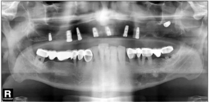
Three months postoperatively, the patient’s dentist adminis- tered a panoramic radiograph and discovered that one of the implants had migrated into the patient’s left antrum.(Fig. 2) The patient’s medical and family history was not remarkable, and intraoral examination showed no signs or symptoms of oroantral fistula. After administering computed tomography imaging to localize the implant, we discovered no foreign body in the maxillary sinus.(Fig. 3) Due to the small size
Fig. 2. Migrated dental implant into the left maxillary sinus.
İbrahim Damlar: Disappearance of a dental implant after migration into the maxillary sinus: an unusual case. J Korean Assoc Oral Maxillofac Surg 2015
Fig. 3. Computed tomography images after the implant disappeared.
İbrahim Damlar: Disappearance of a dental implant after migration into the maxillary sinus: an unusual case. J Korean Assoc Oral Maxillofac Surg 2015 Fig. 4. Six-month follow-up radiograph.
İbrahim Damlar: Disappearance of a dental implant after migration into the maxillary sinus: an unusual case. J Korean Assoc Oral Maxillofac Surg 2015
J Korean Assoc Oral Maxillofac Surg 2015;41:278-280
280
and life-threatening conditions.
In conclusion, foreign bodies in the maxillary sinus should be retrieved as soon as possible to avoid infections, maxillary sinusitis, and the potential risk of foreign body aspiration.
Conflict of Interest
No potential conflict of interest relevant to this article was reported.
References
1. Atef M, Hakam MM, ElFaramawey MI, Abou-ElFetouh A, Ekram M. Nongrafted sinus floor elevation with a space-maintaining tita- nium mesh: case-series study on four patients. Clin Implant Dent Relat Res 2014;16:893-903.
2. Kim JH, Kim YK, Bae JH. Retrospective clinical study on sinus bone graft and tapered-body implant placement. J Korean Assoc Oral Maxillofac Surg 2013;39:77-84.
3. Galindo-Moreno P, Padial-Molina M, Sánchez-Fernández E, Hernández-Cortés P, Wang HL, O'Valle F. Dental implant migra- tion in grafted maxillary sinus. Implant Dent 2011;20:400-5.
4. González-García A, González-García J, Diniz-Freitas M, García- García A, Bullón P. Accidental displacement and migration of endosseous implants into adjacent craniofacial structures: a review and update. Med Oral Patol Oral Cir Bucal 2012;17:e769-74.
5. Chiapasco M, Felisati G, Maccari A, Borloni R, Gatti F, Di Leo F.
The management of complications following displacement of oral implants in the paranasal sinuses: a multicenter clinical report and proposed treatment protocols. Int J Oral Maxillofac Surg 2009;38:
1273-8.
6. Kitamura A, Zeredo JL. Migrated maxillary implant removed via semilunar hiatus by transnasal endoscope. Implant Dent 2010;19:
16-20.
7. Tilaveridis I, Lazaridou M, Dimitrakopoulos I, Lazaridis N, Charis C. Displacement of three dental implants into the maxillary sinus in two patients: report of two cases. Oral Maxillofac Surg 2012;16:
311-4.
III. Discussion
Although complications are uncommon, dental implant migration into the maxillary sinus can occur due to inexpe- rienced operators, high pneumatisation of the sinus, and low bone density in the posterior maxilla6. Although implant mi- gration into the maxillary sinus is usually symptomless, it can cause infections that affect the paranasal sinuses and oroan- tral communications. Implants that have migrated into the pa- ranasal sinuses may also cause sinusitis and adverse reactions to foreign bodies7. Migrated dental implants can primarily be diagnosed using three-dimensional computed tomography but can also be observed on panoramic radiographs and lateral or frontal cephalograms.
Retrieval of migrated implants can be achieved via stan- dard Caldwell-Luc operations, transoral functional endo- scopic sinus surgeries (FESS), and transnasal FESS4. Some advantages of endoscopic removal of foreign bodies from the atrium include low morbidity, rapid recovery periods, and possible treatment of the affected paranasal cavities4. For instance, Kitamura and Zeredo6 describe a case report where a migrated dental implant was removed endoscopically via semilunar hiatus. In their report, access to the maxillary sinus via semilunar hiatus was cited to be undemanding.
To our knowledge, this is the first report of a dental implant that migrated into the maxillary sinus and left the sinus cav- ity without further treatment. Because the patient claimed to have experienced no sensation of swallowing the implant, we believe that the dental implant may have left the antrum via the ostium while the patient was sleeping. Therefore, this complication may create the risk of foreign body aspiration