Aspergillosis is a fungal disease caused by Aspergillus species and is characterized as non-invasive and invasive forms. Non- invasive aspergillosis is the most prevalent form, appearing either as an allergic reaction or cluster of fungal hyphae. Inva- sive aspergillosis is more extensive infection and mostly com- mon in immunosuppressed patients.1
Clinical manifestations of aspergillosis vary depending on the immune status of the host and the presence of tissue damage.1 Sometimes low-grade sinus infection results a mass of fungal hyphae, called aspergilloma.2Most patients with aspergilloma present non-specific symptom. When symptoms occur, they are usually long lasting and not much different from those of chronic sinusitis.3
The maxillary sinus is the most common site for the formation of an aspergilloma, followed by the sphenoid sinus.3The fre- quent infection of maxillary sinus, compared to other sinuses, can be related to the increased chance of fungi moving into the maxillary sinus during the extraction of tooth of maxillary posterior dentition and the endodontic treatment.4,5
This report presents a patient showing mild symptoms of suspected sinusitis with fungal infection based on radiographic examination.
Case Report
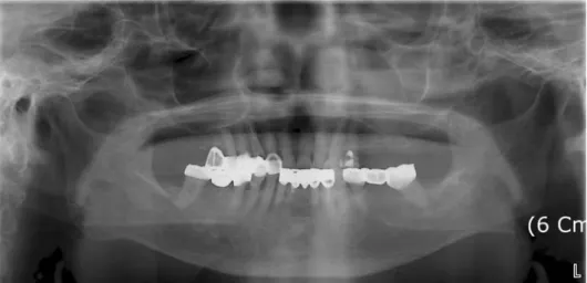
An 86-year-old female was referred to our dental hospital complaining of intermittent pain on the left maxillary first premolar area for one year. She had extracted the left maxillary first molar about two years before. She had diabetes mellitus and hypertension, and was on medication to control these con- ditions. Intraoral examination revealed only tenderness to palpa- tion on the vestibule of the left maxillary first premolar, and no other significant pathologic finding was observed. Intraoral and panoramic radiographic examinations showed no periapi- cal lesion on the left maxillary first premolar area, and radio- pacification of the left maxillary sinus was found (Figs. 1 and 2). For further evaluation of radiolucency on left maxillary molar area, cone-beam computed tomography (CBCT) was performed. CBCT images revealed the complete radiopacifica- tion of the maxillary sinus, and scattered inhomogeneous and irregular shaped radiopaque foci within the lesion (Fig. 3). It was suspected as maxillary sinusitis with fungal infection on the radiographic examinations. Biopsy was performed on the left maxillary sinus under local anesthesia. Macroscopic exam- ination of specimen showed friable brown muddy material and microscopic examination revealed fungal mass and fungal hyphae with acute angulation (Fig. 4). Also, periodic-acid- schiff (PAS) and Gomory or Grocott methenamine silver stain for fungus were both positive. It was diagnosed as aspergilloma based on the histopathologic findings.
─ 187 ─
Aspergilloma of the maxillary sinus: report of a case
Ju-Han Kang, Jae-Jung Yu*, Gyu-Tae Kim, Yong-Suk Choi, Eui-Hwan Hwang
Department of Oral and Maxillofacial Radiology, School of Dentistry, Kyung Hee University
*Department of Oral and Maxillofacial Radiology, Kangdong Sacred Heart Hospital, Hallym Medical Center ABSTRACT
Aspergilloma of the paranasal sinus is a non-invasive form of aspergillosis, most often in the maxillary sinus. This case presents an 86-year-old female with aspergilloma of the left maxillary sinus. The patient’s chief complaint was intermittent pain on the left maxillary first premolar area. A radiopacification of the left maxillary sinus was observed on the panoramic radiograph. Cone-beam computed tomography revealed complete radiopacification of the left maxillary sinus and scattered multiple radiopaque mass inside the lesion. Biopsy was performed under local anesthesia. On microscopic examination, numerous fungal hyphae, which branch at acute angle, were observed.
The diagnosis was made as an aspergilloma based on the histopatholgic examination. (Korean J Oral Maxillofac Radiol 2010; 40 : 187-9)
KEY WORDS : Aspergillosis; Paranasal Sinus Disease; Maxillary Sinus
Received October 18, 2010; Revised November 5, 2010; Accepted November 11, 2010 Correspondence to : Prof. Eui-Hwan Hwang
Department of Oral and Maxillofacial Radiology, School of Dentistry, Kyung Hee University, 1 Hoegi-dong, Dongdaemun-gu, Seoul 130-701, Korea
Tel) 82-2-958-9405, Fax) 82-2-965-1256, E-mail) [email protected]
Korean Journal of Oral and Maxillofacial Radiology 2010; 40 : 187-9
Discussion
The genus Aspergillus, which causes Aspergillosis, is ubiqui- tous organisms, present in soil, decaying vegetation and all types of organic debris. They release resistant spores into the air and inhalation of these spores may result in an opportunistic fungal infection.2Aspergilloma of the paranasal sinus is mostly
found in older individuals with an average age of 64 (range 14-90).6Female seems to be more predisposed than male.7Pre- disposing systemic factors (ie, diabetes mellitus, asthma, and lymphoproliferative diseases) combined with aspergilloma have been reported, but the association between them remains unclear.3Only about 4% of the patients have diabetes mellitus.3 Dental care can be considered as another predisposing factor.
─ 188 ─ Aspergilloma of the maxillary sinus: report of a case
Fig. 1.Panoramic radiograph shows cloudy opacity of the left maxillary sinus.
Fig. 2.Intraoral radiographs show no periapical lesion on left maxillary first premolar. The maxillary sinus floor is intact.
Fig. 3. CBCT images show the opacification and inner increased radiodense foci of the left maxillary sinus. There is sclerotic change and no destruction of the maxillary sinus floor or wall.
It has been reported that zinc-containing dental material which has been intruded into the maxillary sinus, could cause an asper- gillus sinusitis and form an aspergilloma.8
Radiographic findings show unilateral partial or complete radiopacification of a single sinus, usually the maxillary sinus.
Since CT scan can give information on the extent and nature of the disease, CT can be the imaging procedure of choice.
The most common CT finding is partial or complete heteroge- neous opacification of the involved sinus, observed in about 90% of the cases.3 Sometimes central hyperdense spots are observed within opacification of sinus.9Those radiodense spots are thought to be formed from the dystrophic calcification in the center of the fungal masses.1Sclerosis of the bony wall of the involved sinus is commonly observed in about 60% of the cases.10Based on the radiographic study, the presence of an opacified sinus and central hyperdense foci strongly suggests the diagnosis of aspergilloma.3However, because radiographic findings are nonspecific and inconclusive, definitive diagnosis should be made from histopathological findings. In this case, although motional unsharpness had occurred, sclerotic change of sinus bony wall could be observed. Also, radiographic find- ings of complete opacification and inner radiopaque foci of the left maxillary sinus on CBCT image strongly suggested fungal infection of sinus, and reinforced the need of biopsy for definitive diagnosis.
Microscopic examination of aspergilloma shows an aggrega- tion of tightly packed hyphae, which branch at acute angle.1
Examinations of this case are consistent with those histopath- ological characteristics of aspergilloma.
For an immunocompetent patient with a non-invasive asper- gillosis, surgical debridement may be all that is necessary. For an immunocompetent patient with localized invasive aspergill- osis, surgical debridement combined with systemic antifungal therapy may be necessary, depending on the severity of the infection.1The prognosis of aspergilloma is usually good and surgical treatment alone has been effective, especially in healthy patients.2If early diagnosis of aspergilloma was made and proper treatment was performed, the infection usually resolved.2 In conclusion, early diagnosis and treatment is important for a good prognosis of aspergilloma. Since the symptom is usually non-specific or absence, radiographic study, especially CT scan, is necessary. Thus, if radiopaque foci were found inside com- plete radiopacification of the maxillary sinus on radiographic study, aspergilloma could be suspected, and biopsy should be performed for the definitive diagnosis.
References
1. Nevile BW, Damm DD, Allen CM, Bouquot JE. Oral and Maxillofacial Pathology. 3rded. Philadelphia: Saunders/Elsevier; 2009. p. 234-5.
2. Shams MG, Motamedi MH. Aspergilloma of the maxillary sinus com- plicating an oroantral fistula. Oral Surg Oral Med Oral Pathol Oral Radiol Endod 2003; 96 : 3-5.
3. Grosjean P, Weber R. Fungus balls of the paranasal sinuses: a review.
Eur Arch Otorhinolaryngol 2007; 264 : 461-70.
4. Burnham R, Bridle C. Aspergillosis of the maxillary sinus secondary to a foreign body (amalgam) in the maxillary antrum. Br J Oral Maxillo- fac Surg 2009; 47 : 313-5.
5. Khongkhunthian P, Reichart PA. Aspergillosis of the maxillary sinus as a complication of overfilling root canal material into the sinus:
report of two cases. J Endod 2001; 27 : 476-8.
6. Dufour X, Kauffmann-Lacroix C, Ferrie JC, Goujon JM, Rodier MH, Karkas A, et al. Paranasal sinus fungus ball and surgery: a review of 175 cases. Rhinology 2005; 43 : 34-9.
7. Horre R, Schumacher G, Marklein G, Kromer B, Wardelmann E, Gilges S, et al. Case Report. Maxillary sinus infection due to Emeri- cella nidulans. Mycoses 2002; 45 : 402-5.
8. Theaker ED, Rushton VE, Corcoran JP, Hatton P. Chronic sinusitis and zinc-containing endodontic obturating pastes. Br Dent J 1995;
179 : 64-8.
9. Stammberger H, Jakse R, Beaufort F. Aspergillosis of the paranasal sinuses. X-ray diagnosis, histopathology, and clinical aspects. Ann Otol Rhinol Laryngol 1984; 93 : 251-6.
10. Ponikau JU, Sherris DA, Kern EB, Homburger HA, Frigas E, Gaffey TA, et al. The diagnosis and incidence of allergic fungal sinusitis.
Mayo Clin Proc 1999; 74 : 877-84.
─ 189 ─
Ju-Han Kang et al.
Fig. 4.Microscopic examination of specimen shows a tight bundles of fungal hyphae, branching at acute angle (Hematoxylin-Eosin stain, ×100).