협부 간극에 발생한 활액막 육종: 증례보고
한지훈
1·윤규호
1·정정권
1·배정호
1·이설옥
1·오영일
1·신재명
2·백지선
2·박관수
1인제대학교 의과대학
1상계백병원 구강악안면외과,
2일산백병원 구강악안면외과
Abstract(J Korean Assoc Oral Maxillofac Surg 2011;37:545-9)
Synovial sarcoma in the buccal space: a case report
Ji-Hoon Han
1, Kyu-Ho Yoon
1, Jeong-Kwon Cheong
1, Jung-Ho Bae
1, Hsueh-Yu Li
1, Young-Il Oh
1, Jae-Myung Shin
2, Jee-Seon Baik
2, Kwan-Soo Park
1Department of Oral and Maxillofacial Surgery,
1Sanggye Paik Hospital, Inje University College of Medicine, Seoul,
2
Ilsan Paik Hospital, Inje University College of Medicine, Goyang, Korea
Synovial sarcoma (SS) is a malignant soft tissue tumor comprising 5-10% of all soft tissue sarcomas. This tumor normally occurs in the para- articular regions of the extremities but is rare in head and neck sites. SS is sometimes difficult to diagnose because it can mimic benign lesions both clinically and radiologically. This paper presents a rare case of a SS of the buccal space of a 25-year old man. The histology examination and immunohistochemistry of the mass led to a diagnosis of synovial sarcoma. The patient was treated primarily with a surgical resection, followed by radiotherapy and chemotherapy. The follow up examination 17-months after surgery showed no signs of tumor relapse or metastasis.
Key words: Synovial sarcoma, Head and neck neoplasms
[paper submitted 2011. 8. 11 / revised 2011. 10. 11 / accepted 2011. 11. 23]
살남성환자의협부간극내에발생한드문활액막육종의 증례를문헌고찰과함께보고하는바이다
.
II. 증례보고
25
세의남성환자가3
개월전부터우측협점막부위에무언가만져진다는주소로
2009
년12
월 본과에내원하였다.
안모에서우측협부의약간융기된소견을보였고
(Fig. 1),
구강내로는우측협점막과후구치부위에약
4 cm
지름의촉진가능한융기된병소가관찰되었다
.(Fig. 2)
병소의색깔 은주위점막과다르지않았고,
표면에궤양은없었으며,
촉 진시단단한느낌이었지만경결감은없었다.
구강내감각 이상이나안면신경기능장애는없었고,
연하곤란,
개구장 애,
호흡곤란,
애성등의증상은관찰되지않았다.
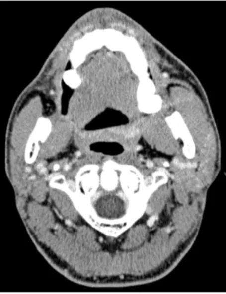
조영증강 된computed tomography (CT)
에서,
우측협부간극내에주 위골침범이없는잘경계지어지고중등도로조영증강된 둥근형태의불균질병소가관찰되었다.(Fig. 3)
타액선초음 파에서도비슷한소견이관찰되었다.(Fig. 4) Transfemoral cerebral angiography (TFCA)
를 추가로시행하였고 뚜렷 한arterial supply
를받는tumor staining
이나동정맥성기형 은관찰되지않았다.
병소는성장양상이느리고환자의임 상증상에특이한소견이없어양성으로의심되었고,
방사I. 서 론
활액막육종은모든연조직육종의약
5-10%
를차지하는악성연조직종양이다1
.
원발활액막육종의호발부위는하 지이고,
청소년이나젊은성인에게잘유발되며,
남성이여 성에비해잘이환된다1,2.
이종양은종양이보고된초기에,
발육중인 활액막조직과유사하다는이유로활액막육종 으로명명되었다.
그러나이종양은실제로활액막이나관 절주위조직이아닌원시미분화된다분화능중배엽성줄 기세포에서기원하는것으로생각되고,
관절주위조직에 한정되지않고활액막과관련없는곳에서도발발한다3.
두경부영역에서활액막육종의발생은드물다
. Jernstrom
4이1954
년에두경부영역에서의활액막육종의발생을처음 보고한이후증례보고가이어지고있지만,
활액막육종의 약9%
정도만이두경부에발생한다5.
우리교실에서는25
박 관 수139-707 서울시 노원구 상계7동 761-1
인제대학교 의과대학 상계백병원 구강악안면외과 Kwan-Soo Park
Department of Oral and Maxillofacial Surgery, Sanggye Paik Hospital, Inje University College of Medicine
761-1 Sanggye 7 dong, Nowon-gu, Seoul 139-707, Korea TEL: +82-2-950-1161 FAX: +82-2-950-1167 E-mail: [email protected]
있는
biphasic tumor
의소견이관찰되었다.(Fig. 6. A)
면역 형광화학법을통해bcl-2
와CD99
에잘염색된방추세포와pancytokeratin
에잘염색된상피세포가관찰되었다.(Figs.
6. B-D) CD34
에는반응하지않았다.
이러한소견에근거하여
biphasic synovial sarcoma (BSS)
로 진단하였다.
조직학 적검사와면역형광화학법에의해synovial sarcoma
의특징 이잘관찰되어유전자분석은시행하지않았다.
선학적으로주위조직에대한침범 없이잘경계지어진병 소가관찰되었으며
,
병소내부에blood flow
가증가된양상 을보이나뚜렷한arterial supply
는 관찰되지않아venous
malformation
으로의심되었다.
이를종합하여Hemangioma
로가진하였다. Fine-needled aspiration
은Hemangioma
의 진단에 결정적이지 않고, blood loss
의 우려가있어 시행하지않았다6
. 2010
년1
월전신마취하에 외과적절제술을시행하였다
.
병소는encapsulation
되어있어잘 절제되었다
.(Fig. 5)
조직학적검사에서,
잘증식된방추형세포가주로분포하고
, gland
를형성하고있는상피세포가산재되어Fig. 1. Pre-operative extraoral photo.
Ji-Hoon Han et al: Synovial sarcoma in the buccal space: a case report. J Korean Assoc Oral Maxillofac Surg 2011
Fig. 2. Pre-operative intraoral photo. Palpable elevated mass on right buccal mucosa and retromolar area.
Ji-Hoon Han et al: Synovial sarcoma in the buccal space: a case report. J Korean Assoc Oral Maxillofac Surg 2011
Fig. 3. Pre-operative computed tomography. Round shaped relatively well-defined mild to moderately enhancing inhomo- geneous mass (3.2 cm×2.8 cm, arrow) with internal high density foci in right buccal space without adjacent bony erosions.
Ji-Hoon Han et al: Synovial sarcoma in the buccal space: a case report. J Korean Assoc Oral Maxillofac Surg 2011
Fig. 4. Pre-operative salivary gland sonography. Ovoid shaped well-defined heterogeneous mass (2.8 cm×2.5 cm×3.9 cm) with increased blood flow in right buccal space.
Ji-Hoon Han et al: Synovial sarcoma in the buccal space: a case report. J Korean Assoc Oral Maxillofac Surg 2011
Fig. 5. Gross finding. Well defined pinkish gray soft tissue (5 cm×
4 cm×3 cm).
Ji-Hoon Han et al: Synovial sarcoma in the buccal space: a case report. J Korean Assoc Oral Maxillofac Surg 2011
Fig. 6. Photomicrographs of synovial sar coma in the buccal space. A. It reveals proliferation of spindle cells and the epithelial components forming gland-like structures (H&E staining,
×200). B. The spindle cells are stained for bcl-2 immunoreactivity (×200).
C. The spindle cells are stained for CD99 immunoreactivity (×200). D. The epithelial components are stained for cytokeratin immunoreactivity (×200).
Ji-Hoon Han et al: Synovial sarcoma in the buccal space: a case report. J Korean Assoc Oral Maxillofac Surg 2011
Fig. 7. Post-operative intraoral photo (15-months after). Scar formation on right retromolar area.
Ji-Hoon Han et al: Synovial sarcoma in the buccal space: a case report. J Korean Assoc Oral Maxillofac Surg 2011
로활액막육종은상피성부분과방추세포부분으로구성 되고두세포의분포비율과분화도에따라네아형으로나
뉜다
. BSS
는뚜렷하게구분되는상피세포와방추세포가혼재되어있는소견을보인다
.
상피성부분은흔히gland
를형성한다
.
크고둥근핵과뚜렷한경계를보이는풍부하면서 옅은세포질이상피세포의특징이고,
입방형이나 원주형으로
gland
주위에분포한다.
방추세포부분은높은핵-
세포질비를보이며일정하면서방향성 있게배열되어있다
.
두세포성분중하나로만구성되었다면monophasic fibrous synovial sarcoma
또는monophasic epithelial synovial sarcoma
로분류할수있다.
조직학적으로미분화양상을보인다면poorly differentiated synovial sarcoma (PDSS)
로분류된다. Monophasic synovial sarcoma (MSS)
와PDSS
의경우조직 학적소견만으로진단하기어렵기때문에이의진단을위 해면역조직화학법이추가적으로요구되고,
때로는세포유 전학적검사가필요하다7,10.
면역조직화학적으로활액막육종의방추세포는
vimentin, B-cell lymphoma 2 (Bcl-2), CD99
에강하게반응하고,
상 피세포는cytokeratin, epithelial membrane antigen
등의표 지자에반응한다7.
상피성표지자는대부분의다른연조직 육종의경우반응하지않지만,
활액막육종에는반응하므 로진단에유용하다. Vimentin, Bcl-2, CD99
은다른신생물 에서도발현되기 때문에감별진단이필요한데 다른연조직육종의경우에양성반응을보이는
CD34
에활액막육종은반응하지않아진단학적가치가있다7
.
이번증례에서는pancytokeratin
에대한면역반응으로상피성분화를확인하였고
,
방추세포에Bcl-2
와CD99
가강하게반응하였다.
또한
CD34
에대한발현은나타나지않아BSS
로진단하였다.
세포유전학적으로활액막육종의
95%
에서특정t(X;18)
(p11.2;q11.2)
염색체전이가나타난다.
이러한전이로분자수준에서
18
번염색체의SYT
유전자와, X
염색체의SSX1, SSX2, SSX4
중하나가결합하여SYT-SSX1, SYT-SSX2
또 는SYT-SSX4
의유전자가만들어진다10.
일반적으로SYT- SSX2
결합은MSS
와연관되고, SYT-SSX1
은BSS
에나타난다11
.
이러한SYT-SSX
의결합전이는활액막육종의결정적진단표지자로높은진단학적가치가있다
.
하지만임 상적,
조직학적,
면역조직화학적으로활액막육종의진단 이확실하다면이러한분자유전학적검사가필요하지않 다12.
두경부영역에서활액막육종의증례가적기때문에이 에대한치료법이명확히제시되지않고있다
.
다른악성종 양과마찬가지로, frozen biopsy
를동반한negative surgical
margin
의외과적절제술이최선의치료법이다.
그러나두경부영역에는 중요한해부학적 구조물들이있기때문에 병소가주위에침윤되었을경우완전한절제가어렵다5
.
따 라서두경부영역의활액막육종의경우외과적절제술후 방사선치료및보조항암화학요법으로구성된multimodal
특별한술 후 합병증은나타나지않았다
.
퇴원후55.8
Gy
의 방사선치료를시행하였고ifosphamide
를사용하여 항암치료를시행하였다.
정기적인임상검사및neck CT, positron emission tomography, chest radiography
촬영을통 해경과관찰한바,
술후21
개월이지난 지금까지병소의재발이나전이는관찰되지않았다
.(Figs. 7, 8)
본교실에서는지속적인주기적경과관찰을시행할예정이다
.
III. 고 찰
활액막육종은 공격적인고악성도의 연조직종양이다
.
두경부영역에서의활액막육종의증례는드물게보고되고 있다.
호발부위로알려진하인두외에악관절부위,
이하 선,
협점막,
상악동,
하악골,
혀,
구강저연구개,
측두하와등 의구강악안면부위에서발생한활액막육종의증례가보 고되었다7.
두경부영역에서활액막육종에 이환된대부분 의환자의경우천천히커지는무통성의촉진가능한종물 을주소로내원한다.
이외에병소부위의통증을호소하거 나,
호흡곤란,
연하곤란,
애성,
개구장애등의증상이나타 나기도한다8.
두경부영역에서발생한활액막육종은조영증강된
CT
에서잘 경계지어진불균질한다방성의병소로관찰된다.
병소내에는석회화,
출혈,
괴사,
낭종의소견이관찰될수 있다. Magnetic resonance imaging
에서는T1
강조영상에서동등신호강도를보이고
, T2
강조영상에서는신호강도가다양하게나타난다
.
이처럼두경부영역에서의발생한활 액막육종의경우방사선학적으로잘 경계된균질한병소 로관찰될경우양성종양으로보일수있다9.
조직학적으Fig. 8. Post-operative computed tomography (15-months after).
Mild fat infiltration and fascial thickening in right retromolar and buccal space.
Ji-Hoon Han et al: Synovial sarcoma in the buccal space: a case report. J Korean Assoc Oral Maxillofac Surg 2011
134 tumors. Cancer 1965;18:613-27.
2. Shmookler BM, Enzinger FM, Brannon RB. Orofacial synovial sarcoma: a clinicopathologic study of 11 new cases and review of the literature. Cancer 1982;50:269-76.
3. Tilakaratne WM. Synovial sarcoma of the mandible. J Oral Pathol Med 2006;35:61-3.
4. Jernstrom P. Synovial sarcoma of the pharynx: Report of a case.
Am J Clin Pathol 1954;24:957-61.
5. Amble FR, Olsen KD, Nascimento AG, Foote RL. Head and neck synovial cell sarcoma. Otolaryngol Head Neck Surg 1992;107:631-7.
6. Capote A, Acero J, García-Recuero I, Rey J, Guerra B, de Paz V. Infratemporal-preauricular-cervical approach for resection of a cavernous intramasseteric hemangioma: a case report. J Oral Maxillofac Surg 2008;66:2393-7.
7. Weiss SW, Goldblum JR. Enzinger and Weiss’s Soft Tissue Tumors. 4th ed. St. Louis: Mosby; 2001:1483-1509.
8. Bukachevsky RP, Pincus RL, Shechtman FG, Sarti E, Chodosh P.
Synovial sarcoma of the head and neck. Head Neck 1992;14:44-8.
9. Rangheard AS, Vanel D, Viala J, Schwaab G, Casiraghi O, Sigal R. Synovial sarcomas of the head and neck: CT and MR imaging findings of eight patients. AJNR Am J Neuroradiol 2001;22:851-7.
10. Bridge JA, Bridge RS, Borek DA, Shaffer B, Norris CW.
Translocation t(X;18) in orofacial synovial sarcoma. Cancer 1988;62:935-7.
11. Koyama S, Morimitsu Y, Morokuma F, Hashimoto H. Primary synovial sarcoma of the kidney: Report of a case confirmed by molecular detection of the SYT-SSX2 fusion transcripts. Pathol Int 2001;51:385-91.
12. Coindre JM, Pelmus M, Hostein I, Lussan C, Bui BN, Guillou L. Should molecular testing be required for diagnosing synovial sarcoma? A prospective study of 204 cases. Cancer 2003;98:2700-7.
13. Miloro M, Quinn PD, Stewart JC. Monophasic spindle cell synovial sarcoma of the head and neck: report of two cases an review of the literature. J Oral Maxillofac Surg 1994;52:309-13.
14. Bilgic B, Mete O, Oztürk SA, Demiryont M, Keles N, Basaran M. Synovial sarcoma: a rare tumor of larynx. Pathol Oncol Res 2003;9:242-5.
15. Eilber FC, Brennan MF, Eilber FR, Eckardt JJ, Grobmyer SR, Riedel E, et al. Chemotherapy is associated with improved survival in adult patients with primary extremity synovial sarcoma. Ann Surg 2007;246:105-13.
16. Moore DM, Berke GS. Synovial sarcoma of the head and neck.
Arch Otolaryngol Head Neck Surg 1987;113:311-3.
17. Dei Tos AP, Dal Cin P, Sciot R, Furlanetto A, Da Mosto MC, Giannini C, et al. Synovial sarcoma of the larynx and hypopharynx.
Ann Otol Rhinol Laryngol 1998;107:1080-5.
18. Meer S, Coleman H, Altini M. Oral synovial sarcoma: a report of 2 cases and a review of the literature. Oral Surg Oral Med Oral Pathol Oral Radiol Endod 2003;96:306-15.
19. Carrillo R, Rodriguez-Peralto JL, Batsakis JG. Synovial sarcomas of the head and neck. Ann Otol Rhinol Laryngol 1992;101:367-70.
20. Torsiglieri AJ Jr, Hendrix RA, Quinn PS. Synovial sarcoma of the jaw. Ear Nose Throat J 1991;70:396-8.
therapy
가 추천된다13,14. 65 Gy
이상선량의 방사선 치료 가재발률을낮추는 것으로알려져있다5.
문헌에의하면ifosphamide
를 사용한 항함화학요법은전이를 억제하고생존율을향상시킨다14,15
.
활액막 육종은 혈행을 통해전 이되고림프절로의전이는드물기때문에임상적,
방사선학적으로림프절 종대가관찰되지 않는다면예방적
neck
dissection
은추천되지않는다16,17.
이러한
multimodal therapy
에도불구하고두경부영역에서발생한활액막육종에이환된환자의
5
년생존율은40-
50%
으로예후가불량하다.
이러한낮은생존율은폐로의전이에기인한다13
.
두경부 영역의활액막육종은20.8%
의재발률과
29.2%
의전이율을 보였는데,
이것은 다른부위에서생긴원발 활액막육종의재발률
(50%)
과전이율(40-
83%)
보다낮았다5,18.
재발은보통수술후2
년내에발생하 지만,
늦게재발하거나전이하는경우도있기때문에장기간의경과관찰이추천된다18,19
.
몇몇문헌에의하면두경부영역에서발생한활액막육 종의경우병소의조기발견이가능하고
,
크기가작고,
상대 적으로낮은 연령에서발견되기때문에다른부위의활액 막육종에비해덜공격적이고좋은예후를보인다고하였 다2,20.
병소의크기가5 cm
이하이고,
광범위한석회화가있 으며,
외과적으로성공적으로절제되고,
미분화 부위나괴 사부위가없으며 유사분열이적을때임상적으로양호한결과를보인다2,5,8
.
이번증례의경우수술후
21
개월이지난지금재발이나 전이의소견은없이양호한결과를보이고있다.
하지만두 경부영역에서발생한활액막육종의경우예후가좋지않 고,
재발의우려가높기때문에본교실에서는지속적인주 기적경과관찰예정이다.
이번증례는
,
비록두경부의활액막육종이드물게발생 하지만,
임상적,
방사선학적으로양성병소와 비슷하기때 문에구강악안면부위에발생한병소의감별진단에활액막 육종이고려되어야함을보여준다.
세침흡인생검등의방 법을통한술전진단이치료계획수립에도움이될수있다.
References
1. Cadman NL, Soule EH, Kelly PJ. Synovial sarcoma: an analysis of