하치조신경 마취 후 전기치수검사기(Electric pulp tester)를 이용한 하순부 감각 변화 평가
구명숙·김진욱·전영훈·권대근·이상한 경북대학교 치의학전문대학원 구강악안면외과학교실
Abstract(J Korean Assoc Oral Maxillofac Surg 2011;37:464-9)
Evaluation of the change of lower lip sensation after inferior alveolar nerve block by using the electric pulp tester
Myong-Suk Ku, Jin Wook Kim, Young Hoon Jeon, Tae Geon Kwon, Sang Han Lee Department of Oral and Maxillofacial Surgery, School of Dentistry, Kyungpook National University, Daegu, Korea
Introduction: As dental implant surgery is becoming increasingly popular, it has become one of the causes for the hypesthesia of the inferior alveolar nerve, along with other surgical procedures, such as a third molar extraction. In addition, it tends to cause legal problems between the operator and patient. Therefore, there must be a proper method that is reliable, objective and economical to assess the nerve impairment. For this reason, an attempt was made to use an Electric Pulp Tester to assess inferior alveolar nerve block anesthesia.
Materials and Methods: Thirty patients were tested. Electric pulp testing of the lower jaw skin was performed at the three different times, before anesthesia, at the onset of sensory changes and after 15 minutes waiting from the onset, and on the 10 points of the chin, which produced 10 sections on the skin area.
Results: Twenty seven patients (90%) could feel the electric stimulus on the chin at all 10 points before local anesthesia and the scores represent the statistical differences between the right and left points except R4 and L4. After anesthesia, the difference between the right and left points (L3-R3, L4-R4, L5-R5) increased significantly with time but two points (L2, R2) showed no significant difference. The scores on the left chin (L3, L4, L5) increased, whereas the other points (R1-R5, L1, L2) showed no significant differences.
Conclusion: This study highlights the potential clinical use of an electric pulp tester for an assessment of inferior alveolar nerve impairment.
Key words: Pulp test, Hypesthesia, Inferior alveolar nerve
[paper submitted 2011. 7. 24 / revised 2011. 9. 2 / accepted 2011. 10. 12]
진술이라는주관적인평가에의존하고있는것이현실이다
.
현재까지하치조신경의 손상정도를평가하기위해사 용되고있는진단방법으로는정적촉각검사,
동적촉각검 사,
두점식별능검사(two point discrimination),
냉온각검 사,
통각검사,
전기생리학적검사(
체성감각유발 전위,
전 기치수진단)
등의방법이사용되고있다1.
이들진단방법 들은객관적인수치로표현하기어렵고,
보다정확한평가 를위해필요한고가의장비등을개인의원에서는갖추기 가힘들다.
따라서감각신경손상에대한객관적이고도쉽 게이용할수있는평가방법에한계가있는실정이다.
전기치수검사기
(electric pulp tester, EPT)
는 치아생활 력평가에서민감도를평가하기위해흔히사용되는기구 로,
낮은자극으로부터자극의강도를서서히증가시키며 피검자의 인지반응으로 치수생활력을 평가하는기구이다
. Certosimo
와Archer
2는이전기치수검사기가국소마취심도를측정하는좋은표지자로사용될수있다고하였고
,
Yikontiola
등3은치아에전기치수검사기로 반응하는수치I. 서 론
하치조신경지각이상은하악제
3
대구치발치,
종양,
골수 염,
골절,
악골성형술등의외과적 처치중에초래되는신 경손상때문에주로발생되지만,
최근치과임플란트의대 중화는하치조신경손상의또 다른원인요소로부각되고 있다.
임플란트로인한하치조신경손상은환자본인의지 각이상으로인한불편함과더불어의원성요인의신경손 상에대한법적인보상문제를야기하고있다.
하지만감각 신경손상의특성상손상받은신경에대한평가가환자의이 상 한
700-705 대구시 중구 달구벌대로 2175
경북대학교 치의학전문대학원 구강악안면외과학교실 Sang Han Lee
Department of Oral and Maxillofacial Surgery, School of Dentistry, Kyungpook National University 2175, Dalgubeoldaero, Jung-gu, Daegu 700-721, Korea TEL: +82-53-600-7561 FAX: +82-53-426-5365 E-mail: [email protected]
하였다
.(Fig. 1)
감각이상을보이지않는 정상적인환자들을대상으로하였으며대상자의평균연령은
31±3.4
세(26-
44
세)
였다. 2. 연구 방법
전기치수검사를시행하는술자가소독용장갑을착용한 상태에서
,
환자에게기구의금속판을잡게하고금속프로 우브에치약을적용하여접촉면적을넓힌다음술자가천 천히 노브를돌려 환자가반응하는수치를기록하는방법 을사용하였으며,
반응시기에따라마취전,
후연조직반응 이온시점과15
분경과후에다시측정하여모두3
회측정 하였다.
1) EPT
0마취전정상환자의전기치수검사기반응수치로프로우 브끝에 치약을바른 다음환자가전기치수검사기의금속 를평가하여 신경감각 이상을평가하는 것이객관적이며
경제적인방법이라고보고하였으나아직도보편적으로이 용되고있지않다
.
하치조신경손상환자들은치아에대한 감각이상보다는하순및이부에대한감각저하및소실로 인한불편함을크게호소하는데,
전기치수검사기에 의한 하순및이부의연조직반응을객관적인평가방법의하나 로이용할수있다면상당한임상적의의를기대할수있을 것이다.
전기치수검사기는 신경이밀집되어있는치수에 역치이상의자극을주었을때통증반응을검사하는기계 로서,
안정성때문에최대전류량이크지않다.
전기치수검 사기로연조직을자극할때반응을나타내기위해서는하순및이부의전류감지역치
(perception threshold)
이상의자극을줄수있어야한다
.
이에이번연구에서는전기치수검사기가 하순및 이부 의감지역치이상의전류를생산할수있는지검사하기위 하여
,
정상인의하악연조직을전기치수검사기를이용하여 자극할때의반응여부와반응의일관성을조사하였으며,
하치조신경전달마취 후마취된부위의 전기치수검사기 반응을관찰하여이러한방법이신경손상환자가지각이상 을호소할때객관적인검사의한방법으로이용될수있는 지알아보고자하였다.
이번연구의목적은전기치수검사기에의한전기적자극 에하악연조직의반응이일정하게나타나는지와 감각변 화와상관된역치변화를감지할수있는지를검사하여향 후지각이상이일어난환자를진단하는데있어서임상에 적용할수있는가능성을확인하고자하였다
.
II. 연구 대상 및 방법
1. 연구 대상
본원에내원한
30
명의환자(
남자16
명,
여자14
명)
를대 상으로전기치수검사기(Gentle-Pulse Pulp Vitality Tester D624MS, Parkell Inc., Edgewood, NY, USA)
로반응을측정Fig. 1. Gentle-Pulse Pulp Vitality Tester D624MS (Parkell Inc., Edgewood, NY, USA).
Myong-Suk Ku et al. Evaluation of the change of lower lip sensation after inferior alveolar nerve block by using the electric pulp tester. J Korean Assoc Oral Maxillofac Surg 2011
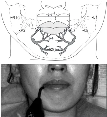
Fig. 2. Schematic diagram of the evaluation landmarks for electric pulp tester included inferior alveolar nerves and photo of measuring patient. ① R1: a center of right mandible posterior 1/3, ② R2: a center of right mandible middle 1/3, ③ R3: the point 5 mm beneath right commissure of the lip (angular branch),
④ R4: one third of lower lip vermilion border: right (inferior labial branch), ⑤ R5: a center of perpendicular line from R4 to chin (mental branch), ⑥ L5: a center of perpendicular line from L4 to chin (mental branch), ⑦ L4: one third of lower lip vermilion border: left (inferior labial branch), ⑧ L3: the point 5 mm beneath left commissure of the lip (angular branch), ⑨ L2: a center of left mandible middle 1/3, ⑩ L1: a center of left mandible posterior 1/3.
Myong-Suk Ku et al. Evaluation of the change of lower lip sensation after inferior alveolar nerve block by using the electric pulp tester. J Korean Assoc Oral Maxillofac Surg 2011
III. 결 과
실험대상자중남자
2
명,
여자1
명을제외한27
명(90%)
에서전기치수검사기에의한전기적자극을모든부위에서 느낄수있었다.
반응이없었던3
명중2
명은1
부위에서(
각 각R1
과R4), 1
명은2
부위(R1
과L2)
에서전기치수검사기 에의한전기적자극을감지하지못하였다.
마취전 정상군
10
부위의전기치수검사기자극시반응 한수치의평균은남자5.71±1.68,
여자5.36±1.58 ( P=0.64)
로 남녀별 차이에서 유의성 있는차이는 보이지 않았다.
좌우 값이동일하게나타나는가를보기위하여좌우차를paired t-test
를이용한결과마취 전30
명의 정상군에서이신경의 하순지
(R4, L4)
를 제외하고 좌우 값이통계적으로유의한차이가있었으며전반적으로우측의역치가더 크게나와우측이더 둔감함을 보였다
.
마취후에하악각과하순사이중간
1/3 (R2, L2)
에서는2
회의실험결과좌우 값이통계적으로동일하게나타났으나하악각 부위에 서는다르게나타났다
.
좌측하치조신경마취후마취된3,
4, 5
부위의좌우값은통계적으로유의한차이를나타내었다
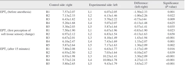
.(Table 1)
마취후 시간대별로좌우값의변화량을비교하면
,
하 치조신경을 마취할때마취가되는부위(L3, 4, 5)
의좌우 차가유의성있게나타났다.
이는하악전달마취가진행되 면서역치가높아짐을나타내는것으로,
사후검증에서도L5-R5
의마취전과 후연조직반응이나타난시점의비교만을제외하고좌우
3, 4, 5
부위모두에서마취후 좌우의전기치수검사기의반응수치가유의성있는차이를보여주 판을잡게하고프로우브를
R1
에서R5, L5
에서L1,
즉①
에서
⑩
의순서로측정할부위에접촉시킨후환자가전기적 인반응을느낄때까지노브를돌려반응시의수치를기록 하였다.
이때환자에게는통증을느낄때까지기다리는것 이아니라전기적인느낌이오면바로손을들것을요구하였다
.(Fig. 2)
만일마취후전기치수검사기의노브눈금(0-
10)
에서반응이없는경우통계적검증을위해마취전,
후의평균적변화량과가장가까운
12
를임의로정하였으며 피부의10
부위를좌우로나누어평균을측정하였다.
2) EPT
1(
마취반응직후)
2%
염산리도케인(1:100,000, 1.8 mL, Huons Inc., Seoul,
Korea) 1
개를이용하여좌측하치조신경전달마취후좌측입술의지각변화가오면
①
에서⑩
의순서로EPT
를이용 하여측정하였다.
3) EPT
2(
마취반응15
분경과)
EPT
1측정후15
분이지난시점에서다시①
에서⑩
의순서로측정하였다
.
1), 2), 3)
을EPT
0, EPT
1, EPT
2로 나누어EPT
에 반응하 는수치의좌우차 변화량과마취시점에따른각측정부위변화량의통계적인유의성을
SPSS ver. 10 (SPSS Inc.,
Chicago, IL, USA)
을 이용하여 대응비교t
검증(paired t-test),
변량분석(ANOVA),
사후분석법(Post-hoc test)
을시 행하였으며P
값0.05
이하를통계적으로유의한수준으로 나타내었다.
Table 1. Difference between the right and the left value evaluated by EPT
Control side: right Experimental side: left Difference:
(left-right) Significance (P-value) EPT0 (before anesthesia)
EPT1 (first perception of soft tissue sensory change)
EPT2 (after 15 minutes)
R1 R2 R3 R4 R5 R1 R2 R3 R4 R5 R1 R2 R3 R4 R5
7.57±2.07 7.13±2.33 6.43±1.82 5.20±1.68 5.30±2.03 7.50±1.90 6.97±2.12 6.67±2.01 6.10±2.47 5.87±2.64 7.80±2.08 6.93±2.50 6.93±1.98 5.73±2.24 5.80±2.65
L1 L2 L3 L4 L5 L1 L2 L3 L4 L5 L1 L2 L3 L4 L5
6.07±2.05 6.13±1.46 5.70±2.22 5.07±2.07 5.87±1.64 6.67±1.96 6.83±1.54 8.10±1.49 7.43±1.69 7.17±1.63 6.63±1.77 7.03±1.64 10.23±1.41 10.00±1.78 9.43±1.79
-1.50±2.33 -1.00±2.26 -0.73±144 -0.13±1.48
0.57±1.41 -0.83±1.90 -0.13±1.63 1.43±1.94 1.33±2.07 1.30±2.09 -1.17±2.49 0.10±2.67 3.30±2.09 4.27±2.13 3.63±2.37
0.001 0.022 0.009 0.625 0.035 0.023 0.658
<0.001 0.001 0.002 0.016 0.839
<0.001
<0.001
<0.001 (EPT: electric pulp tester, R1: a center of right mandible posterior 1/3, R2: a center of right mandible middle 1/3, R3: the point 5 mm beneath right commissure of the lip (angular branch), R4: one third of lower lip vermilion border: right (inferior labial branch), R5: a center of perpendicular line from R4 to chin (mental branch), L1: a center of left mandible posterior 1/3, L2: a center of left mandible middle 1/3, L3: the point 5 mm beneath left commissure of the lip (angular branch), L4: one third of lower lip vermilion border: left (inferior labial branch), L5: a center of perpendicular line from L4 to chin (mental branch))
Values are presented as mean±standard deviation. Statistical significance tested by paired t-test.
Myong-Suk Ku et al. Evaluation of the change of lower lip sensation after inferior alveolar nerve block by using the electric pulp tester. J Korean Assoc Oral Maxillofac Surg 2011
자자신에의한증상호소
(
자각검사청취),
마비범위표시,
정적촉각검사,
방향식별도,
두 점식별능,
유해자극인지 도,
냉·
온각검사등피검자의지각에의존하는경우와,
체 성감각 유발전위(somatosensory evoked potentials)
와이 순-
목반사검사(mento-blink reflex)
등의전기생리학적검 사,
전산화적외선체열검사(digital infrared thermographic
imaging)
등의객관적인자료를볼수있는방법등다양하다5-8
.
정적 촉각검사는Frey
촉모, SW
지각검사(Semmes- Weinstein test),
솜,
붓,
탐침등에의한접촉시험방법등이있다
. SW
지각 검사방법중하나인3
점법은이신경의종었다
.(Table 2)
시간에따른
3
회의실험에서좌우와상관없이각점의변 화량에서마취심도와연관하여일관성있는수치를보여주는가를비교하기위하여
,
각점의시간에따른ANOVA test
를시행하였다
.
그결과전달마취에의하여마취되지않는R1, R2, R3, R4, R5, L2, L1
에서시간의변화에따른유의성있는차이를볼수없었으나
,
전달마취후직접적으로마취되는부위인
L3, L4, L5
에서감각소실의정도에따라유의성있는차이를보여전기치수검사기가일관성있는 수치 를나타내며감각이상의정도가클수록그수치가증가함 을보여주었다
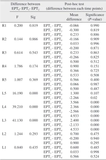
.(Table 3)
IV. 고 찰
하치조신경이충분히 마취되면마취된쪽의하악 대구 치
,
소구치,
견치와전치의치수와치주조직및이신경이분 포되어있는하순의점막과이부피부에감각저하및소실 이일어나게된다4.
신경손상시손상정도를정확히파악 하고예후를예측하는것은매우중요하다.
신경손상은환Table 2. Verification of statistical difference among the test periods EPT0- EPT1- EPT2
Difference between EPT0- EPT1- EPT2
Post-hoc test
(difference between each time points)
F Sig Mean
difference
Significance (P-value) L1-R1
L2-L3
L3-R3
L4-R4
L5-R5
0.656
2.029
35.983
40.938
19.309
0.521
0.138
0.000
0.000
0.000
EPT1-EPT0
EPT1-EPT2 EPT2-EPT0
EPT1-EPT0
EPT1-EPT2 EPT2-EPT0
EPT1-EPT0
EPT1-EPT2
EPT2-EPT0
EPT1-EPT0
EPT1-EPT2
EPT2-EPT0 EPT1-EPT0
EPT1-EPT2
EPT2-EPT0
0.667 0.333 0.333 0.867 -0.233 1.100 2.167 -1.867 4.033 1.467 -2.933 4.400 0.733 -2.333 3.067
0.489 0.835 0.835 0.293 0.913 0.142
<0.001 0.001
<0.001 0.011
<0.001
<0.001 0.334
<0.001
<0.001 (EPT: electric pulp tester, R1: a center of right mandible posterior 1/3, R2: a center of right mandible middle 1/3, R3: the point 5 mm beneath right commissure of the lip (angular branch), R4: one third of lower lip vermilion border: right (inferior labial branch), R5: a center of perpendicular line from R4 to chin (mental branch), L1: a center of left mandible posterior 1/3, L2: a center of left mandible middle 1/3, L3:
the point 5 mm beneath left commissure of the lip (angular branch), L4:
one third of lower lip vermilion border: left (inferior labial branch), L5: a center of perpendicular line from L4 to chin (mental branch)) Statistical significance tested by ANOVA and Post-hoc analysis with Tukey test.
Myong-Suk Ku et al. Evaluation of the change of lower lip sensation after inferior alveolar nerve block by using the electric pulp tester. J Korean Assoc Oral Maxillofac Surg 2011
Table 3. Comparison of the EPT value of each evaluation points over time (EPT0- EPT1- EPT2) and Post-hoc test of the difference of each evaluation landmarks
Difference between EPT0- EPT1- EPT2
Post-hoc test
(difference between each time points)
F Sig Mean
difference
Significance (P-value) R1
R2
R3
R4
R5
L5
L4
L3
L2
L1
0.200
0.144
0.614
1.786
1.007
16.190
39.210
41.130
1.244
0.840
0.819
0.866
0.543
0.174
0.369
0.000
0.000
0.000
0.293
0.435
EPT1 - EPT0
EPT1 - EPT2 EPT2 - EPT0
EPT1 - EPT0
EPT1 - EPT2
EPT2 - EPT0
EPT1 - EPT0
EPT1 - EPT2
EPT2 - EPT0 EPT1 - EPT0
EPT1 - EPT2
EPT2 - EPT0 EPT1 - EPT0
EPT1 - EPT2
EPT2 - EPT0
EPT1 - EPT0
EPT1 - EPT2
EPT2 - EPT0
EPT1 - EPT0 EPT1 - EPT2
EPT2 - EPT0
EPT1 - EPT0 EPT1 - EPT2
EPT2 - EPT0
EPT1 - EPT0
EPT1 - EPT2
EPT2 - EPT0
EPT1 - EPT0
EPT1 - EPT2 EPT2 - EPT0
-0.066 -0.300 0.233 -0.166 0.033 -0.200 0.233 -0.266 0.500 0.900 0.366 0.533 0.566 0.066 0.500 1.300 -2.266 3.566 2.366 -2.566 4.933 2.400 -2.133 4.533 0.700 -0.200 0.900 0.600 0.033 0.566
0.990 0.819 0.886 0.909 0.996 0.871 0.863 0.825 0.512 0.151 0.725 0.508 0.400 0.987 0.487 0.107 0.02 0.000 0.000 0.000 0.000 0.000 0.000 0.000 0.475 0.940 0.295 0.485 0.998 0.524 (EPT: electric pulp tester, R1: a center of right mandible posterior 1/3, R2: a center of right mandible middle 1/3, R3: the point 5 mm beneath right commissure of the lip (angular branch), R4: one third of lower lip vermilion border: right (inferior labial branch), R5: a center of perpendicular line from R4 to chin (mental branch), L1: a center of left mandible posterior 1/3, L2: a center of left mandible middle 1/3, L3:
the point 5 mm beneath left commissure of the lip (angular branch), L4:
one third of lower lip vermilion border: left (inferior labial branch), L5: a center of perpendicular line from L4 to chin (mental branch)) Myong-Suk Ku et al. Evaluation of the change of lower lip sensation after inferior alveolar nerve block by using the electric pulp tester. J Korean Assoc Oral Maxillofac Surg 2011
McDaniel
등13은마취의심도를측정하기위해과도한전기 적자극에대해서치수에조직학적인반응을가져오지않 으며안전하고효과적인방법이라고보고하였다.
깊이마 취가되는시간에대해서Mikesell
등14과Vreeland
등15은 하악은15
분,
상악은5
분이걸린다고보고하였으며이번실 험에서도하악연조직이상감각을느낀후15
분뒤다시측 정하여마취에의한오차를줄이고자하였다.
전기치수검사기는 정전압 방식으로 노브를 돌릴수록 전압이천천히증가하게되는데치아는수백
KΩ
에서수MΩ
의저항이형성되어치아에흐르는전류는20-50 μA
의전류에서생활치가자극을느끼게되고치아의상태에따
라최대
150 μA
에서자극을느끼게된다16. 60 Hz
에서피부의전류감지역치는남자
1.1 mA,
여자0.7 mA
로치아에비해감지역치가매우높으나치아보다
100-1,000
배정도저항이 낮은수천
Ω
의저항이흘러전기치수검사기에의해 전류감지역치까지자극이인지되므로하악피부에서도전 기적자극을느낄수있으리라생각한다.
그러나안전을위 해 전기적자극의 전류량이제한적이며0-10
까지의측정 폭이넓지않아,
일반적으로감지역치가높게나타나는남 자,
나이가많은환자,
향정신성의약품등의약물을복용하 는환자에게사용하기에는제한적인한계가있다.
주로감 지역치가낮은환자에있어서피부에지각이상을알수있 는진단적기구로사용가능할것으로보이며치아에대한 측정수치와함께사용할경우매우유용하리라생각한다.
이번연구는국소마취를 시행하고그에따른지각변화 를전기치수검사기로측정하여신경손상에의한하치조신 경감각이상을완전히재현한것은아니므로
,
분석에한계 가 있었다.
이러한 한계에도불구하고전기치수검사기가 하악연조직부위에정상적인감지역치이상의자극을줄 수있고,
감각의변화가없는부위에서는일관된반응을보 이며,
마취후역치의변화를객관적인수치로나타낼수있 다는것을확인하였으며,
이는전기치수검사기가감각변화 의객관적인평가 방법으로사용될가능성을보여주는것 으로생각한다.
V. 결 론
마취를하거나신경손상이일어나면먼저나타나는증상 으로하순의지각이상을 호소하게된다
.
이번연구에서는 전기치수검사기를이용하여하순의감각정도를객관적으 로감지해내는가능성을검증하고자,
하순연조직에대하 여전기치수검사기에의한전기적자극의인지 여부를평 가하였다.
이를위하여본원에내원한30
명의건강한환자(
남자16
명,
여자14
명,
평균연령31±3.4
세)
를대상으로전 기치수검사기를이용하여마취전,
마취중연조직반응이 나타난시점,
연조직반응시점으로부터15
분후에10
부위 에서전류감지시해당하는수치를각각측정,
비교하였다.
말지인하순지,
구각지,
이지의주행을고려하여좌우구각사이를
3
등분한적순이행부(
하순지),
구각에서5 mm
하방의점
(
구각지),
이부하단으로내린수직선의중점(
이지), 3
정점을검사하고
1.65
번에서6.65
번의개량된나일론단사를사용하여환자의감각을측정하는방법으로
,
저자는이 번실험에서3
점법에해당하는부위를마취시이신경이지배하는부위
3, 4, 5
번으로정하여측정하였다1.
전기생리학적검사중에 전기치수진단이포함되는데 정적촉각검사
,
방향식별도,
두점식별능,
유해자극인지 도,
냉·
온각검사등과같이환자가 감각을느낄때반응하 는주관적인반응이기는하나,
기구를사용하므로술자들 간의다양성이적고환자가느끼는정도를숫자를이용하 여나타낼수있으므로보다객관적이다. Yikontiola
등3은 양측하악지시상분할골단술실시후하치조신경감각이상 을평가하였는데기존의정적촉각검사,
두점식별능검사,
냉·
온각검사와함께하악치아를전기치수검사기로측정 하였으며,
환자의주관적인증상과가장일치하는방법이 전기치수검사임을보고하였다.
이번실험에서마취전좌
·
우측을전기치수검사기로 측정하였을때
,
좌우하순지(R4, L4)
를제외하고는좌우가다르게나타나좌우의역치가같지않음을보였다
. Lerner
등9은이공부위에서삼차신경의정량적인감각신경전달역치 의평가에서좌우이부의역치가다르며이는해부학적신 경학적인다양성에서기인한다고보고하였다
.
마취후,
마 취되지않은2
부위의 전기치수검사기에반응하는좌우값 에서의미있는차이가없이 일관성있는 반응을보였으며마취되는부위인
3, 4, 5
부위에서전기치수검사기에의한전류감지역치가증가하는경향을뚜렷이보여주었다
.
흔히사용되는치수측정기구로는Parkell
의D624, D626
이주로사용되고있는데
D624
는자극의세기를수동으로노브를돌려증가시키는방식이며
, D626
은디지털방식으 로버튼을이용하여 자극의세기를증가시킨다. Kim
등10 의연구에서같은치아를대상으로그 치아의임피던스에 따른전류를측정하였을 때D624
는60 μA
까지측정되나D626
은45 μA
까지측정되었다.
치아보다높은역치가필요한피부를측정하여야하므로보다 높은전류를흐르게
할수있는
D624
를이번실험에사용하였다.
전기치수검사기를연조직에사용한보고는거의드물다
.
다만
Dal Santo
등11은전기치수검사기의일관성있는반응을연구하기위해남녀
20
명의수복되지않은절치,
소구치,
구치부및연조직인잇몸을전기치수검사기로3
회씩2
차례 에걸쳐자극하여반응한수치에서통계적으로유의한차 이가없음을보고하였다.
전기치수검사기는현재 치수의생활력측정 이외에도 다양하게활용되는데치과마취의 심도를평가하는 비교 적정확한기구로인정받고있으며치아의민감도를측정 하기도하고12신경이상을평가하는데사용되기도한다2,8
.
3. Yikontiola L, Kinnunen J, Oikarinen K. Comparison of different tests assessing neurosensory disturbances after bilateral sagittal split osteotomy. Int J Oral Maxillofac Surg 1998;27:417-21.
4. Korean Dental Society of Anesthesiology. Dental Anesthesiology.
1st ed. Seoul: Koonja Publishing; 2005.
5. Ghali GE, Epker BN. Clinical neurosensory testing: practical applications. J Oral Maxillofac Surg 1989;47:1074-8.
6. Ghali GE, Jones DL, Wolford LM. Somatosensory evoked potential assessment of the inferior alveolar nerve following third molar extraction. Int J Oral Maxillofac Surg 1990;19:18-21.
7. Jääskeläinen SK, Teerijoki-Oksa T, Forssell H. Neurophysiologic and quantitative sensory testing in the diagnosis of trigeminal neuropathy and neuropathic pain. Pain 2005;117:349-57.
8. Kim YW, Kim MR. Diagnostic efficacy of DITI (Digital Infrared Thermographic Imaging) for the dysesthesia of the lower lip &
chin. J Korean Assoc Oral Maxillofac Surg 2002;28:53-60.
9. Lerner TH, Goldstein GR, Hittelman E. Quantitative sensory nerve conduction threshold (sNCT) evaluation of the trigeminal nerve at the mental foramen area. J Prosthet Dent 2000;84:103-7
10. Kim JS, Nam KC, Kim SC, Lee SJ, Kim DW. Development of an electric pulp tester with constant current source. IEEK 2004;
41:123-30.
11. Dal Santo FB, Throckmorton GS, Ellis E 3rd. Reproducibility of data from a hand-held digital pulp tester used on teeth and oral soft tissue. Oral Surg Oral Med Oral Pathol 1992;73:103-8.
12. Walline BW, Wagner JG, Marx DB, Reinhardt RA. Comparison of methods for measuring root and mucogingival sensitivity. Oral Surg Oral Med Oral Pathol Oral Radiol Endod 2000;90:641-6.
13. McDaniel KF, Rowe NH, Charbeneau GT. Tissue response to an electric pulp tester. J Prosthet Dent 1973;29:84-7.
14. Mikesell A, Reader A, Beck M, Meyers W. Analgesic efficacy of volumes of lidocaine in human maxillary infiltration. J Endod 1987;13:128.
15. Vreeland DL, Reader A, Back M, Meyers W, Weaver J. An evaluation of volumes and concentrations of lidocaine in human inferior alveolar nerve block. J Endod 1989;15:6-12.
16. Nam KC, Ahn SH, Kim SC, Kim DW, Lee SJ. Measurement of the excessive stimulus time after the sensory threshold level during electric pulp testing. J Korean Acad Cons Dent 2004;29:226-32.
1. 30
명중27
명(90%)
에서10
부위모두전기치수검사기의전기적자극에반응하였으며검사중통증이나불 편감은느끼지않았다
.
2.
전기치수검사기에의해나타나는수치의좌우차이를볼때마취전
R4, L4 (
이신경의하순지)
를제외하고는좌우가유의성있는차이를나타냈으며주로우측의 역치가더높아더둔감함을보였다
.
마취 후두번의측정에서모두
R2, L2 (
하순과하악각사이우측1/3)
에서는유의성있는차이는나타나지않아 마취의영 향을받지않고좌우가비슷하게나오는양상을보였
으며마취된
3, 4, 5
에서좌우의차이가유의성있게증가하였다
.
3.
마취전과후각각의측정부위수치를비교하였을때마취가되지않는
R1-R5, L1, L2
는유의성있는변화가나타나지않았으며마취부위인
L3, 4, 5
에서는수치가증가하여
,
마취에의한감각저하를객관적으로 나타낼수있음을보여주었다.
이번연구결과는전기치수검사기를이용하여하순의감 각저하를평가하는것이지각마비정도를평가하는하나의 객관적인자료로이용될수있다는것을나타내는것으로
,
임상적적용의가능성을시사한다고할수있다.
References
1. Noma H, Sasaki K. Disturbance and regeneration of the inferior alveolar nerves. 1st ed. Tokyo: Ishiyaku Publishing; 2001.
2. Certosimo AJ, Archer RD. A clinical evaluation of the electric pulp tester as an indicator of local anesthesia. Oper Dent 1996;21:25-30.