Roles of Steroid Receptor Coactivator-3 and TTF-1 in Lung Development and Lung Cancer
Inseok Kwak*
Department of Biological Science, Silla University, Busan 617-738, Korea
Received November 16, 2015 /Revised December 10, 2015 /Accepted December 12, 2015
Steroid receptor coactivators (SRC) are transcriptional coactivators. Among SRCs, SRC-3 is the most studied in relation to different types of tumors. However, the role of SRC-3 in early lung development and lung cancer has not been well studied. The expression profiles of SRC-3 showed that SRC-3 con- tributed to bronchial and alveolar development in embryonic lung development. SRC-3 was strongly expressed in Clara cells and type II alveolar cells during fetal lung development (E17.5- E18.5), and SRC-3 was expressed in both cell types in the adult lung. TTF-1 was expressed in the lungs of hetero- zygote SRC-3 mice and Clara cell-specific-CCSP-TAg tumor mice, along with SRC-3 expression. The expression of TTF-1 was localized at transformed Clara cells and multifocal adenocarcinomas in lung cancer mice. However, SRC-3 was not expressed in the multifocal adenocarcinomas, suggesting that SRC-3 might not be involved in the invasiveness of lung cancer. Cotransfection of TTF-1 in Clara cell-specific mtCC cell lines resulted in significant activation of CCSP expression. However, co- transfection of SRC-3 had no significant effects on transient transfection. These in vivo and in vitro results suggest that SRC-3 does not play a significant role in lung tumor progression. In conclusion, SRC-3 is involved in bronchial and alveolar development in fetal and adult lungs, but it does not play an important role in the progression of Clara cell-derived lung cancer.
Key words :
CCSP, lung cancer, lung development, SRC-3, TTF-1
*Corresponding author
*Tel : +82-51-999-6307, Fax : +82-51-999-5176
*E-mail : [email protected]
This is an Open-Access article distributed under the terms of the Creative Commons Attribution Non-Commercial License (http://creativecommons.org/licenses/by-nc/3.0) which permits unrestricted non-commercial use, distribution, and reproduction in any medium, provided the original work is properly cited.
Journal of Life Science 2015 Vol. 25. No. 12. 1439~1444 DOI : http://dx.doi.org/10.5352/JLS.2015.25.12.1439
Introduction
According to lung cancer fact sheet by the American Lung Association lung cancer is the leading causes of cancer-re- lated death in the United States. The main primary lung can- cer types are small-cell lung carcinoma and non-small-cell lung carcinoma (NSCLC) [1]. Most primary lung cancers are carcinomas that derive from epithelial cells and ad- enocarcinoma is neoplasia of epithelial tissue that has glan- dular origin [18]. Adenocarcinoma, a subclass of NSCLC, is one of the leading causes of lung cancers in the United States [10, 18]. Pulmonary adenocarcinoma might arise from Clara cells from epithelium of airways of the lung [7, 11, 18] and Clara cells are constantly exposed to the external toxic chem- icals and carcinogens [8, 10]. In order to investigate mouse model for lung adenocarcinoma originated from Clara cells, the Clara cell-specific oncogenic mice was previously devel-
oped [4]. This mouse developed Clara cell-specific tumor by inserting the SV40-T antigen in the promoter region of the mouse Clara cell secretory protein (CCSP) gene [4, 13].
CCSP, also known as CC-10 or uteroglobin, is produced in non-ciliated epithelial cells of conducting airways [14] and CCSP could function as a differentiation marker of Clara cell of the lung [15, 23]. This CCSP- specific oncogenic mouse model is resembling with a NSCLC of human lung cancer and provides tools for the study of molecular interaction with other protein involved in the process of lung cancer [11, 12].
The steroid receptor coactivator-3 (SRC-3) interacts with steroid receptor and several other transcriptional factors [16, 21]. SRC-3, also known as AIB1 (amplified in breast cancer), was reported originally in breast cancer in which gene am- plification was occurred frequently and the expressions of SRC-3 were observed in several human tumors including breast cancer [2, 5, 6, 9]. Temporal and spatial expressions of SRC-3 are coincident with CCSP expression pattern in the Clara cells of the lung [12] and in which CCSP could play an important role in the early lung development [22].
However, the role of SRC-3 has not been studied in the early
lung development and correlation of expression profiles of
SRC-3 and CCSP might suggest the functional role of SRC-3
in early lung development.
Thyroid transcription factor-1 (TTF-1), also known as a NK2 homeobox 1 (NKX2.1), is a homeodomain transcription factor and plays a role in the regulation of the genes which are expressed in the lung [3, 24]. TTF-1 is expressed in the epithelial cells of the developing lungs [17, 19] and TTF-1 is predominately expressed in pulmonary adenocarcinoma [20] and it suggests that TTF-1 can serve as a differentiation marker protein in early lung development and lung cancer.
In addition, TTF-1 could serve as a major regulator of CCSP gene expression [17, 24], and it has been reported that TTF-1 could interact with the SRC [22]. Thus, TTF-1 might play important role in the lung development and lung tumor pro- gression in combination with SRC-3.
However, little is known for the role of SRC-3 in combina- tion with TTF-1 in early lung development and in lung cancer. In order to further study the role of the SRC-3 in early lung development, the expression of endogenous SRC-3 was examined in the early embryonic mouse lung from SRC-3 heterozygotes. The role of SRC-3 in combination with TTF-1 in lung cancer progression were examined in Clara cell-specific lung cancer model in vivo. In addition, functional roles of SRC-3 and TTF-1 for the expression of CCSP gene in lung cancer were also examined in vitro using Clara cell specific mouse transformed Clara cells by transient transfection assays.
Materials and Methods
Animals and histochemical staining
Lung samples were collected at various days of embry- onic (E) mouse (E11.5, E12.5, E13.5, E15.5, E17.5, and E18.5) and adult mouse lung. The expression profiles of SRC-3 in the lung during embryonic development and in adult were analyzed by the expression of LacZ of the heterozygous SRC-3 mice, in which contain the lacZ reporter gene driven by the endogenous mouse SRC-3 gene [21]. Bi-transgenic mice were generated previously, which express the SV40 T antigen driven by mouse CCSP promoter (CCSP-TAg) and SRC-3 knockout background [4, 12, 13, 21]. The lung tissue samples embedded in paraffin from above mice were kindly provided by Dr. Francesco J. DeMayo at Baylor College of Medicine, Houston, TX. Mouse lung tissue samples were fixed in 4% paraformaldehyde and washed with 10, 15 and 20% sucrose in Hanks' balanced salt solution (HBSS) at 4
0C for 24 hr, consecutively. Histochemical staining for X-gal for
the expression of LacZ in the lung of SRC-3 was performed in the lung of heterozygotes of SRC-3 mice [12. 21]. After perfusion, lung samples were fixed in 4% paraformaldehyde overnight at 4
0C. Lung samples at various stage of develop- ment in embryonic mouse (E11.5, E12.5, E13.5, E15.5, E17.5 and E18.5) and adult lung embedded in paraffin were sec- tioned at 5 µm thick. Samples were stained for β -galactosidase activity with X-gal activity at room temperature. Histochemical staining for TTF-I was per- formed in the lung tissue samples from ten week old mice of SRC-3 heterozygote and CCSP-TAg. Lung samples were inflated with 10% buffered formalin, then dehydrated in 70%
ethanol. Fixed lung tissues were embedded in paraffin, then, cut into 5 µm sections and histochemical staining for TTF-1 from lung sections of SRC-3 heterozygotes and CCSP-TAg mice were performed using anti-sera against TTF-1 (1:5,000) at room temperature.
Cell culture and transient transfection assays Mouse transformed Clara Cells (mtCC) were used for transient transfection assays [4, 12, 14]. MtCC cells were cul- tured at 37
oC in a humidified atmosphere with 5% CO2 in Dulbecco’s modified Eagle’s medium (DMEM) containing 10% fetal calf serum (FCS), penicillin (100 IU/ml), and strep- tomycin (0.1 mg/ml). DMEM and FCS were purchased from Gibco BRL (Gaithersburg, MD, USA). Trypsin, and anti- biotic-antimycotic (ABAM) were obtained from Sigma Chemical Co. (St. Louis, MO, USA).
MtCC cells were grown to 60-70% confluency on 24 well culture dishes and transfected with a mixture of 0.5 µg CCSP- Luciferase reporter plasmids and 5 ul of the Superfect transfection reagent (QIAGEN, Valencia, CA, USA) as rec- ommended by the manufacturer. For transfection assays with SRC-3 or TTF-1, the cells were transfected with a 50 ng of expression vector and empty eukaryotic expression vector was used as a control in co-transfection studies.
Transfected cells were incubated for 3 hr and then washed with DMEM to remove the transfecting agent. Cells were then fed with DMEM with 10% FCS and incubated for 24 hr at 37
0C. The cells were harvested, centrifuged for 5 mi- nutes, and re-suspended in 10 μl of passive cell-lysis buffer (Promega, Madison, WI, USA). The cell debris was cleared by centrifugation and protein concentration was measured using Bradford reagent (Bio-Rad, Hercules, CA, USA).
Luciferase activities were measured by luminescent signals
using a commercial kit (Promega, Madison, WI, USA) ac-
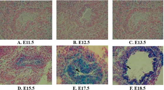
Fig. 1. Expression of SRC-3 in the embryonic lung during development in mice. The fetal lung samples were collected at various days of embryos (E) at (A): E11.5, (B): E12.5, (C): E13.5, (D): E15.5, (E): E15.5 and (F): E18.5. Expression profile of SRC-3 in embryonic lung was analyzed by the expression of LacZ using heterozygous SRC-3 mice. X-gal staining was performed for the LacZ reporter gene expression driven by the endogenous mouse SRC-3 gene. The black arrow indicates the Clara cell and the open arrow indicates the type II alveolar epithelial cell (Fig. 1. E-F).
cording to the manufacturer's protocol and normalized per μg of the protein. All transfection experiments were carried out in replicates of three and repeated at least three times.
Plasmid for the expression of the CCSP gene for transient transfection assays
The promoter region of mouse CCSP gene was cloned and ligated to the firefly luciferase (Luc) reporter gene, pGL3-Basic Luc (Promega, Madison, WI, USA) [12, 14, 17].
This CCSP-Luc plasmid was used to examine the effect of SRC-3 and TTF-1 for the expression of the CCSP gene in
vitro by transient transfection assays. The expression vectorsof SRC-3 and TTF-1 were provided by Dr. Francesco J.
DeMayo (Baylor College of Medicine, Houston, TX).
Results and Discussion
Little is known regarding the role of SRC-3 in early lung development. In order to investigate the role of SRC-3 in early development of the lung in mice, the temporal and spatial expressions of the SRC-3 were examined using SRC-3 heterozygous mice carrying SRC-3 driven LacZ expression.
Lung samples at various stage of development of embryos (E) at E11.5, E12.5, E13.5, E15.5, E17.5 and E18.5 were exam- ined for the LacZ expression. Weak expression of SRC-3 was observed as early as the day of the embryo at E11.5 in the
pulmonary cells of the lung (Fig. 1A) and maintained at low levels of expression until E13.5 (Fig. 1B-C). Weak expression of SRC-3 was observed in the pulmonary parenchyma at ear- lier developmental time points (E11.5-E13.5, Fig. 1A-C). The expression of SRC-3 gradually localized to epithelial cells lin- ing of the upper airways (E15.5, Fig. 1D) and greater ex- pressions of SRC-3 were observed in the Clara cells of the airway from the later stage of lung development (E15.5, Fig.
1D). A strong expression of SRC-3 was observed in the Clara cells from the day of the embryo (E17.5-E18.5, Fig. 1E-F) and the expression of SRC-3 in the Clara cells were continuously observed throughout the adult stage (Fig. 2A). Positive cor- relation of expression patterns of SRC-3 in the Clara cell spe- cific manner in early lung development and in the pro- liferation of adult lung suggests SRC-3 plays important roles in the differentiation and proliferation of the Clara cells of the bronchiolar epithelium of the lung.
In addition, the expressions of SRC-3 were observed in the type II alveolar epithelial cells at later times of develop- ment (E17.5-E18.5, Fig. 1E-F) and strong positive signals for SRC-3 were localized in type II cells of the adult lung (Fig.
2A). These results suggests that SRC-3 plays additional roles
on the development of type II alveolar epithelial cells in em-
bryonic lung and on the proliferation of adult lung. Fetal
lung maturation is influenced by many factors including ste-
roid hormones and steroid hormone acts through steroid re-
A B
C D
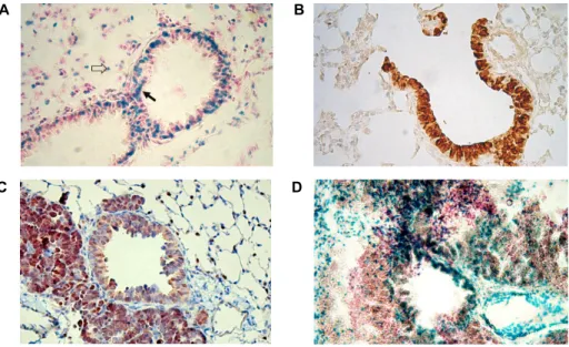
Fig. 2. Expression of SRC-3 and TTF-1 in adult lung and lungs with CCSP-TAg induced cancer mice. (A): Histochemical staining of LacZ expression was performed in adult lung of SRC-3 heterozygous mice for the SRC-3 expression. (B): Histochemical staining for TTF-1 from lung of SRC-3 heterozygous adult mice was performed using anti-sera against TTF-1 (1:5,000). (C and D): Histology and histochemical staining for TTF-1 (C) and for SRC-3 (D) were performed from the adult lung samples of CCSP-TAg induced lung cancer mice and paraffin embedded samples were sectioned at 5 μm thick. The black arrow indicates the Clara cell and the open arrow indicates the type II alveolar epithelial cell (Fig. 2A).
ceptor [13] and thus SRC-3 interacts with steroid receptors and several other transcriptional factors as coactivator [22].
It suggests that SRC-3 plays a role as coactivator for the tran- scriptional factors involved in the process of development of Clara cells and type II cells in embryonic lung. In con- clusion, SRC-3 expression was localized to epithelial cells lining the upper airways at later time points of embryonic lung and adult lung, and SRC-3 plays important roles in the bronchial- and alveolar development and proliferation of the lung.
The thuspositive pptemporal and spatial expression of SRC-3 (Fig. 2A) in adult lung are consistence with previous reports of the CCSP expression profiles in Clara cells [12, 17], in which CCSP could function as a differentiation mark- er protein of Clara cells. The expression of SRC-3 was clearly observed mainly in the nuclei, not in the cytoplasm of Clara cells of adult lung (Fig. 2A). In contrast, most of expressions of SRC-3 were localized in the cytoplasm and small samples showed nuclear localization of SRC-3 in human breast can- cers study [9]. In order to further elucidate the role of the SRC-3 in our Clara cell-specific lung cancer model, the tem- poral and spatial expressions of the thyroid transcription fac- tor -1 were analyzed in along with SRC-3 expression profiles in the lung of CCSP-TAg tumor mice. Strong positive stain- ing of TTF-1 was observed in normal Clara cells as well as
type II alveolar epithelial cells in the heterozygotes of SRC-3 mice lung (Fig. 2B). Strong expression of TTF-1 was also ob- served in transformed Clara cells and multifocal ad- enocarcinoma areas (Fig. 2C). Since TTF-1 might serve as a major regulator of CCSP expression by interacting with SRC in lung cancer model [17, 20, 22], these results strongly suggest that TTF-1 plays important roles in tumor pro- gression and invasiveness in our Clara cell-specific lung tu- mor model. However, the expression of SRC-3 was not ob- served in the area of multifocal adenocarcinoma and SRC-3 was not expressed in transformed Clara cells in the lung of CCSP-TAg with SRC-3 heterozygous bi-genic mice (Fig.
2D). Although expression of SRC-3 was observed in the areas of non-transformed Clara cells, expression of SRC-3 was not observed in tumor foci in oncogenic mice (Fig. 2D).
This observation suggests that SRC-3 might not be involved in the invasiveness of the lung tumor progression, in agree- ment with previous report [12].
Since TTF-1 might serve as a major regulator of CCSP
expression by interacting with SRC in lung cancer model
[17, 20, 22], TTF-1 could provide a good tool for the study
of interaction with SRC-3 involved in the process of lung
cancer. In order to further examine the role of SRC-3 and
TTF-1 in CCSP gene expression in the Clara cell-specific
manner, mouse transformed Clara cells were used for in vitro
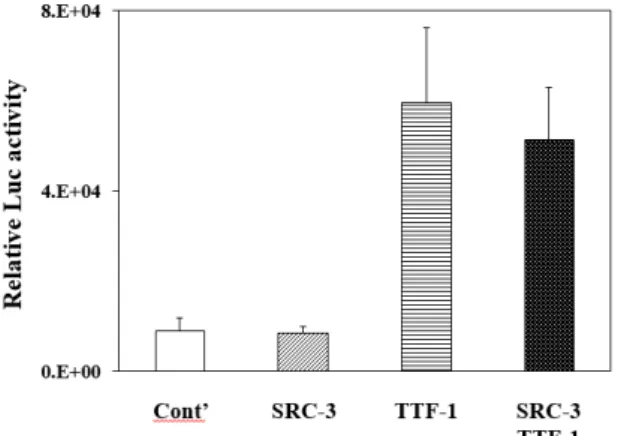
Fig. 3. Effects of SRC-3 and TTF-1 on the expression of CCSP gene. Mouse transformed Clara cells were used for tran- sient transfection assays for the expression of the CCSP gene and the CCSP gene expression was measured by the activities of the CCSP-Luc reporter plasmid. Co- transfection with SRC-3 or TTF-1 or with the combina- tion of both were performed and empty expression vec- tor was used as a control (cont’).
study. Co-transfection of SRC-3 exhibited no significant ef- fects on the expression of the CCSP gene in the mtCC (Fig.
3) and co-transfection of SRC-1 or SRC-2 in the mtCC also exhibited no significant effects (data not shown). In addition, co-transfection of SRC-1, SRC-2, or SRC -3 has no significant effects on the CCSP gene expression in H441, human lung adenocarcinomas originated from Clara cell-like cell lines (data not shown). However, co-transfection of TTF-1 resulted in a significant activation of CCSP expression in the mtCC.
It suggests that TTF-1 plays important roles in the expression of CCSP gene in vitro lung cancer model. However, syner- gistic effect was not observed by the addition of both TTF-1 and SRC-3 in the mtCC (Fig. 3). These in vitro data demon- strate that SRC-3 does not play a significant role in the ex- pression of CCSP in vitro in relation with lung cancer and these results are in agreement with our results in vivo [12].
These results suggest that SRC-3 might not play a critical role in lung cancer progression in vivo and in vitro. In con- clusion, SRC-3 plays roles in early bronchial- and alveolar development, however, SRC-3 does not play important role in lung cancer progression.
Acknowledgement
Author deeply thanks to Dr. Francesco J. DeMayo at Baylor College of Medicine, Houston, TX for providing tis- sues blocks and expression vectors used in this research.
References
1. Alberg, A. J. and Samet, J. M. 2003. Epidemiology of lung cancer. Chest 123, 21S-49S.
2. Anzick, S. L., Kononen, J., Walker, R. L., Azorsa, D. O., Tanner, M. M., Guan, X. Y., Sauter, G., Kallioniemi, O. P., Trent, J. M. and Meltzer, S. 1997. AIB1, a steroid receptor coactivator amplified in breast and ovarian cancer. Science 277, 965-968.
3. Bingle, C. D. 1997. Thyroid transcription factor-1. Int. J.
Biochem. Cell Biol. 29, 1471-1473.
4. DeMayo, F. J., Finegold, M. J., Hansen, T. N., Stanley, L.
A., Smith, B. and Bullock, D. W. 1991. Expression of SV40 T antigen under control of rabbit uteroglobin promoter in transgenic mice. Am. J. Physiol. 1261, L70-76.
5. Glaeser, M., Floetotto, T., Hanstein, B., Beckmann, M. W.
and Niederacher, D. 2001. Gene amplification and ex- pression of the steroid receptor coactivator SRC3 (AIB1) in sporadic breast and endometrial carcinomas. Horm. Metab.
Res. 33, 121-126.
6. Gnanapragasam, V. J., Leung, H. Y., Pulimood, A. S., Neal, D. E. and Robson, C. N. 2001. Expression of RAC 3, a steroid hormone receptor co-activator in prostate cancer. Br. J.
Cancer 85, 1928-1936.
7. Hicks, S. M., Vassallo, J. D., Dieter, M. Z., Lewis, C. L., Whiteley, L. O., Fix, A. S. and Lehman-McKeeman, L. D.
2003. Immunohistochemical analysis of Clara cell secretory protein expression in a transgenic model of mouse lung carcinogenesis. Toxicology 187, 217-228.
8. Hong, K. U., Reynolds, S. D., Giangreco, A., Hurley, C. M.
and Stripp, B. R. 2001. Clara cell secretory protein-express- ing cells of the airway neuroepithelial body microenviron- ment include a label-retaining subset and are critical for epi- thelial renewal after progenitor cell depletion. Am. J. Respir.
Cell Mol. Biol. 24, 671-681.
9. Iwase, H., Omoto, Y., Toyama, T., Yamashita, H., Hara, Y., Sugiura, H. and Zhang, Z. 2003. Clinical significance of AIB1 expression in human breast cancer. Breast Cancer Res. Treat.
80, 339-345.
10. Jemal, A., Murray, T., Samuels, A., Ghafoor, A., Ward, E.
and Thun, M. J. 2003. Cancer statistics, CA. Cancer J. Clin.
53, 5-26.
11. Kauffman, S. L., Alexander, L. and Sass, L. 1979. Histologic and ultrastructural features of the clara cell adenoma of the mouse lung. Lab. Invest. 40, 708-716.
12. Kwak, I. S. 2012. Roles of the Steroid Receptor Coactivator-3 in Lung Cancer. Cancer Prev. Res. 17, 344-349.
13. Kwak, I. S, Tsai, S. Y. and DeMayo, F. J. 2004. Genetically engineered mouse models for lung cancer. Annu. Rev.
Physiol. 66, 647-663.
14. Margraf, L. R., Finegold, M. J., Stanley, L. A., Major, A., Hawkins, H. K. and DeMayo, F. J. 1993. Cloning and tis- sue-specific expression of the cDNA for the mouse Clara cell 10 kD protein: comparison of endogenous expression to rabbit uteroglobin promoter-driven transgene expression.
Am. J. Respir. Cell Mol. Biol. 9, 231-238.
초록:폐의 분화와 폐암에서 SRC-3와 TTF-I의 역할
곽인석*
(신라대학교 의생명과학대학 생명과학과)
Steroid Receptor Coactivator (SRC)는 스테로이드 수용체 전사 활성화 단백질로, 이중에서 SRC-3는 많은 종류 의 종양과 관련하여 연구되었다. 그러나 현재 배아에서의 폐의 분화와 폐암 진행과정에서 SRC-3의 기능적 역할에 대한 연구는 제한적이다. 본 연구는 SRC-3가 생쥐 배아의 폐 분화과정에서 기관지와 폐포의 분화에 중요한 역할 을 함을 보여준다. 높은 레벨의 SRC-3 유전자 발현이 클라라 세포와 type II 세포에서 배아발달 말기 시기인 E17.5 - E18.5에서 관찰되었으며, 성체 생쥐의 폐에서도 클라라 세포와 type II 세포에서 SRC-3 유전자 발현이 관찰되었 다. SRC-3의 폐암에서의 역할을 연구하기 위하여 클라라 세포 특이적 폐암 생쥐 모델을 이용하여 관찰한 결과, SRC-3 잡종 생쥐의 폐와 클라라 세포 특이적 종양 생쥐의 폐에서 TTF-1 유전자와 SRC-3 유전자는 공동 발현되었 다. 위 모델에서 TTF-1 유전자 발현은 클라라 세포 유래 종양부위와 다발성 선암 영역에서 선명하게 관찰되었지 만, SRC-3 유전자 발현은 다발성 선암 부위에서는 관찰되지 않았다. 이 결과로 SRC-3가 폐암 진행과정 중 침윤성 에는 중요한 역할을 수행하지 않음을 확인하였다. SRC-3와 TTF-1의 폐암에서 역할을 클라라 세포 특이적 암 세포 주인 mtCC 세포를 사용하여 transient transfection 분석한 결과, TTF-1는 클라라 세포 특이적 단백질인 CCSP 유 전자 발현을 현저하게 활성화하였으나, SRC-3는 CCSP 유전자 발현의 활성화에 중요하게 관여하지 않음을 확인하 였다. 이 결과는 SRC-3가 폐암 진행에 필수적인 역할을 수행하는 단백질이 아님을 제시한다. 결론적으로, SRC-3 는 생쥐 배아와 성체 생쥐에서 기관지와 폐포의 분화에 중요한 역할을 수행하지만, 클라라 세포 유래의 폐암 진행 과정에서는 SRC-3는 중요한 역할을 수행하지 않는다.
15. Nord, M., Cassel, T. N., Braun, H. and Suske, G. 2000.
Regulation of the Clara cell secretory protein/uteroglobin promoter in lung. Ann. N. Y. Acad. Sci. 923, 154-165.
16. O'Malley, B. W. 2007. Coregulators: from whence came these "master genes". Mol. Endocrinol. 21, 1009-1013.
17. Ray, M. K., Chen, C. Y., Schwartz, R. J. and DeMayo, F.
J. 1996. Transcriptional regulation of a mouse Clara cell-spe- cific protein (mCC10) gene by the NKx transcription factor family members thyroid transciption factor 1 and cardiac muscle-specific homeobox protein (CSX). Mol. Cell. Biol. 16, 2056-2064.
18. Schiller, J. H. 2001. Current standards of care in small-cell and non-small-cell lung cancer. Oncology 61, Suppl. 1, 3-13.
19. Stahlman, M. T., Gray, M. E. and Whitsett, J. A. 1996.
Expression of thyroid transcription factor-1(TTF-1) in fetal and neonatal human lung. J. Histochem. Cytochem. 44, 673- 678.
20. Tan, D., Li, Q., Deeb, G., Ramnath, N., Slocum, H. K., Brooks, J., Cheney, R., Wiseman, S., Anderson, T. and Loewen, G. 2003. Thyroid transcription factor-1 expression prevalence and its clinical implications in non-small cell lung cancer: a high-throughput tissue microarray and im-
munohistochemistry study. Hum. Pathol. 34, 597-604.
21. Xu, J., Liao, L., Ning, G., Yoshida-Komiya, H., Deng, C. and O'Malley, B. W. 2000. The steroid receptor coactivator SRC-3 (p/CIP/RAC3/AIB1/ACTR/TRAM-1) is required for nor- mal growth, puberty, female reproductive function, and mammary gland development. Proc. Natl. Acad. Sci. USA 97, 6379-6384.
22. Yi, M., Tong, G. X., Murry, B. and Mendelson, C. R. 2002.
Role of CBP/p300 and SRC-1 in transcriptional regulation of the pulmonary surfactant protein-A (SP-A) gene by thy- roid transcription factor-1 (TTF-1). J. Biol. Chem. 277, 2997- 3005.
23. Zemke, A. C., Snyder, J. C., Brockway, B. L., Drake, J. A., Reynolds, S. D., Kaminski, N. and Stripp, B. R. 2009.
Molecular staging of epithelial maturation using secretory cell-specific genes as markers. Am. J. Respir. Cell Mol. Biol.
40, 340-348.
24. Zhou, L., Lim, L., Costa, R. H. and Whitsett, J. A. 1996.
Thyroid transcription factor-1, hepatocyte nuclear fac- tor-3beta, surfactant protein B, C, and Clara cell secretory protein in developing mouse lung. J. Histochem. Cytochem.
44, 1183-1193.