Anti-proliferative and Pro-apoptotic Effects by Lees Extracts of Ehwa Makgeolli Containing Oriental Herbs
Min-Jeong Kwon1, Seung Hoon Lee1, Chung Wook Chung1, Ho-Yong Sohn2, Woo-Chang Shin3 and Jong-Sik Kim1*
1Department of Biological Sciences, Andong National University, Andong 760-749, Korea
2Department of Food and Nutrition, Andong National University, Andong 760-749, Korea
3Research Institute, Kooksoondang Brewery Co., Ltd., Gyeonggi 462-160, Korea
Received September 11, 2015 /Revised November 7, 2015 /Accepted November 9, 2015
In the present study, ethanol extracts and their subsequent organic solvent fractions were extracted from the lees of Ehwa Makgeolli containing oriental herbs, a commercialized traditional Korean rice wine, and the prepared lees samples were designated as from KSD-E3-1 to KSD-E3-5. First, their ef- fects on cell viability and on the expression of pro-apoptotic ATF3 and NAG-1 genes in human color- ectal HCT116 cells were investigated. Among the treated lees samples, the hexane fraction (KSD-E3-2) and the ethyl acetate fraction (KSD-E3-3) of lees extracts from Ehwa Makgeolli significantly reduced cell viabilities, in a dose dependent manner. The treatment with KSD-E3-2 and KSD-E3-3 also in- creased the expression of pro-apoptotic NAG-1 and ATF-3 genes and their proteins, which were de- tected with RT-PCR and Western blot analysis, respectively. In addition, poly-(ADP-ribose) polymer- ase (PARP) cleavage was detected by treatment with the fraction KSD-E3-3, indicating that KSD-E3-3 could induce apoptosis in HCT116 cells. Interestingly, this PARP cleavage was recovered by trans- fection of NAG-1 small interfering RNA. The results indicate that NAG-1 is one of the genes respon- sible for apoptosis induced by the fraction KSD-E3-3 from Ehwa Makgeolli. Overall, the findings may help in understanding the molecular mechanisms of the anti-proliferative and pro-apoptotic activities mediated by the lees of Ehwa Makgeolli.
Key words : Anti-proliferation, apoptosis, Ehwa Makgeolli, lees, NAG-1
*Corresponding author
*Tel : +82-54-820-5798, Fax : +82-54-820-7705
*E-mail : [email protected]
This is an Open-Access article distributed under the terms of the Creative Commons Attribution Non-Commercial License (http://creativecommons.org/licenses/by-nc/3.0) which permits unrestricted non-commercial use, distribution, and reproduction in any medium, provided the original work is properly cited.
Journal of Life Science 2016 Vol. 26. No. 2. 241~246 DOI : http://dx.doi.org/10.5352/JLS.2016.26.2.241
서 론
대장암은 초기 증상이 거의 없어 조기진단이 어렵다고 알려 져 있으며, 현재 우리나라 암 사망률 4위를 차지하고 있다.
대장암은 낮은 확률의 유전적 요인과 함께 환경적 요인으로 인해 발병한다고 보고되고 있다[15]. 최근 대장암을 비롯한 여 러 가지 암 예방을 위한 다양한 천연물 발굴 및 식품 개발을 위한 연구가 활발하게 진행되고 있다[4, 16]. 식물에서 분리한 생리활성 물질들이 항암 활성이 높은 것들이 많으며, 암세포 의 사멸을 유도하는 대표적인 물질로는 안토시아닌(anthocya- nin) [19], 아세토제닌(acetogenin) [2] 그리고 탁솔(taxol) [1]
등이 있다.
이화주는 우리나라 전통 발효 술로 일반적인 막걸리와는 달리 맛이 달고 걸쭉한 것이 특징이며, 배꽃이 필 때 쌀가루로
만든 누룩과 쌀을 반죽하여 구멍 떡을 만들어 여름에 빚는 술로 알려져 있다. 최근에는 피부 기능활성 평가에서 일본식 koji에 비해 우수한 피부 미백 효과 및 노화 억제 활성이 보고 되었다[13]. 또한, 한국의 한방 약재의 발효 추출물은 각각 고 유한 생리활성을 가지고 있어[3, 20] 현대에서도 다양한 건강 식품 및 건강 보조 식품에 첨가되어 널리 활용되고 있다. 본 연구에 사용한 한방 이화주는 일반 이화주의 발효과정에 다양 한 생리활성이 보고된 4가지 약재인 감초, 구기자, 상백피와 삼백초를 첨가한 것으로서[13], 유기용매를 이용한 분획물을 획득하여 인간 대장암 세포주에서 암 세포의 성장 및 사멸에 미치는 영향을 연구하고자 하였다.
주박(lees)은 우리나라 막걸리를 비롯한 전통 주 제조 시 생성되는 부산물로서 쌀, 물, 누룩, 효모 등이 함께 발효되는 과정에서 생성되는 다양한 유효성분이 함유되어 있는 것으로 알려져 있다. 최근 주박이 당뇨[11] 및 알레르기 체질 개선[6]
에 효과가 있고, 심혈관계 질환을 예방[10]하며, 항균[9] 및 미 용 효과[14]를 나타내는 등 다양한 효능을 가지고 있는 것으로 보고되었다. 그러나, 주박은 다양한 효능을 가지고 있음에도 불구하고 식품위생법상 식품 및 식품 첨가 제한 원료로 지정 되어 있어서 폐기물로 처리하거나 일부 사료나 비료로 활용하 고 있는 정도이다. 따라서, 주박 및 주박에 함유되어 있는 핵심
- Note -
Table 1. List of organic solvent fractions of lees extracts from Ehwa Makgeolli
Source of lees Sample name Extraction solvent Sample name Extract or Fraction
Ehwa
Makgeolli KSD-E3 80% EtOH
KSD-E3-1 Extract
KSD-E3-2 Hexane fraction KSD-E3-3 Ethyl Acetate fraction KSD-E3-4 Butanol fraction KSD-E3-5 Water rediue 물질들에 의한 다양한 생리활성을 연구함으로써 주박의 활용
성을 증대시키는 연구가 필요하다고 판단된다.
본 연구에서는 한방이화주 주박 추출물 및 이의 순차적 유 기용매 분획물을 제조하고, 대장암 세포주 HCT116에서 이들 시료에 의한 항 성장 활성, 세포사멸 및 기전을 연구하였다.
이러한 연구결과는 전통주 제조 시 발생되는 부산물인 주박이 새로운 천연물 유래의 암 예방을 위한 생리활성 물질의 자원 으로 활용될 수 있음을 제시한다.
재료 및 방법
한방이화주 주박 추출물 및 분획물의 제조
한방 이화주 주박은 ㈜국순당(Seongnam, Korea)에서 제공 받아 사용하였으며, 제조방법은 다음의 절차에 따라 진행하였 다. 주박의 에탄올 추출물은 시료 1.5 kg에 95% 에탄올 6 L를 가하여 상온에서 3일간 2회 추출 하였고, 여과한 후 60℃ 에서 감압 농축하여 분말로 제조하였다. 분물 4 g을 물에 현탁한 후, n-hexane, ethyl acetate 및 butanol을 이용하여 순차적으로 분획하고 최종적으로 물 잔류물을 회수하였다. 이렇게 준비된 시료를 각각 KSD-E3-1 ~ KSD-E3-5으로 명명하였다(Table 1).
추출물과 분획물은 dimethyl sulfoxide (DMSO, Sigma, St Louis, MO, USA)에 녹여 -20℃에서 보관하며 실험에 사용하 였다.
세포배양 및 재료
대장암 세포주 HCT116는 American Type Culture Collec- tion (ATCC, Rockville, MD, USA)에서 구입하였다. 세포주 배양은 10 % FBS (Fetal Bovine Serum, Gibco, Grand Island, NY, USA), 1% penicillin 및 streptomycin (WelGene, Gyeong- san, Korea)을 첨가한 Dulbecco’s Modified Eagle Medium (DMEM, Gibco)을 사용하였다. 세포 배양은 37℃, 5% CO2 하 의 배양기에서 실시하였다.
세포 성장률 연구
대장암 세포주 HCT116에서 한방이화주 주박 추출물 및 유 기용매 분획물 처리가 세포 생존율에 미치는 영향을 연구하기 위해 cell viability assay를 수행하였다. 즉, 96 well plate에 1×10⁵ cells/well의 세포를 접종하고, 24시간 후에 한방이화주
주박 시료를 각각 1 mg/ml 이하의 농도로 처리하였다. 대조구 로는 용매인 DMSO를 각 실험군 중 가장 많은 양으로 처리된 샘플의 양과 동일하게 처리하였다. 시료를 24시간 동안 처리 하고 MTS (3-(4,5-dimethylthiazol-2-yl)5-(3-carboxy-methox- yphenyl)-2-(4-sulfophenyl)-2 H-tetrazolium) 용액 (Promega, Madison, WI, USA)을 각 well당 20 μl씩 첨가한 후 4시간 동안 반응시켰다. 반응이 끝난 후 NanoQuant PlateTM (Tecan Trading AG, Switzerland) 를 사용하여 580 nm에서 흡광도를 측정하였다. 실험 결과 수치는 5개의 독립적인 well에서 수행 한 값을 Sigma plot program 10.0을 이용하여 분석한 후 그래 프로 나타내었다.
Western blot analysis
세포배양용 60 mm dish에 HCT116세포를 24시간 동안 배 양한 후 한방이화주 주박 시료를 조건에 따라 처리하였다. 수 확한 세포는 4X RIPA (Cell Signaling, Beverly, MA, USA) 용액을 첨가하여 sonication을 한 후 원심분리 하여 total pro- tein을 추출하였다. 추출한 단백질은 Bradford protein assay kit (Bio-Rad, Hercules, CA, USA)을 이용하여 정량 하였다.
총 단백질 20 μg을 전기영동 한 후, 전기적으로 membrane으 로 transfer 한 후 Western blot에 사용하였다. 1차 항체로는 Actin, ATF3, cleaved PARP (89 kDa), PARP (116 kDa), NAG-1 항체를 사용하였다. NAG-1 항체는 미국 NIEHS (National Institute of Environmental Health Sciences, RTP, NC, USA)의 Dr. Eling group으로부터 제공받았으며 NAG-1 을 제외한 모든 항체는 Santa Cruz사 (Santa Cruz, CA, USA) 혹은 Cell Signaling사(Beverly, MA, USA)에서 구입하여 사용 하였다. 2차 항체로는 HRP-conjugated anti-mouse, HRP-con- jugated anti-rabbit 항체를 사용하였다. 2차 항체를 1시간 동안 처리한 후 enhanced chemiluminescence kit (ECLTM, Amer- sham, Pittsburgh, PA, USA)를 이용하여 검출 반응시킨 후 C-DIGIT (LI-COR, Lincoln, NE, USA)를 이용하여 단백질 발 현을 확인하였다.
Reverse Transcription-Polymerase Chain Reaction (RT-PCR)
수확한 HCT116 세포주로부터 RNeasy mini kit (Qiagen, Valencia, CA, USA) 을 이용하여 제조사의 매뉴얼에 따라 to-
Table 2. Sequences of oligonucleotide primers of human ATF3, NAG-1 and GAPDH genes
Gene name GenBank Acc. No. Sequence
ATF3 NM_001040619 F: 5'-TGGTGTTTGAGGATTTTGCT-3'
R: 5'-ATTTCTTTCTCGTCGCCTCT-3'
NAG-1 NM_004864 F: 5'-CTCTCAGATGCTCCTGGTGT-3'
R: 5'-GAATCTTCCCAGCTCTGGTT-3'
GAPDH NM_002046 F: 5'-CTGACCTGCCGTCTAGAAA-3'
R: 5'-GAGCTTGACAAAGTGGTCGT-3'
tal RNA를 추출하였다. 추출한 total RNA 2 μg을 주형으로 PrimeScriptTM RT-PCR Kit (TaKaRa, Japan)를 이용하여 cDNA를 합성하였다. 합성된 cDNA를 주형으로 하여 유전자 특이적인 oligonucleotide primer (Table 2)를 이용하여 PCR (Polymerase Chain Reaction) 과정을 수행하였다. PCR 반응 은 94℃에서 5분 denaturation 시키고, 94℃에서 30초, 58 ℃ 에서 30초, 72℃에서 30초의 cycle을 총 30번 반복한 뒤, 마지막 으로 72℃에서 10분간 반응시켰다. 최종적인 PCR product는 전기영동 후 Gel Image Analysis System (CoreBio, Seoul, Korea)을 이용하여 사진 촬영하였다.
siRNA transfection
인간 대장암 세포주인 HCT116 세포주를 60 mm dish에 접종 후, 약 24시간 뒤에 lipofectamine 2000 reagent (Invitro- gen, Carlsbad, CA, USA)를 이용하여 siRNA를 transfection 하였다. 대조구로 사용한 Negative Control (NC) siRNA (SN-1003, Bioneer, Daejeon, Korea)와 NAG-1 siRNA (SN- 1061367, Bioneer) construct을 serum free DMEM 배지 50 μl 에 lipofectamin 10 μl와 6 μg의 siRNA를 각각 첨가한 뒤 혼합 하여 transfection시켰다. 4시간 후 FBS가 첨가된 DMEM 배지 로 교환한 후, 24시간 동안 추가 배양한 다음 주박 시료를 조건 에 따라 처리하였다. 주박 시료를 24시간 동안 처리하고 세포 를 수확하였다.
통계처리
모든 실험은 최소한 3회 이상 반복실험을 실시 하였으며, 실험결과는 평균±표준편차로 나타내었고, 각 실험결과의 유 의성 검토는 시료가 포함되지 않은 대조구와 비교하여 stu- dent’s t-test에 의해 판정하였으며 p<0.05 일 때 유의성이 있다 고 판단하였다.
결과 및 고찰
한방 이화주 주박 추출물의 유기용매 분획물이 세포 성장에 미치는 영향
본 연구그룹은 선행연구에서 8종의 주박과 3종의 누룩으로 부터 총 85종의 추출물 및 분획물을 제조하고, 이들의 암세포
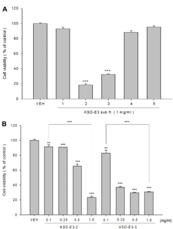
항 성장 활성을 보고한 바 있다[7]. 본 연구에서는 Table 1에 나타낸 바와 같이 1차 선별된 주박 추출물 중 한방이화주 주박 (KSD-E3)으로부터 제조한 80% 에탄올 추출물(KSD-E3-1), 이 의 순차적 유기용매 분획물 3종(KSD-E3-2, KSD-E3-3, KSD- E3-4)과 최종적으로 물 잔류물(KSD-E3-5)이 HCT116 세포주 의 성장에 미치는 영향을 연구하였다. 그 결과 1차 선별 결과 와 유사하게 KSD-E3-2 분획물과 KSD-E3-3 분획물에 의해 가 장 높게 세포생존율이 감소됨을 확인할 수 있었다(Fig. 1A).
현저한 암세포 성장률 감소를 보여준 KSD-E3-2 분획물과 KSD-E3-3 분획물의 농도에 따른 처리가 HCT116세포 생존율 에 미치는 영향을 연구하였다. 즉, HCT116 세포주에 분획물 2종을 0.1, 0.25, 0.5, 그리고 1.0 mg/ml의 농도로 24시간 처리 한 후 세포 생존율을 측정하였다. 그 결과, KSD-E3-2 분획물을 처리한 경우 0.5 mg/ml의 농도 처리 군부터 cell viability가 감소됨을 확인할 수 있었고, KSD-E3-3 분획물을 처리한 경우 0.25 mg/ml의 농도 처리 군부터 급격하게 cell viability가 감 소됨을 확인하였다(Fig. 1B). 이러한 결과에 근거하여 추후 실 험을 위한 처리 농도를 KSD-E3-2 분획물은 0.5 mg/ml로 정하 고, KSD-E3-3 분획물은 0.1 mg/ml로 정하였다.
유기용매 분획물에 의한 ATF3와 NAG-1 유전자 및 단백 질의 발현 증가
한방 이화주 주박의 분획물 KSD-E3-2 (hexane 분획물)와 KSD-E3-3 (ethyl acetate 분획물)이 pro-apoptotic 유전자인 ATF3와 NAG-1의 발현에 미치는 영향을 연구하였다. KSD-E3- 2 분획물은 0.5 mg/ml, KSD-E3-3 분획물은 0.1 mg/ml의 농도 로 각각 24시간 동안 처리한 다음, NAG-1과 ATF3의 유전자의 발현을 확인하였다. Fig. 2A에서 보는 바와 같이 두 분획물의 처리에 의해 ATF3와 NAG-1 유전자의 발현이 모두 증가되었 다. 또한, 동일한 분획물 처리 조건하에서 ATF3와 NAG-1 단 백질의 발현을 확인하기 위하여 Western blot analysis을 수행 하였다. 그 결과, Fig. 2B에서 보는 바와 같이 두 분획물 처리에 의해 ATF3와 NAG-1 단백질 모두 발현이 증가되었으며, KSD-E3-3 분획물 처리군의 경우 KSD-E3-2 분획물 처리군 보 다 낮은 농도임에도 불구하고 ATF3와 NAG-1의 단백질의 발 현을 훨씬 높게 증가시켰다. Activating transcription factor-3 (ATF-3) 유전자는 많은 천연물 유래 생리활성 물질에 의해 발
A
B
Fig. 1. Effects of organic solvent fractions of lees extracts from Ehwa Makgeolli on HCT116 cell viabilities. (A) HCT 116 cells were plated at 1×10⁵ cells/well in a 96-well plate and incubated with five different samples from lees of Ehwa Makgeolli for 24 hr. And then, cell via- bility was measured using MTS proliferation assay.
(B) Cells were treated with five different concen- trations of KSD-E3-2 and KSD-E3-3. **p<0.01, ***p<
0.001.
A
B
Fig. 2. Up-regulation of ATF3 and NAG-1 by the treatment of KSD-E3-2 and KSD-E3-3. HCT116 cells were in- cubated with KSD-E3-2 and KSD-E3-3 for 24 hr. And then, total RNAs or proteins were prepared from treated cells. (A) Total RNA was used for RT-PCR with NAG-1 and ATF3 gene specific primers. (B) Western blot was carried out by using ATF-3, NAG- 1, and Actin antibodies.
현이 유도되며, 이러한 생리활성물질에 의한 세포사멸 및 항 암 활성과 관련이 깊은 것으로 보고되었다[8, 12]. Non-ster- oidal anti-inflammatory gene-1 (NAG-1) 유전자는 TGF-beta superfamily의 하나로서 최근 다양한 천연물에 의한 항암 활 성의 중요한 중재자의 역할에 대한 총설이 보고되었다[21].
NAG-1 siRNA의 도입에 의한 세포 사멸의 회복 KSD-E3-3 (ethyl acetate 분획물)분획물에 의한 세포생존율 의 감소의 원인이 apoptosis와 관련이 있는지 확인하기 위하 여, apoptosis의 지표인자 중의 하나인 poly (ADP-ribose) pol- ymerase (PARP) 단백질의 cleavage를 확인하였다. PARP 는 DNA repair 단백질로 세포에서 분해되지 않았을 때 제 기능 을 하며, 세포사멸 유도 시 116 kDa의 poly (ADP-ribose) poly- merase (PARP) 는 미토콘드리아 경로를 통해 활성화된 cas- pase-3 에 의해 89 kDa으로 절단되는 특성이 있어 apoptosis의 지표의 하나로서 이용되고 있다[5, 17, 18]. Fig. 3A에서 보는
바와 같이 KSD-E3-2와 KSD-E3-3 분획물을 처리한 경우 KSD- E3-3 분획물의 처리에 의해서만 89 kDa으로 잘려진 PARP가 관찰되었다. 이러한 연구결과는 상대적으로 낮은 농도인 0.1 mg/ml의 KSD-E3-3분획물의 처리에 의해 apoptosis가 유도 되는 반면, 0.5 mg/ml의 KSD-E3-2분획물의 처리에 의해서는 apoptosis가 유도되지 않음을 나타낸다. KSD-E3-3 분획물의 처리에 의해 유도되는 apoptosis가 pro-apoptotic 유전자인 NAG-1 의 발현 증가와 관련이 있는지 확인하기 위하여 NAG- 1 siRNA를 이용하여 증명하였다. 즉, HCT116 세포주에 대조 구인 Negative Control (NC) siRNA와 NAG-1 siRNA를 trans- fection한 후, 24시간 후에 KSD-E3-3 분획물을 0.1 mg/ml 농 도로 처리 혹은 DMSO를 처리하였다. Fig. 3B에서 보는 바와 같이 NC siRNA를 transfection 한 후 KSD-E3-3 분획물을 처 리한 경우 NAG-1 단백질이 현저하게 증가되었고, 또한 PARP cleavage도 관찰됨으로써 apoptosis가 정상적으로 일어남을 확인하였다. 반면, NAG-1 siRNA를 transfection 후 KSD-E3-3 분획물을 처리한 경우, NAG-1 단백질의 발현이 현저하게 감 소하였으며, PARP cleavage 또한 현저하게 감소됨을 확인하 였다(Fig. 3B). 이러한 연구결과는 한방이화주 주박 추출물의 ethyl acetate 분획물인 KSD-E3-3의 처리에 의해 apoptosis가 유도되며, 이러한 apoptosis는 pro-apoptotic 유전자인 NAG-1 의 발현증가와 직접적인 관련성이 있음을 시사한다. 향후, KSD-E3-3 분획물로부터 apoptosis와 직접적인 관련성이 있는
A
B
Fig. 3. Induction of apoptosis by the treatment of KSD-E3-3 and recovery of apoptosis by NAG-1 siRNA trans- fection. (A) HCT116 cells were treated with 0.5 mg/
ml KSD-E3-2 or 0.1 mg/ml KSD-E3-3 for 24 hr, and treated cells were collected for Western blot analysis using PARP, cleaved PARP, and Actin antibodies. (B) Either Negative Control (NC) siRNA or NAG-1 siRNA was transfected into HCT116 cells. After 24 hr, cells were treated with DMSO (vehicle) or 0.1 mg/ml KSD-E3-3. After 24 hr treatment, cells were collected for Western blot analysis using NAG-1, PARP, cleaved-PARP, and Actin antibodies.
핵심물질을 찾는 연구가 지속되어야 할 것으로 판단된다.
감사의 글
본 연구는 2012년도 농림수산식품부 고부가가치식품기술 개발사업(과제번호 112073-3)에 의해 수행되었으며, 이에 감 사 드립니다.
References
1. Cao, L., Chen, S., Zhang, C., Chen, C., Lu, N., Jiang, Y., Cai, Y., Yin, Y. and Xu, J. 2015. ING4 enhances paclitaxel's effect on colorectal cancer growth in vitro and in vivo. Int.
J. Clin. Exp. Pathol. 8, 2919-2927.
2. Han, B., Wang, T. D., Shen, S. M., Yu, Y., Mao, C., Yao, Z. J. and Wang, L. S. 2015. Annonaceous acetogenin mimic AA005 induces cancer cell death via apoptosis inducing fac- tor through a caspase-3-independent mechanism. BMC Cancer 15, 139.
3. Hong, E. S., Ahn, G. W. and Jo, B. K. 2008. The study on the potential anti-aging properties of Prunella vulgaris ex- tracts in vitro and in vivo. J. Soc. Cosmet. Sci. 34, 129-135.
4. Jaganathan, S. K., Vellayappan, M. V., Narasimhan, G. and Supriyanto, E. 2014. Role of pomegranate and citrus fruit juices in colon cancer prevention. World J. Gastroenterol. 20, 4618-4625.
5. Jang, S. Y., Hong, D., Jeong, S. Y. and Kim, J. H. 2015.
Shikonin causes apoptosis by up-regulating p73 and down-regulating ICBP90 in human cancer cells. Biochem.
Biophys. Res. Commun. 15, 30362-30374.
6. Kang, Y. J., Park, S. J., Bae, K. H., Yoo, J. M., Pyo, H. B., Cho, J. H. and Kim, T. J. 2011. Ethyl acetate extract of Korean rice wine lees inhibits IgE-mediated degranulation in rat ba- sophilic leukemia RBL-2H3 cells and passive cutaneous ana- phylaxis in mice. J. Life Sci. 21, 1364-1369.
7. Kang, H. T., Lee, S. H., Kim, S. Y., Kim, M. S., Shin, W.
C., Sohn, H. Y. and Kim, J. S. 2014. Anti-proliferative activ- ities of solvent fractions of lees extracts in human colorectal HCT116 cells. J. Life Sci. 24, 967-972.
8. Kim, K. J., Lee, J., Park, Y. and Lee, S. H. 2015. ATF3 medi- ates anti-cancer activity of trans-10, cis-12-conjugated lino- leic acid in human colon cancer cells. Biomol. Ther. (Seoul).
23, 134-140.
9. Kim, M. S., Lee, Y. S., Kim, J. S., Shin, W. C. and Sohn, H. Y. 2014. Anti-microbial and Anti-thrombosis activities of lees of Sweet Potato Soju. J. Life Sci. 42, 258-266.
10. Kim, M. S., Lee, Y. S., Kim, J. S., Shin, W. C. and Sohn, H. Y. 2015. Evaluation of in-vitro Anti-thrombosis and an- ti-oxidation activity of lees of Takju (Wookukseng). J. Life Sci. 25, 425-432.
11. Kim, S. M. and Cho, W. K. 2006. Effects of Takju (Korean turbid rice wine) Lees on the serum glucose levels in strep- tozotocin-induced diabetic rats. Kor. J. Dietary Culture 21, 638-643.
12. Lee, J. R., Lee, M. H., Eo, H. J., Park, G. H., Song, H. M., Kim, M. K, Lee, J. W. and Jeong, J. B. 2014. The contribution of activating transcription factor 3 to apoptosis of human colorectal cancer cells by protocatechualdehyde, a naturally occurring phenolic compound. Arch. Biochem. Biophys. 564, 203-210.
13. Lee, S. J., Kwon, Y. Y., Cho, S. W., Kwon, H. S. and Shin, W. C. 2013. Effects of Ehwa Makgeolli containing oriental herbs on skin whitening and wrinkles. J. Kor. Soc. Food. Sci.
Nutr. 42, 550-555.
14. Lee, S. M., Lee, S. J., Kwon, Y. Y., Baek, S. H., Kim, J. S., Sohn, H. Y. and Shin, W. C. 2014. Skin whitening and an- ti-wrinkle effects of extract from jubak of oriental herbal liquor. J. Kor. Soc. Food. Sci. Nutr. 43, 1695-1700.
15. Li, Y., Beckman, K. B., Caberto, C., Kazma, R., Lum-Jones, A., Haiman, C. A., Marchand, L. L., Stram, D. O., Saxena, R. and Cheng, I. 2015. Association of genes, pathways, and haplogroups of the mitochondrial genome with the risk of colorectal cancer: The Multiethnic Cohort. PLoS One doi:
10.1371/journal.pone.0136796.
16. Luo, Y., Wang, S. X., Zhou, Z. Q., Wang, Z., Zhang, Y. G., Zhang, Y. and Zhao, P. 2014. Apoptotic effect of genistein on human colon cancer cells via inhibiting the nuclear fac- tor-kappa B (NF-κB) pathway. Tumour Biol. 35, 11483-11488.
초록:한방이화주 주박 추출물에 의한 암세포 항성장 및 세포사멸 기전 연구 권민정1․이승훈1․정정욱1․손호용2․신우창3․김종식1*
(1국립안동대학교 생명과학과, 2국립안동대학교 식품영양학과, 3㈜국순당 연구소)
본 연구에서는 한국 전통 막걸리 중 하나인 한방 이화주의 주박으로부터 에탄올 추출물과 그것의 순차적인 유 기용매 분획물을 획득하고, 이들 시료들이 인간 대장암 세포인 HCT116 세포주의 cell viability와 pro-apoptotic 유전자인 ATF-3와 NAG-1의 발현에 미치는 영향을 연구하였다. 처리된 유기용매 시료 가운데 KSD-E3-2 분획물 (hexane 분획물)과 KSD-E3-3 분획물 (ethyl acetate 분획물)의 처리에 의해 현저하게 세포생존율이 감소하였으며, 두 분획물에 의해 농도의존적으로 세포 생존율이 감소하였다. 또한, 두 분획물의 처리에 의해 pro-apoptotic 유전 자인 NAG-1과 ATF-3의 유전자와 단백질의 발현이 증가함을 확인하였다. 특히, KSD-E3-3 분획물 0.1 mg/ml의 처리에 의해 NAG-1 단백질의 발현이 현저하게 증가하였으며, 또한 apoptosis의 지표인 PARP cleavage가 관찰되 었다. 이러한 KSD-E3-3 분획물에 의해 유도된 apoptosis는 NAG-1 siRNA transfection에 의해 회복됨을 확인하였 다. 따라서, KSD-E3-3 분획물에 의한 NAG-1 단백질의 발현증가와 apoptosis 간의 직접적인 관련성이 있음을 시 사한다. 종합적으로 본 연구 결과는 한방이화주 주박에 함유된 생리활성물질의 암세포 항 성장 활성 및 apoptosis 기전을 이해하는데 도움을 줄 것으로 생각된다.
17. Sui, C. G., Meng, F. D., Li, Y. and Jiang, Y. H. 2015. Antipro- liferative activity of rosamultic acid is associated with in- duction of apoptosis, cell cycle arrest, inhibition of cell mi- gration and caspase activation in human gastric cancer (SGC-7901) cells. Phytomedicine 22, 796-806.
18. Xu, M. Y. and Kim, Y. S. 2014. Antitumor activity of glycyrol via induction of cell cycle arrest, apoptosis and defective autophagy. Food Chem. Toxicol. 74, 311-319.
19. Xu, J., Su, X., Lim, S., Griffin, J., Carey, E., Katz, B., Tomich, J., Smith, J. S. and Wang, W. 2015. Characterization and sta-
bility of anthocyanins in purple-fleshed sweet potato P40.
Food Chem. 186, 90-96.
20. Yang, H. J., Ahn, Y. J., Kim, J. H. and Park, S. N. 2008.
Antioxidative activity and component analysis of Quercus glauca leaf extracts. J. Soc. Cosmet. Sci. 34, 189-200.
21. Yang, M. H., Kim, J., Khan, I. A., Walker, L. A. and Khan, S. I. 2014. Nonsteroidal anti-inflammatory drug activated gene-1 (NAG-1) modulators from natural products as an- ti-cancer agents. Life Sci. 100, 75-84.