대두 발효물이 인간 유래 피부세포의 증식 및 성장에 미치는 영향
김은주1, 한명륜2, 이소영1, 김애정1*
1경기대학교 일반대학원 대체의학과, 2혜전대학교 제과제빵과
Effect of Fermented Soybean on the Proliferation and Growth in HaCaT and Fibroblast Cell
Eun-Joo Kim1, Myung-Ryun Han2, So-Young Lee1, Ae-Jung Kim1*
1Department of Alternative Medicine Graduate School Kyonggi University
2Department of Baking Science & Technology Hyejeon University
요 약 본 연구는 대두 단백질을 효소(pepsin) 분해 및 미생물(
Lactobacillus Rhamnosus ) 발효를 통해 생성된 저분자 생리활성 펩티드로 제조하여 인간 유래 피부세포에서 세포활성에 미치는 효과를 확인하고, 천연유래 기능성 미용소재로 서의 이용 가능성을 확인하고자 하였다. 연구를 수행하기 위해 대두발효물을 제조하였고, 대두발효물에 대해 LC-MS 분석을 하였으며, WST-1 assay의 방법을 통해 cell viability를 측정하였다. 세포독성을 측정한 결과, 0~2,000 μg/mL 의 모든 농도 구간에서 세포독성이 나타나지 않았다. 특히 800 μg/mL의 농도에서는 대조군에 비해 160~180 %, 양성 대조군으로 사용한 EGF(epidermal growth factor)에 비해 120 %에서는 세포증식 효과가 나타나 피부노화 억제 및 피부세포 재생 소재로서의 뛰어난 가능성을 보여주었다. 또한 대두발효물과 그 분획물의 상처 치유 능력을 확인하기 위 해 세포이동 실험을 수행한 결과, 대두발효물과 분핵물 중 F4와 F5가 대조군에 비해 우수한 세포이동능을 보여주었고, EGF와는 유사하거나 그 이상의 효과를 보여주었다. 결론적으로 대두발효물과 분핵물(F4 and F5)의 피부개선효과가 유 사하게 나타나 피부미용 소재 및 피부 질환 치료의 소재로의 활용이 기대된다.
Abstract This study was undertaken to determine the effect of fermented soybean extract and its
fractions on skin cell proliferation and growth. The extract was procured by the pepsin and Lactobacillus rhamnosus fermentation of soybean. LC-MS analysis was performed subsequent to soybean fermentation, and cell viability was measured by the WST-1 assay. Cell proliferation was observed to increase after exposing cells to the fermented soybean extract and its fractions at all concentrations tested (0~2,000 μg/mL). In particular, compared to the normal control group and 120 % proliferation of the EGF (epidermal growth factor) positive control group, 160~180 % cell proliferation was achieved at 800 μ g/ml, indicating the excellent potential as an application material for skin aging inhibition and skin cell regeneration. In addition, we also examined the effects of fermented soybean extract and its fractions on wound healing ability, in HaCaT cells and fibroblasts. Our results indicate excellent cell migration abilities after treatment with fermented soybean extract and its fractions, as compared to the control treatment. Similar cell migration abilities were observed in the positive control group (EGF). Taken together, our results indicate that fermented soybean extract and its fractions (F4 and F5) exert amelioratory effects as a natural material for skin.
Keywords : Fermented Soybean Extract, Keratinocyte, Fibroblast, Proliferation, Migration
*Corresponding Author : Ae-Jung Kim(Kyonggi Univ.) email: [email protected]
Received December 18, 2020 Revised January 5, 2021
Accepted February 5, 2021 Published February 28, 2021
1. 서론
최근 피부 건강에 대한 피부 장벽의 기능과 중요성이 강조되면서 항산화 기전을 중심으로 피부 장벽의 기능에 대해 손상 회복 및 조절에 관한 연구가 다양하게 진행되 어 왔다[1-2]. 피부의 구성은 표피, 진피 및 피하조직의 3개의 층으로 구성되어 있다. 병원균과 알레르기 물질 등 을 포함하는 외부 감염원의 침입 및 외부 자극으로부터 피부를 보호하는 물리적 장벽의 기능이 있다[3-5]. 그리 고, 체내 수분을 유지하는 보습 장벽은 천연 보습인자 (NMF: Natural Moisturizing Factor)를 통해 이루어 지며, 면역기능을 담당하는 항균 펩티드(antimicrobial peptide) 및 호중구, 비만세포 등의 면역세포가 화학적 장벽의 기능을 동시에 담당하고 있다[6,7]. 보습 장벽의 기능을 수행하는 hyaluronic acid, phospholipid, free fatty acid, ceramide, filaggrin, urea, amino acid, peptide 등의 다양한 보습인자들은 피부표피 기저 층에서 각질을 형성하는 세포의 각질화 과정에서 분해되 거나 섬유아세포에 의해 합성되어 세포외기질에 축적됨 으로써 피부의 다양한 생리 활성 기능을 수행한다[8]. 또 한, 물리적·화학적 자극에 의해 피부가 손상되면 섬유아 세포는 다양한 성장인자, 사이토카인, 기질단백질 등을 분비하며 증식 및 상처 부위를 수축시키고, 세포외기질을 합성한다[9,10]. 각질형성세포는 사이토카인 등의 인자 들을 분비하여 기저막 성분을 생성하며, 손상 부위로 이 동(migration)하는 동시에 세포의 증식(proliferation)을 유도한다[11]. 따라서 피부조직의 재 상피화 및 상처 치 유에 각질 형성 세포와 섬유아세포의 증식은 매우 중요 하다. 각질 형성 세포 및 섬유아세포의 기능 저하로 천연 보습인자의 합성이 저하되거나 생성률이 낮아지게 되면 피부 장벽의 결함이 발생하게 된다. 이는 피부의 수분량 감소, 표피의 위축 및 과각질화, 아토피피부염, 피부의 탄 력성 저하, 주름 형성과 같은 피부 노화 및 질병을 유발 한다[12-16].
대두(Glycine max L.)는 콩과(Leguminosae)에 속 하는 1년생 초본 또는 그 열매로 단백질이 약 40 %, 탄 수화물이 약 30 %, 지방이 약 20 % 함유되어 있으며, 이 소플라본(isoflavone), 레시틴, 식물성 스테롤과 같은 기 능성 성분을 함유하고 있어 영양학적 가치가 뛰어나다 [17,18]. 또한, 항산화, 미백, 항노화, 항염증, 여드름 개 선 등의 생리활성이 입증되었고, 각종 피부 개선 기능성 소재로 개발되고 있다[19-22]. 이러한 천연물을 기능성 소재로 활용하기 위한 다양한 연구가 지속적으로 수행되
고 있는데 특히, 천연물의 성분이나 소재의 활용성을 증 진시키기 위해 유산균 발효를 통해 발효물의 생리활성을 증진시켜 맛과 향, 조직감을 개선하고, 새로운 생리활성 성분을 생성하는 등의 긍정적인 영향을 주는 것으로 보 고되어 있다[23,24]. 따라서 다양한 천연물을 이용한 발 효물의 연구가 진행되고 있으며, 과학적으로 밝혀진 생리 활성을 통해 기능성 화장품 소재 연구가 활발히 진행되 고 있다. 그러나 Lactobacillus Rhamnosus에 의한 피 부 세포증식 및 영향에 대한 연구는 미비한 실정이다.
따라서 본 연구에서는 Lactobacillus Rhamnosus의 균주를 이용하여 대두단백질을 발효시켜 대두발효물 (Bio- Peptone
®)을 제조하고, 생성된 대두발효물 및 그 분획물이 각질형성세포(human epidermal keratinocytes) 와 진피형성세포(human dermal fibroblasts)의 성장 및 증식에 미치는 효과를 확인하여 피부미용 소재 및 피 부 질환 치료의 소재로서의 이용 가능성을 알아보았다.
2. 본론 2.1 재료 및 방법
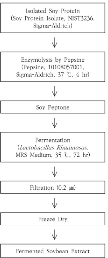
2.1.1 대두발효물의 제조
본 실험에 사용된 대두발효물(Bio-Peptone
®)은 Fig.
1의 공정으로 제조되었다. Isolated soy protein(Sigma Aldrich, USA)에 pepsin(Sigma Aldrich, USA)을 혼합 하여 37 ℃에서 4시간 동안 효소 분해시켜 얻은 soy peptone에 MRS medium에서 종균배양(seed culture) 된 락토바실러스 람노서스(Lactobacillus Rhamnosus) 균종을 넣고 혼합한 후, 35 ℃에서 72시간 이상 배양하 여 발효시켰다. 발효 후 발효액을 여과하고, 유산균을 제 거한 후, 대사산물이 함유된 여과액을 감압, 농축, 동결건 조한 뒤 발효 분말을 제조하여 사용하였다[1].
2.1.2 대두발효물의 LC-MS 분석
제조된 대두발효물은 LTQ Orbit-rap XL 질량분석
기(Thermo Fisher Scientific, Bremen, Germany)와
Accela UPLC(Thermo Fisher Scientific, San Jose,
CA, USA)를 사용하여 성분 및 분자량을 확인하였다. 분
석에 사용한 column은 Acquity BEC C18 column
(1.7 μm, 150×2mm, Waters, Dublin, Ireland)을 사
용하였고, 0.1 % formic acid(Sigma Aldrich, USA)가
포함된 증류수(Burdick & Jackson, Korea)와 아세토
니트릴(Sigma Aldrich, USA) 용매를 용출 용매로 사용 하였다. 시험물질의 분자량 확인을 위해서 LS-MS분석은 positive ion([M+H]+) 모드를 이용하였으며, 분자량 범 위를 115∼1,500의 범위로 분석하였다.
Isolated Soy Protein (Soy Protein Isolate, NIST3236,
Sigma-Aldrich)
Enzymolysis by Pepsine (Pepsine, 10108057001, Sigma-Aldrich, 37 ℃, 4 hr)
Soy Peptone
Fermentation ( Lactobacillus Rhamnosus , MRS Medium, 35 ℃, 72 hr)
Filtration (0.2 ㎛)
Freeze Dry
Fermented Soybean Extract
Fig. 1. Production of a fermented soybean extract with
Lactobacillus Rhamnosus
.2.1.3 대두발효물을 이용한 분획물의 획득
대두발효물에 대한 생리활성 지표 성분 분석 및 생리 활성 검정용 분획 시료의 제조를 위해 Waters 사의 Delta Prep LC300 분취용 크로마토그래피(Prep LC, Milford, MA, USA)와 Kromasil 100 C18 컬럼(10 μ m, 250×30 mm, Eka Chemicals AB, Bohus, Sweden)을 이용하여 분획을 수행하였다. 1 g의 대두발 효물을 컬럼에 주입하고, 증류수(Burdick & Jackson, Korea)와 아세토니트릴(Sigma Aldrich, USA) 용매를 이용하여 50분, 0∼100 % 아세토니트릴 용매 기울기 방 법으로 분획을 수행하였다. 각 분획물에 대한 성분 프로 파일 패턴 분석을 위해 LC-MS 분석을 진행한 후, LC-MS 분석결과 성분들의 프로파일 패턴을 고려하여 6 개의 분획물 시료를 제조하였다.
2.1.4 세포주 및 세포 배양
인간유래 각질형성 세포주인 HaCaT 세포 (human epidermal keratinocytes)와 인간유래 진피형성 세포 주인 Fibroblast 세포(human dermal fibroblast)는 Innoprot사(Bizkaia, Spain)에서 분양받아 10 % fetal bovine serum(FBS, Welgene, Daegu, Korea)이 포함 된 Dulbecco’s modified Eagle’s medium(DMEM, Welgene, Daegu, Korea)을 사용하여 37 ℃, 5 % CO
2세포 배양기에서 배양하였다[26]. 배양액은 3일 마다 교 체하였으며, 80 % 이상 세포의 수가 성장하면 계대배양 을 하였다. 2주 동안 세포의 상태가 안정되면 실험을 진 행하였다.
2.1.5 세포 독성(Cell Viability)
대두발효물이 HaCaT 세포와 fibroblast 세포에 독성을 미치는지 확인하기 위해, cell proliferation reagent인 WST-1을 이용하여 WST-1 assay를 진행하였다.
96-well plate의 well 당 1×10
4cells/well을 200 μL 씩 분주한 후 37 ℃, 5 % CO
2세포 배양기에서 24시간 동안 배양시켰다. 배양 후 FBS가 함유되지 않은 DMEM 배지와 시험물질인 대두발효물을 농도별로 처리하고 24 시간 뒤에 진행하였다. 각각의 well에 WST-1 용액(Cell Proliferation Reagent WST-1, Cat no. 05 015 944 001, Roche)을 전체의 10 %가 되도록 20 μL를 넣은 뒤 37 ℃, 5 % CO
2세포 배양기에서 2시간 동안 반응시켰다.
흡광도 측정은 VERSA max microplate reader(Molec -ular Devices, Sunnyvale, CA)를 이용하였으며, 450 nm, 620 nm의 구간에서 흡광도를 측정하였다. 세포의 생존비는 대조군을 기준으로 계산하였다[27].
2.1.6 세포의 이동능 측정
HaCaT 세포 또는 Fibroblast 세포를 6-well plate
에 1×10
6cells/well로 2 mL 씩 분주한 후 세포 배양기
(37 ℃, 5 % CO
2)에서 24시간 동안 배양하였다. 세포의
수가 80∼90 % 정도 채워졌을 때 FBS가 포함되지 않은
배지로 교체하고, 각각의 well에 200p tip으로 배지 바
닥을 긁어 주어 세포에 스크래치하여 떨어져 나간 세포
를 PBS로 세척 제거하였다. FBS를 포함하지 않은 새로운
배지를 넣은 후 현미경(phase contrast microscope,
OLYMPUS, Tokyo, Japan)을 통해서 100배율로 5구역
의 범위의 이미지를 캡쳐하였다. 이후 PBS로 3회 세척한
후 대조군 그룹은 10 % FBS가 있는 조건으로 나머지 그
룹은 10 % FBS와 epidermal growth factor(EGF human, SRP-3027, Sigma-Aldrich) 20 μg/mL, 10
% FBS와 시험물질인 대두발효물을 각각의 농도로 처리 한 후 37 ℃, 5 % CO
2incubation 조건에서 24시간 동 안 배양하였다. 24시간이 지난 후 배지를 제거하고 PBS 로 세척한 후, 현미경(phase contrast microscope, OLYMPUS, Tokyo, Japan)을 통해서 100배율로 5구역 의 범위를 캡쳐하였으며, wound healing의 정도를 Image J 프로그램(NIH)을 이용하여 계산하였다[28].
2.1.7 통계분석
본 연구의 모든 실험은 동일한 조건에서 각각 독립적 으로 3회 이상 실시하였으며 실험결과는 SPSS window version 17.0(SPSS Inc., Illinois, USA)을 이용하여 분 석하였다. 실험결과는 평균과 표준편차를 구하였고, 대조 군과 비교군의 두 집단 간의 검정은 독립표본 t-test를 실시하였으며, 유의성 검증은 5% 수준에서 실시하였다.
2.2 결과 및 고찰
2.2.1 대두발효물의 LC-MS분석
본 연구의 시험물질인 대두발효물에 포함된 성분을 확 인하기 위해, LC-MS 장비를 이용하여 질량분석 크로마 토그램의 패턴 분석과 주요 성분에 대한 피크의 분자량 을 확인하였다. Fig. 2a에 제시한 바와 같이 positive mode 질량분석 주요 피크들의 분자량을 확인한 결과, 6 개의 m/z 182.0815 ([M+H]+), m/z 166.0865([M+H]+), m/z 223.1708 ([M+H]+), m/z 231.1708([M+H]+), m/z 245.1867 ([M+H]+), m/z 279.1711([M+H]+) 피 크들이 확인되었다(Fig. 2b).
2.2.2 대두발효물의 분획
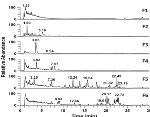
Fig. 2에서 확인된 다양한 성분들에 대한 생리활성 비 교를 위해 분획물을 제조하였다. 우선 Prep-LC(Waters, Milford, MA, USA)를 이용하여 280 nm에서 피크를 모 니터링한 후(Fig. 3), 이를 바탕으로 각 분획의 성분을 확 인하기 위해 LC-MS 프로파일 분석을 수행하였다(Fig.
4). 분획한 시료는 크로마토그램을 참고하여 6개의 구역 으로 나누어 분취한 후, Table 1에서 보이는 것처럼, 각 각의 분획으로 활성검색용 시료를 제조하였다.
(a)
(b)
Fig. 2. LC-MS profile analysis of a fermented soybean extract. MS chromatogram (a) and MS spectrum of six major peaks (b) in MS chromatogram at retention time of 1.61, 3.82, 7.44, 10.46, 15.82, and 20.26 min.
Fig. 3. Prep-LC chromatogram of a fermented soybean extract at 280 nm.
Fig. 4. LC-MS chromatogram of six Prep-LC fraction samples.
(a) (b)
Fig. 5. Effects of soybean-derived low molecular weight bioactive peptide on viability of HaCaT and fibroblast cells.
(a) HaCaT cells were treated with fermented soybean extract at indicated concentration for 24 hr (b) Fibroblast cells were treated with fermented soybean extract at indicated concentration for 24 hr. Cell viabilities were determined as described in materials and methods. Values represent normalized cell viability value (mean±S.D., n=3). The value of untreated cells was taken as 100 %. * p <0.05; ** p <0.01. All experiments were repeated at least three times.
(a) (b)
Fig. 6. Effects of fractions (F1, F3, F4, F5, and F6) on viability of HaCaT cells and fibroblast cells.
(a) HaCaT cells were treated with fermented soybean extract and its fractions (F1, F3, F4, F5, and F6) at 800 μg/mL for 24 hr (b) Fibroblast cells were treated with fermented soybean extract and its fractions (F1, F3, F4, F5, and F6) at 800 μg/mL for 24 hr. Cell viabilities were determined as described in materials and methods. Values represent normalized cell viability value (mean±S.D., n=3). The value of untreated cells was taken as 100 %. * p <0.05; ** p <0.01. All experiments were repeated at least three times.
Table 1. Six Prep-LC fraction samples of a fermented soybean extract
Fraction No. F1 F2 F3 F4 F5 F6
Weight(mg) 287.76 73.28 57.44 14.059 162.59 121.78 Solution
(1,000 μg/mL)
2.2.3 대두발효물의 세포 독성
대두발효물의 세포 실험 적정 농도를 확인하기 위해, HaCaT 세포와 fibroblast 세포에 대두발효물을 0∼
2,000 μg/mL로 농도별로 처리한 후 24시간 동안 배양 하여 WST-1 assay 방법에 따라 세포 증식 변화를 확인 하였다. Fig. 5에 제시된 바와 같이 HaCaT 세포(Fig.
5a)와 fibroblast 세포(Fig. 5b)의 전 농도구간(0∼
2,000 μg/mL)에서 세포 독성이 나타나지 않았을 뿐 아
니라 세포의 증식이 활발하게 일어남을 확인하였다. 피부
세포에 성장을 촉진시키는 표피 성장인자로 알려진 양성
대조군으로 사용한 EGF(epidermal growth factor)보
다도 강한 세포 증식 효과가 나타났다. 대두발효물은
800 μg/mL의 농도에서 가장 뛰어난 세포 증식 효과를
보여주었는데, 모든 농도 구간에서 EGF와 유사하거나
(a) (b)
(c) (d)
Fig. 7. Effects of fraction 4 (F4) and fraction 5 (F5) on viability of HaCaT and fibroblast cells.
(a) HaCaT cells were treated with F4 at indicated concentration for 24 hr (b) HaCaT cells were treated with F5 at indicated concentration for 24 hr (c) Fibroblast cells were treated with F4 at indicated concentration for 24 hr (d) Fibroblast cells were treated with F5 at indicated concentration for 24 hr. Cell viabilities were determined as described in materials and methods. Values represent mean±S.D. (n=3), of normalized cell viability value. The value of untreated cells was taken as 100 %. * p <0.05; ** p < 0.01. All experiments were repeated at least three times.
더 높은 세포증식 효과가 나타나 피부 노화 억제 및 피부 세포 재생 소재로서의 뛰어난 응용 가능성을 보여주었다.
HaCaT 세포와 fibroblast 세포에서 대두발효물이 800 μg/mL의 농도에서 대조군에 비해 약 1.7배 이상 증가하 였고, 이 농도에서 가장 뛰어난 세포 증식 효과를 보여주 었기 때문에 이후 진행하는 분획물에 대한 세포 독성 실 험은 800 μg/mL의 농도로 진행하였다.
2.2.4 대두발효물과 분획물의 세포 독성
대두발효물로 부터 분획 된 6개의 분획물 중, 용해되 지 않는 F2 분획물을 제외하고 총 5개의 분획물 (F1, F3, F4, F5, F6)에 대한 세포 독성 실험을 800 μg/mL의 농 도에서 진행하였다. 그 결과 Fig. 6에 제시된 바와 같이 5개의 모든 분획물 모두 세포 독성은 나타나지 않았다.
그 가운데 F4 분획물(Fig. 6a)과 F5 분획물(Fig. 6b)은 대두발효물과 유사하거나 더 높은 세포증식 효과가 나타 났고, HaCaT 세포에서는 대조군 대비 2배 이상의 세포 증식효과가 나타났다(p<0.01, Fig. 6a). 따라서 F4와 F5 분획물만을 선정하여 HaCaT 세포(Fig. 7a & b)와 fibroblast 세포(Fig. 7c & d)를 0∼2,000 μg/mL로 각
각 농도별로 처리하고, 24시간 배양을 하여 각 세포의 생 존비를 확인한 결과 (Fig. 7) 모든 농도 구간에서 세포에 대한 독성은 없었으며, F4 분획물과 F5 분획물 역시 대 두발효물과 동일하게 800 μg/mL 부근에서 세포의 증식 효과가 가장 높았다.
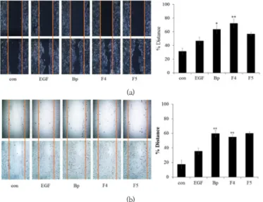
2.2.5 대두발효물의 상처 치유 효과
진피에서 지속적으로 증식하여 표피로 이동하는 피부 세포의 이동능은 각피세포로 분화하기 위한 일반적인 생 리활성기능이며, 상처로 인한 피부 재생을 위해 매우 중 요한 세포 활성 기능 중의 하나이다[29]. EGF의 성장인 자는 세포의 성장, 증식, 그리고 분화를 촉진 시키는 아미 노산 53개가 결합 된 단백질이다[30]. 피부 속에 침투된 EGF가 각질형성 세포의 성장과 분화를 촉진시키도록 신 호를 보내고, 각질층의 복원을 통해 빠른 재생 주기를 가 지게 된다[31]. 대두올리고펩타이드는 사람의 피부 손상 을 보호하는 것으로 알려져 있다[28]. 따라서 피부세포의 이동능에 대두발효물이 미치는 효과를 알아보기 위해 세 포이동 실험을 진행한 결과(Fig. 8) 대두발효물을 0∼
1,000 μg/mL로 각각의 농도에 따라 처리하였을 때,
(a)
(b)
Fig. 8. Effects of a fermented soybean extract on migration of HaCaT and fibroblast cells.
(A) HaCaT cells were treated with epidermal growth factor (EGF) as positive control and fermented soybean extract at indicated concentration for 24 hr (B) Fibroblast cells were treated with epidermal growth factor (EGF) as positive control and fermented soybean extract at indicated concentration for 24 hr. Wound healing assay was performed as described in materials and methods. Phase contrast images were taken at 24 hr after scratching to determine healed distance. Values represent mean±S.D. (n=3). * p <0.05; ** p <0.01. All experiments were repeated at least three times.
(a)
(b)
Fig. 9. Effects of a fermented soybean extract and fractions (F4 and F5) on migration of HaCaT cells and fibroblast cells.
(a) HaCaT cells were treated with epidermal growth factor (EGF) as a positive control and fermented soybean extract and fractions (F4 and F5) at indicated concentration for 24 hr (b) Fibroblast cells were treated with epidermal growth factor (EGF) as a positive control and fermented soybean extract and fractions (F4 and F5) at indicated concentration for 24 hr.
Wound healing assay was performed as described in materials and methods. Phase contrast images were taken at 24 hr
after scratching to determine healed distance. Values represent mean±S.D. (n=3), * p <0.05; ** p <0.01. All experiments were
repeated at least three times.
HaCaT(Fig. 8A)와 fibroblast(Fig. 8B)의 피부 세포 이 동능이 대조군에 비해 약 1.5배 이상 향상된 것이 확인되 었다(p<0.01). 또한, EGF보다도 높은 효과가 나타나 피 부 손상을 효과적으로 치유하고 피부 재생을 돕는 물질 로의 활용이 가능할 것으로 판단된다.
2.2.6 대두발효물과 분획물(F4, F5)의 상처 치유 효과 대두발효물과 함께 F4와 F5 분획물의 피부세포 이동 능을 확인한 결과(Fig. 9), HaCaT 세포(Fig. 9a)와 fibroblast 세포(Fig. 9b) 모두 800 μg/mL의 농도 구간 에서 대조군보다 유의하게 향상된 이동능을 확인할 수 있었다. 또한, 대두발효물과 분획물이 F4와 F5가 EGF와 유사하거나 그 이상의 효과를 나타내 상처 치유 효과가 우수함을 확인할 수 있었다.
3. 결론
본 연구는 대두 단백질을 효소(pepsin) 분해 및 미생 물(Lactobacillus Rhamnosus) 발효를 통해 생성된 저 분자 생리활성 펩티드로 제조하여 인간 유래 피부세포에 서 세포활성에 미치는 효과를 확인하고, 천연유래 기능성 미용소재로서의 이용 가능성을 확인하고자 하였다.
HaCatT 세포 및 fibroblast 세포에 대한 대두단백질 과 그 분획물의 cell viability를 WST-1 assay를 수행한 결과, 0-2,000 μg/mL의 각각의 농도 구간 모두 세포의 독성이 나타나지 않았다.
농도 구간 중 800 μg/mL의 농도에서는 대조군 대비 160-180%, EGF(epidermal growth factor) 대비 120%로 세포증식의 증가가 확인되었다.
세포 이동 실험으로 대두발효물과 그 분획물의 상처 치유 능력을 확인한 결과 대두발효물과 분획물(F4와 F5) 모두 EGF와 유사하거나 그 이상의 효과가 확인되었다.
결론적으로 대두발효물과 분핵물(F4와 F5)의 피부개 선 효과가 유사하게 나타나 경제성을 고려해야 하는 미 용 산업계에서는 대두발효물을 피부미용 소재 및 피부 질환 치료의 소재로 활용하는 것이 유리할 것으로 사료 된다.
References
[1] D. Agyei, “Bioactive proteins and peptides from
soybeans”,
Recent Patents on Food, Nutrition &Agriculture
, Vol.7, No.2, pp.100–107, Agu. 2015.
DOI: https://doi.org/10.2174/221279840766615062913414 [2] S. P. Hong, U. H. Choi, S. G. An, “Skin barrier and antimicrobial activity”,
The Journal of Skin Barrier Research, Vol.11, No.1, pp.55-63, Jun. 2009.
[3] H. Y. Yu, I. J. Yang, V. R Lincha, I. S. Park, D. U. Lee, H. M. Shin, “The Effects of the Fruits of Foeniculum vulgare on Skin Barrier Function and Hyaluronic Acid Production in HaCaT Keratinocytes”,
Journal of life science, Vol.25, No.8, pp.880-888, Agu. 2015.
DOI: http://dx.doi.org/10.5352/JLS.2015.25.8.880 [4] H. S. Jung, M. Y. Song, H. S. Kim, H. H. Seo, J. H. Lee,
K. R. Lee, I. Hong, S. H. Moh. “Development of Anti-Wrinkle Materials using Galloyl-Peptide Derivatives”,
Journal of the Korea Academia-Industria, Vol.16, No.8, pp.5452-5457, Agu. 2015.
DOI: http://dx.doi.org/10.5762/KAIS.2015.16.8.5452 [5] M. A. Ruiz, B. Clares, M. E. Morales, V. Gallardo.
“Evaluation of the anti-wrinkle efficacy of cosmetic formulations with an anti-aging peptide (Argireline®)”,
ARS Pharmaceutica, Vol.50, No.4, pp.168-176, Nov. 2009.
[6] B. F. Gibbs, A. Zougman, R. Masse, C. Mulligan.
“Produc-tion and characterization of bioactive peptides from soy hydrolysate and soy-fermented food”,
Food Research International, Vol.37, No.2, pp.123–131, Mar. 2004.
DOI: https://doi.org/10.1016/j.foodres.2003.09.010 [7] W. Wang, N. A. Bringe, M. A. Berhow, E. Gonzalez de
Mejia. “β-Conglycinins among Sources of Bioactives in Hydrolysates of Different Soybean Varieties That Inhibit Leukemia Cells in Vitro”,
Journal of Agricultural and Food Chemistry, Vol.56, No.11, pp.4012–4020, Mar. 2008.
DOI: https://doi.org/10.1021/jf8002009
[8] E. J. Na, J. S. Moon. “Studies on the Biological Activi-ties of the Bioconversioned Soybean Extracts”,
Jounal of The Korean Society of cosmetology, Vol.21, No.1, pp.82-92, Feb. 2015.
[9] A. Kannan, N. S. Hettiarachchy, J. O. Lay, R. Liyanage.
“Human cancer cell proliferation inhibition by a pen-tapeptide isolated and characterized from rice bran”,
Peptides, Vol.31, No.9, pp.1629–1634, Sep.
2010.
DOI: https://doi.org/10.1016/j.peptides.2010.05.018 [10] B. F. Gibbs, A. Zougman, R. Masse, C. Mulligan.
“Production and characterization of bioactive peptides from soy hydrolysate and soy-fermented food”,
Food Research International, Vol.37, No.2, pp.123–131, Mar. 2004.
DOI: https://doi.org/10.1016/j.foodres.2003.09.010 [11] M. Förster, M. A. Bolzinger, H. Fessi, S. Briançon.
“Topical delivery of cosmetics and drugs. Molecular
aspects of percutaneous absorption and delivery”,
European Journal of Dermatology, Vol.19, No.4,
pp.309-323, Agu. 2009.
DOI: https://doi.org/10.1684/ejd.2009.0676
[12] B. Forslin. “A domain mosaic model of the skin barrier”,
Acta Dermato-venereologica, Vol.74, No.1, pp.1-6, Jan. 1994.
[13] Z. Nemes, P. M. Steinert. “Brick and mortar of the epidermal barrier”,
Experimental & Molecular Medicine, Vol.31, No.1, pp.5-19, Mar. 1999.
DOI: https://doi.org/10.1038/emm.1999.2
[14] S. Ioannis, F. Demosthenes, V. Katerina, G. T. Andreas, K. Valentinos, B. Evangelos. “Cyanobacterial cyclopeptides as lead compounds to novel targeted cancer drugs”,
Marine Drugs, Vol.8, No.3, pp.629-657, Mar. 2010.
DOI: https://doi.org/10.3390/md8030629
[15] J. K. Kim, J. H. Lee, I. H. Bae, D. B. Seo, S. J. Lee.
“Beneficial Effect of a Collagen Peptide Supple-ment on the Epidermal Skin Barrier”,
Korean Journal of Food Science and Technology, Vol.43, No.4, pp.458-463, Agu. 2011.
DOI: https://doi.org/10.9721/KJFST.2011.43.4.458 [16] S. Sakai, T. Sayo, S. Kodama, S. N. Inoue.
“N-Methyl-L-serine stimulates hyaluronan production in human skin fibroblasts”, Skin Pharmacol. Appl”,
Skin Phar-macology and Physiology, Vol.12, No.5, pp.276-283, Oct. 1999.
DOI: https://doi.org/10.1159/000066253
[17] J. C. Lee, E. Lee, Y. C. Lee, H. Oh, H. S. Yoon, T. K.
Ha, E. H. Hong. “Effects of Fructus foeniculi extract on recovering liver function”,
The Korea Journal of Herbology, Vol.22, No.4, pp.213-218, Dec. 2007.
[18] M. C. Kwon, C. H. Kim, H. S. Kim, A. Q. Syed, B. Y.
Hwang, H. Y. Lee. “Anti-wrinkle Activity of Low Molecular Weight Peptides Derived from the Collagen Isolated from Asterias amurensis”,
Korean Journal of Food Science and Technology, Vol.39, No.6, pp.625-629, Oct. 2007.
[19] S. J. Lee, H. R. Choi, J. C. Lee, H. J. Park, H. K. Lee, J. T. Jeong, T. B. Lee. “The Anti-aging Effects of Various Berries in the Human Skin Keratinocyte (HaCaT) Cells”,
Korean Journal of Food Science and Technology, Vol.46, No.2, pp.198-204, Apr. 2014.
DOI: https://doi.org/10.9721/KJFST.2014.46.2.198 [20] A. V. Rawlings, C. R. Harding. “Moisturization and skin
barrier function”,
Dermatologic Therapy, Vol.17, No.1, pp.43-48, Jan. 2004.
DOI: https://doi.org/10.1111/j.1396-0296.2004.04S1005.x [21] F. J. Smith, A. D. Irvine, A. Terron-Kwiatkowski, A.
Sandilands, L. E. Campbell, Y. Zhao, H. Liao, A. T.
Evans, D. R. Goudie, S. Lewis-Jones, G. Arseculeratne, C. S. Munro, A. Sergeant, G. O'Regan, S. J. Bale, J. G.
Compton, J. J. DiGiovanna, R. B. Presland, P.
Fleckman, W. H. McLean. “Loss-of-function mutations in the gene encoding filaggrin cause ichthyosis vulgaris”,
Nature Genetics, Vol.38, No.3, pp.337-342, Jan. 2006.
DOI: https://doi.org/10.1038/ng1743
[22] R. L. Gallo, M. Murakami, T. Ohtake, M. Zaiou.
“Biology and clinical relevance of naturally occurring antimicro-bial peptides”,
Journal of Allergy and Clinical Immu-nology, Vol.110, No.6, pp.823-831, Sep.
2002.
DOI: https://doi.org/10.1067/mai.2002.129801 [23] J. S. Kim, E. J. Bak, B. C. Lee, Y. S. Kim, J. B. Park, I.
G. Choi. “Neuregulin induces HaCaT keratinocyte migration via Racl-mediated NADPH-oxidase activation”,
Journal of Cellular Physiology, Vol.226, No.11, pp.3014-3021, Feb. 2011.
DOI: https://doi.org/10.1002/jcp.22649
[24] G. M. Marcela, R. G. Eva, R. M. Del Carmen, M. E.
Rosalva. “Evaluation of the Antioxidant and Anti-proliferative Effects of Three Peptide Fractions of Germinated Soybeans on Breast and Cervical Cancer Cell Lines”,
Plant Foods for Human Nutrition, Vol.71, No.4, pp.368-374, Jul. 2016.
DOI: https://doi.org/10.1007/s11130-016-0568-z [26] Y. Nzengue, R. Steiman, C. Garrel, "Oxidative stress
and DNA damage induced by cadmium in the human keratinocyte HaCaT cell line: role of glutathione in the resistance to cadmium", Toxicology, Vol.243, pp.193-206, Jan. 2008.
DOI: https://doi.org/10.1016/j.tox.2007.10.005 [27] J. Lopez-Garcia, M. Lehocky, P. Humpolicek, P. Saha,
"HaCaT keratinocytes response on antimicrobial a telocollagen substrates: extent of cytotoxicity, cell viability and proliferation", J Funct Biomater, Vol.5, No.2, pp.43–57, Apr. 2014.
DOI: https://doi.org/10.3390/jfb5020043
[28] J. Jonkman, J. Cathcart, F. Xu, M. Bartolini, J. Amon, K.
Stevens, P. Colarusso, "An introduction to the wound healing assay using live-cell microscopy", Cell Adhes.
Migr, Vol.8, pp. 440–451, Apr. 2014
[29] H. Y. Yu, I. J. Yang, V. R. Lincha, I. S. Park, D. U. Lee, H. M. Shin. “The Effects of the Fruits of Foe-niculum vulgare on Skin Barrier Function and Hya-luronic Acid Production in HaCaT Keratinocytes”,
Journal of Life Science, Vol.25, No.8, pp.880-888, Agu. 2015.
DOI: http://dx.doi.org/10.5352/JLS.2015.25.8.880 [30] B. McKnight, R. Seidel, R. Moy. “Topical Human
Epidermal Growth Factor in the Treatment of Senile Purpura and the Prevention of Dermatoporosis”,
Journal of Drugs in Dermatology, Vol.14, No.10, pp.1147-1150, Oct. 2015.
[31] K. Kaufmann, G. Thiel. “Epidermal growth factor and thrombin induced proliferation of immortalized human keratinocytes is coupled to the synthesis of Egr-1, a zinc finger transcriptional regulator”,
Journal of Cellular Biochemistry, Vol.85, No.2, pp.381-391, Mar. 2002.
DOI: https://doi.org/10.1002/jcb.10145
김 은 주(Eun-Joo Kim) [정회원]
• 1995년 2월 : 서울과학기술대학교 환경공학과 (환경공학학사)
• 1997년 2월 : 서울과학기술대학교 일반대학원 환경공학과 (환경공학 석사)
• 2021년 2월 : 경기대학교 일반대 학원 대체의학과 (대체의학박사)
• 2009년 12월 ~ 현재 : ㈜제니트리 대표
<관심분야>
기능성식품, 식문화
한 명 륜(Myung-Ryun Han) [정회원]
• 2000년 2월 : 단국대학교 식품공 학과(공학사)
• 2002년 8월 : 단국대학교 식품공 학과(공학석사)
• 2008년 8월 : 단국대학교 식품공 학과(공학박사)
• 2008년 3월 ~ 2019년 2월 : 혜전 대학교 식품영양과 교수
• 2019년 3월 ~ 현재 : 혜전대학교 제과제빵과 교수
<관심분야>
식품공학, 기능성식품, 식문화
이 소 영(So-Young Lee) [정회원]
• 2002년 2월 : 영남대학교 가정관 리학과 (생활과학학사)
• 2004년 2월 : 영남대학교 일반대 학원 가정학과 조리및주거학전공 (생활과학석사)
• 2017년 8월 : 경기대학교 대체의 학대학원 식품치료전공 (대체의학
• 2020년 8월 : 경기대학교 일반대학원 대체의학과 (대체의석사) 학박사)
<관심분야>
임상영양, 기능성식품, 식문화
김 애 정(Ae-Jung Kim) [정회원]
• 1986년 2월 : 숙명여자대학교 식 품영양학과 (가정학학사)
• 1988년 8월 : 숙명여자대학교 자 연과학대학원 식품영양학과 (가정 학석사)
• 1992년 8월 : 숙명여자대학교 자 연과학대학원 식품영양학과 (이학
• 1993년 3월 ~ 2011년 8월 : 혜전대학교 식품영양과 교수 박사)
• 2011년 9월 ~ 현재 : 경기대학교 대체의학대학원 교수
<관심분야>
임상영양, 기능성식품, 식문화