J. Exp. Biomed. Sci. 12 (2006) 249–254
The Effect of Epidermal Growth Factor on Cell Proliferation and Its Related Signal Pathways in Pig Hepatocytes
Dong-il Kim, Ho-jae Han and Soo-hyun Park
†Bio-therapy Human Resources Center, Department of Veterinary Physiology, College of Veterinary Medicine, Chonnam National University, Gwangju 500-757, Korea
It has been reported that liver is a very important organ to xenotransplantation. Pig is known to be a most suitable species in transplantation of human organs. However, the physiological function of pig hepatocytes is not clear elucidated.
Epidermal growth factor (EGF) is known to be a mitogen in various cell systems. Thus, we examined the effect of EGF on cell proliferation and its related signal cascades in primary cultured pig hepatocytes. EGF stimulates cell proliferation in a dose (> 1 ng/ml) dependent manner. EGF-induced increase of [
3H]-thymidine incorporation was blocked by AG 1478 (10
-6M, an EGF receptor antagonist) genistein and herbymycin A (tyrosine kinase inhibitors, 10
-6M), suggesting the role of activation and tyrosine phosphorylation of EGF receptor. In addition, EGF-induced increase of [
3H]-thymidine incorporation was prevented by neomycin (10
-4M), U73122 (10
-5M) (phospholipase C [PLC] inhibitors), staurosporine (10
-8M), or bisindolylmaleimide I (10
-6M) (protein kinase C [PKC] inhibitors), suggesting the role of PLC and PKC.
Moreover, EGF-induced increase of [
3H]-thymidine incorporation was blocked by PD 98059 (a p44/42 mitogen activated protein kinase [MAPK] inhibitor), SB 203580 (a p38 MAPK inhibitor), and SP 600125 (a JNK inhibitor). EGF increased the translocation of PKC from cytosol to membrane fraction and activated p42/44 MAPK, p38 MAPK and JNK. In conclusion, EGF stimulates cell proliferation via PKC and MAPK in cultured pig hepatocytes.
Key Words: Epidermal growth factor, Protein kinase C, Mitogen activated protein kinase, Pig hepatocytes
서 론
인간에게서 이종 장기이식은 최근 화두로 떠오르고 있다.
인간과 면역학적 유사성 때문에 영장류가 가장 적절한 장기 원이지만, 윤리학적 문제와 더불어 대단위 육성 및 신속한 성장의 장애 때문에 이들의 사용은 호의적이지 않고 있다.
장기를 인간에게 이식할 경우 가장 적합한 동물은 insulin 생성, 포도당 조절이 인간과 유사하고 신속한 성장이 된다 는 점에서 돼지가 가장 각광을 받고 있다 (Saif et al., 1996;
Miller & Ullrey, 1987). 돼지 태아의 췌장세포를 당뇨병 환자 에게 이식 보고 및 돼지의 간세포를 간질환 환자에게 이식했 다는 보고 역시 이를 뒷받침 해주고 있다 (Groth, 1995; Tector et al., 2001). 최근 돼지의 간세포 배양을 통한 인공장기의 구 축이 필요하다고 하는 보고 역시 간이식에 있어서 간세포가
중요성을 갖는다고 하겠다 (Li et al., 2005).
간세포의 성장은 간의 이식 등에 있어서 세포 손상에 대한 보상기전으로 이용되고 있다 (Fausto & Riehle, 2005). 이러한 간세포의 성장에는 다양한 세포성장인자들이 관여하는 것으 로 보고되고 있다 (Michalopoulos, 1990). 간세포에서 Insulin like growth factors (IGFs) 및 hepatocyte growth factor (HGF)가 세포성장을 증가 시킨다고 보고되었다 (Kimura & Ogihara, 1997b; 1998). EGF 역시 간세포의 기능에 영향을 미치는 것 으로 보고되고 있다 (Kong et al., 2000; Tsai et al., 2001). EGF 는 수용체에 결합하여 이량화를 유도하여 tyrosine kinase 잔 기에 자가인산화가 되어 EGF 수용체 복합체의 활성을 야기 하여 세포기능에 관여하는 것으로 알려져 있지만 (Yarden &
Sclessinger, 1987), 돼지의 간세포에서 EGF의 세포성장을 비 롯한 세포기능에 대한보고는 이루어지고 있지 않고 있다.
일반적으로 세포에 대한 성장 효과는 다양한 신호전달계 가 관여한다. 이들 중 protein kinase C (PKC)는 phospholipase C (PLC)의 PIP2 분해에 의한 DAG 활성에 의해 활성화 되며 세포성장 등의 다양한 세포내 반응 등에 관여하는 것으로 알려져 있다 (Musashi et al., 2001). Mitogen activated protein kinases (MAPKs)는 일반적으로 p44/42 MAPK, p38 MAPK 및
*논 문 접 수: 2006년 8월 18일 수정재접수: 2006년 8월 28일
†교신저자: 박수현, (우) 500-757, 광주광역시 북구 용봉동 300, 전남대학교 수의과대학 생리학교실
Tel: 062-530-2832, Fax: 062-530-2809 e-mail: [email protected]
SAPK/JNK으로 구분이 되며 세포의 성장 및 스트레스 등의 다양한 반응에 관여하는 것으로 보고되고 있다 (Bogatcheva et al., 2003). 이러한 보고들은 PKC 및 MAPK의 활성이 EGF 에 의한 간세포 성장에 신호전달 매개체로 작용할 수 있음 을 말해 주고 있다. 그러나 지금까지 이에 대한 연구는 미약 한 실정에 있다. 따라서 본 실험에서는 돼지의 간세포를 초 대 배양하여 EGF가 세포성장에 미치는 효과와 이와 관련된 신호전달계에 대해 알아보았다.
재료 및 방법
1. 재 료
Dulbecco's Modified Eagle's Medium (D-MEM)/Ham's nutrient mixture F-12 (D-MEM/F-12)와 Class IV collagenase는 Life Technologies (Grand Island, NY, USA)로부터 구입하였다. D- glucose, staurosporine, H-7, PD 98059는 Sigma Chemical Com- pany (St. Louis, MO, USA)로부터 구입하였다. Bisindolylma- leimide I, SB 203580 및 SP 600125는 Calbiochem (La Jolla, CA, USA)에서 구입하였다. 그 외 실험에 이용된 모든 시약 들은 고품질의 시약을 사용하였다.
2. Hepatocyte의 초대 배양
간세포는 2~3 kg 돼지를 이용하여 분리하였다. Li 등 (2005)에 의해 발표된 두 단계의 collagenase 관류 방법이 이 용되었다. 간단히 기술하면 isoflurane/02 마취를 시도한 후 복정중선을 따라 개복을 실시하였다. 이후 문맥의 아랫부분 을 결찰한 후 cathether를 삽입한 후 30 mM HEPEs가 들어 있는 Mg2+/Ca2+이 없는 Hanks buffer로 37℃에서 약 10분 동 안 관류 시켰다. 이후 0.05% type I collagenase가 있는 Eagle's 배지로 교환하였다. 이후 분리된 간을 petri dish에 놓고 Glisson 피막을 벗긴다. 세포수는 5×10⁵ 정도의 세포로 배양 시켰다. 세포의 생존율은 trypan blue exclusion 방법을 이용하 여 실시하였으며 생존율이 90%가 넘는 경우에만 실험에 사 용하였다. 실험에 사용된 돼지들은 실험동물사에서 사육되었 고 실험 기간 동안 사육 환경은 23±2℃, 상대습도 55±10%
를 유지하였고, 인공조명 (12시간 점등, 12시간 소등)하에서 사육하였으며, 사료와 정제수는 자유로이 급여하였다. 모든 실험은 서울대학교 실험동물연구지원센터 동물실험윤리위원 회 (Institutional Animal Care and Use Committee, IACUC)의 동 물실험 표준작업지침서 (Standard Operation Procedures [SOP]) 에 따라 수행되었다.
3. [3H]-thymidine incorporation
간세포들이 50~60% 성장하여 subconflent를 형성하였을 때, 배지를 교환하였다. [3H]-thymidine incorporation 실험은
Brett 등 (1993)에 의해 기술된 대로 실시하였다. 세포들에 1 µCi의 [3H]-thymidine을 투여 후 4시간 후에 시료를 처리하 여 8시간 배양 시킨 후 실험에 사용하였다. 세포들은 phosp- hate buffered saline (PBS)로 2번 세척한 후 10% trichloroacetic acid (TCA)에서 15분간 고정시켰다. 이후 5% TCA를 5분간 2번 세척을 하였다. 산에 불용성인 부분을 실온에서 2 N NaOH를 처리하여 Liquid scintillation counting에 의하여 방 사성을 측정하였다. 실험결과는 대조군의 %로 보정하였다.
4. Western immunoblotting
배지를 제거한 간세포를 PBS로 2번씩 세척한 후, 각기 150 µl의 lysis buffer (10× PBS, 1% NP-40, 20% SDS, 0.5 M EDTA, 0.01 M PMSF, 10 mg/ml Leupeptin, 1 mg/ml pepstatin A) 를 처리하여 균질화를 시켰다. 균질화된 세포를 tube에 옮긴 후, 15,000 rpm에서 10분간 원심 분리하여 상층액을 새로운 tube에 저장하였다. Bradford 단백질 정량법을 이용하여 각각 60 µg의 sample들을 8% SDS-PAGE 전기영동을 시킨 후, polyvinylidine difluoride membrane에 transfer하였다. Membrane 은 5% skim milk에 1시간 동안 차단을 시켰고, 각각의 항체 (CDK-2, CDK-4, p27 kip1, p21 WAF1/Cip1, p44/42 MAPK, p38 MAPK, pJNK)를 1% skim milk에 1,000배 희석하여 4℃에서 18시간 이상 배양하였다. 그 후, membrane을 0.1% Tween-20/
1× TBS에 10분 간격으로 3번 세척하였고, membrane을 1%
skim milk에 5,000배 희석된 horseradish-peroxidase labeled 2 차 항체에 1시간 동안 배양한 후, 3번 세척을 거쳐서 ECL 시약을 1분간 처리한 다음 X-ray 필름에 30초간 노출시켜 현 상하였다.
5. 통계처리
실험결과의 통계처리는 Student's t test 및 Analysis of Variance (ANOVA)로 하였으며, P 값 <0.05를 유의한 차이의 한계로 하였고, 실험결과의 표현은 means ± S.E로 하였다.
결 과
1. EGF가 간세포 성장 및 세포성장 조절 단백질에 미치는 효과
EGF가 간세포의 세포성장에 미치는 효과를 알아보기 위 하여 EGF를 농도별로 (0~1,000 ng/ml) 처리한 후 thymidine incoporation 실험을 실시하였다. 실험결과 0.1 ng/ml에서는 대조군과 유의성 있는 차이는 인정되지 않았으나 1 ng/ml 이 상에서 유의성 있는 증가 작용이 인정되었으며 이러한 작 용은 100 ng/ml에서 가장 큰 효과를 보였다 (Fig. 1). 이러한 EGF가 세포성장 조절 단백질들과 어떠한 관련이 있는지를 western immunoblotting을 실시하여 알아보았다. Figure 2에서
보이듯이 100 ng/ml의 EGF를 처리한 결과 CDK-2 및 CDK-4 의 발현은 증가 시켰으나, p27 Kip1 및 p21WAF1/Cip1의 발 현은 감소 시키는 것으로 나타났다 (Fig. 2).
2. EGF에 의한 세포성장에 있어서 PKC 및 MAPK 활성 과의 관련성
EGF에 의한 세포성장 효과가 수용체 특이적인지를 알아 보기 위하여 EGF 수용체 길항제인 AG1478 (10-6 M)을 EGF 처리 30분 전에 처리하였다. 실험결과 EGF에 의한 세포성장 효과는 선택적으로 차단되는 것으로 나타났다 (Fig. 3). 아울 러, 어떠한 신호전달경로를 통하여 작용하는지를 알아보기 위하여 tyrosine kinase 억제제인 genistein (10-6 M) 및 herby- mycin A (10-6 M)를 EGF 처리 30분 전에 처리하였다. Figure
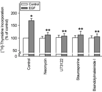
3에서 보이듯이 genistein 및 herbymycin A는 EGF에 의한 세 포성장 효과를 선택적으로 차단하였다 (Fig. 3). EGF에 의한 세포성장이 PLC/PKC 경로와 관여하는지를 알아보기 위하여 PLC 억제제들인 neomycin 및 U73122 및 PKC 억제제들인 staurosporine 및 bisindolylmaleimide I을 처리하여 실험한 결 과 EGF에 의한 세포성장 효과는 선택적으로 차단되는 것으 로 나타났다 (Fig. 4). 실제로 EGF 처리 시 세포질에서 세포 막으로의 PKC의 이동을 볼 수 있었다 (Fig. 5). MAPK와의 관련성을 알아보았다. EGF에 의한 세포성장 촉진 작용은 PD
Fig. 3. Effect of AG1478, herbimycin A and genistein on EGF- induced cell proliferation. Pig hepatocytes were incubated with AG1478 (10-6 M), herbimycin (10-6 M) and genistein (10-6M) for 30 min prior to the treatment of EGF (100ng/ml) for 8 hr. Values are means ± S.E. of 9 separate experiments performed on 3 di- fferent cultures. *P<0.05 vs. control, **P<0.05 vs. EGF alone.
Fig. 2. Effects of EGF on CDK-2, CDK-4, p27 kip1, and p21 WAF1/Cip1. Pig hepatocytes were incubated with EGF (100 ng/
ml) for 8 hr. CDK-2, CDK-4, p27, and p21 were detected as de-
scribed in "Materials and Methods". Fig. 4. Effects of PLC and PKC inhibitors on EGF-induced cell proliferation. Pig hepatocytes were incubated with neomycin (10-5 M), U73122 (10-6 M, PLC inhibitors), sturosporine (10-7M), and bisindolylmaleimide I (10-6M) for 30 min prior to the treatment of EGF (100 ng/ml) for 8 hr. Values are means ± S.E. of 9 separate experiments performed on 3 different cultures. *P<0.05 vs. control,
**P<0.05 vs. EGF alone.
Fig. 1. Dose response curve of EGF on cell proliferation. Pig hepatocytes were incubated with different dosage of EGF (0 to 10-6 g/ml). Values are means ± S.E. of 9 separate experiments per- formed on 3 different cultures. *P<0.05 vs. control.
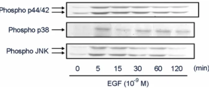
98059 (p44/42 MAPK 억제제), SB 203580 (p38 MAPK 억제 제) 및 SP 600125 (JNK/SAPK 억제제) 처리 시 차단되는 것 으로 나타났다 (Fig. 6). 실제적으로 MAPKs의 활성을 측정하 여 본 결과 EGF 처리 시 p44/42 MAPK, p38 MAPK 및 JNK 활성 모두 5분에서 증가하는 것으로 나타났으며 이후 점차 적으로 감소하는 것으로 나타났다 (Fig. 7).
고 찰
최근 여러 연구자들에 의해서 이종 장기이식에 있어서 간의 중요성을 인식하고 돼지 간세포의 생리학적 기능을 알아보고자 돼지의 간세포 배양법 연구를 시도하고 있다 (Jasmund et al., 2002; Chen et al., 2002). EGF는 세포 손상 등 에 의한 간세포의 재생에 있어 중요한 역할을 한다 (Yama- moto, et al., 2005; Oosthuizen & Lambrechts, 2004). 본 연구결 과에서는 EGF 처리 시 돼지 간세포에서 CDK-2 및 CDK-4 의 발현이 증가하였으며 p21 및 p27의 경우는 감소하는 것 으로 나타났다. 이는 세포주기 중 G1기의 단축을 통해 EGF 가 세포성장을 증가 시킨다는 것을 의미하고 있다. 이러한 결 과는 Thevananther 등 (2004)의 결과에서 EGF 처리 시 cyclin D1이 증가한다는 보고에 근거하여 볼 때 cyclin D는 CDK-4 및 CDK-6와 복합체를 형성하여 세포내의 세포주기에 관여
하기 때문에 본 연구결과에서는 cyclin D1은 측정은 하지 않았지만 일맥상통하는 것으로 판단된다. 최근 Barone 등 (2006)은 간세포에서 세포성장 촉진 작용에는 cyclin D1, p21 및 p27 등의 단백질들의 변화를 통한 G1기를 단축시켜 작용 한다고 하여 본 연구결과를 뒷받침해 주고 있다.
EGF는 세포막의 EGF 수용체에 결합을 하여 작용을 나타 낸다. 이들 수용체는 tyrosine 잔기를 가지고 있으며 이들의 기능이 중요한 역할을 하는 것으로 일반적으로 알려져 있다 (Hsieh & Conti, 2005; Jorissen et al., 2003). 본 실험에서도 역 시 EGF에 의한 세포성장 시에 tyrosine 잔기의 활성이 필수 적인 것으로 나타났다. Tyrosine kinase 수용체를 함유하고 있 는 EGF는 간세포의 성장을 위하여 PLC-γ의 직접적인 활성 으로 인하여 phosphatidyl inositol (1,4,5) triphosphate 및 PKC 활성제인 diacylglycerol 같은 2차 전달 물질을 생산하는 것으 로 보고되었다 (Berridge, 1993). 이러한 보고와도 일치하게 본 실험에서도 PLC 억제제 및 PKC 억제제에 의해서 간세포 의 세포성장 효과는 유의성 있게 차단되어 PLC 및 PKC의 활성이 EGF에 의한 세포성장 촉진 작용에 관여된다는 것을 볼 수 있었다. 본 실험에서는 EGF 처리 시 세포질에서 세포 막으로의 PKC 활성이 유도된다는 사실을 증명하였고 나아 가 이러한 PKC의 활성이 세포성장에 영향을 미친다는 것까 지 증명하였다. 비록 본 실험에서는 PKC의 isoforms들에 대 해서는 살펴보지 않았다. 이에 대한 연구는 향후 연구에서 밝혀져야 할 것으로 사료된다.
MAPKs는 몇몇 세포외 자극에 의해서 활성화 되며 다양 한 세포내 신호전달경로에서 중요한 역할을 하는 것으로 알 려지고 있다 (MacCorkle & Tan, 2005). Gao 등 (2004)은 초대 배양한 랫트의 간 위성세포에서 p44/42 MAPK 활성이 세포 성장인자에 의한 세포성장 효과에 관여하는 것을 보고하여 간세포에서도 이러한 p44/42 MAPK 활성이 세포성장 촉진 효과에 미칠 수 있다는 가능성을 보여 주었다. 본 실험에서는 EGF에 의한 세포성장은 p44/42 MAPK 억제제인 PD 98059 에 의해서 차단되는 것으로 나타났다. 이러한 결과는 세포는 다르지만 최근 Heo 등 (2006)에 의한 마우스 배아세포에서 Fig. 5. Effect of EGF on PKC activation. Pig hepatocytes were
treated with EGF (100 ng/ml) for 8 hr. Then, cytosolic and particu- late fraction was separated as described in "Materials and Methods".
Fig. 6. Effects of MAPKs inhibitors on EGF-induced cell proli- feration. Pig hepatocytes were preincubated with SB 203580, PD 98059, and SP 600125 (10-6 M) for 30 min prior to the treatment of EGF (100 ng/ml) for 8 hr. Values are means ± S.E. of 9 separate experiments performed on 3 different cultures. *P<0.05 vs. control,
**P<0.05 vs. EGF alone.
Fig. 7. Effects of EGF on MAPKs activation. Pig hepatocytes were treated with EGF (100 ng/ml) for different time (0 to 120 min). Then, phosphorylated p38, JNK, p44/42 MAPKs were de- tected as described in "Materials and Methods".
p44/42 MAPK의 활성이 EGF에 의한 세포성장 효과에 관여 한다는 결과와 일치하였다. Choi 등 (2004)은 ERK1/2 이외 에도 JNK/SAPK 활성이 세포성장에 함께 관여할 수 있다고 하였으며, 간세포에도 세포성장의 효과에 JNK 및 p38 MAPK 활성이 중요한 역할을 하는 것으로 보고되었다 (Thevananther et al., 2004; Awad et al., 2000). 본 실험에서는 흥미롭게도 p44/42 MAPK 이외에도 JNK/SAPK 및 p38 MAPK 활성 모 두가 EGF에 의한 세포성장 촉진 효과에 주요한 역할을 하 는 것으로 나타났다. 본 연구결과는 간세포의 EGF에 세포성 장 효과 규명뿐만 아니라 향후 이종 장기 영역에서 돼지에 대한 생리학적 자료가 매우 부족한 관계로 인하여 돼지 간세 포의 기능성 연구에 기초자료로 활용될 것으로 사료된다. 결 론적으로 돼지의 간세포에서 EGF는 PKC 및 MAPK 활성을 통해 세포성장을 유도하는 것으로 나타났다.
감사의 글
본 연구는 농림부 바이오장기 생산 연구사업의 연구비 (번호 200503010302) 및 BK21 바이오치료 산업인력 양성사 업팀의 지원을 받아 수행되었으며 이에 깊이 감사드립니다.
REFERENCES
Awad MM, Enslen H, Boylan JM, Davis RJ, Gruppuso PA.
Growth regulation via p38 mitogen-activated protein kinase in developing liver. J Biol Chem. 2000. 275: 38716-38721.
Barone M, Ladisa R. Di Leo A, Spano D, Francioso D, Aglio V, Amoruso A, Francavilla A, Iolascon A. Estrogen-induced proliferation in cultured hepatocytes involves cyclin D1, p21(Cip1) and p27(Kip1). Dig Dis Sci. 2006. 51: 580-586.
Berridge MJ. Inositol trisphosphate and calcium signalling. Nature 1993. 361: 315-325.
Bogatcheva NV, Dudek SM, Garcia JG, Verin AD. Mitogen- activated protein kinases in endothelial pathophysiology. J Investig Med. 2003. 51: 341-352.
Brett CM, Washington CB, Ott RJ. Interaction of nucleoside analogues with the sodium-nucleoside transport system in brush border membrane vesicles from human kidney. Phar- macol Res. 1993. 10: 423-426.
Chen Z, Ding Y, Zhang H. Morphology, viability and functions of suckling pig hepatocytes cultured in serum-free medium at high density. Dig Surg. 2002. 19: 184-191.
Choi J, Park SY, Joo CK. Hepatocyte growth factor induces pro- liferation of lens epithelial cells through activation of ERK1/2 and JNK/SAPK. Invest Ophthalmol Vis Sci. 2004. 45: 2696 -2704.
Fausto N, Riehle KJ. Mechanisms of liver regeneration and their clinical implications. J Hepatobiliary Pancreat Surg. 2005.
12: 181-189.
Gao R, Ball DK, Perbal B, Brigstock DR. Connective tissue growth factor induces c-fos gene activation and cell proliferation through p44/42 MAP kinase in primary rat hepatic stellate cells. J Hepatol. 2004. 40: 431-438.
Groth CG. Transplantation of porcine fetal pancreas to diabetic patients. Lancet 1995. 345: 735.
Heo JS, Lee YJ, Han HJ. EGF stimulates proliferation of mouse embryonic stem cells: involvement of Ca2+ influx and p44/42 MAPKs. Am J Physiol Cell Physiol. 2006. 290: C123-C133.
Hsieh M, Conti M. G-protein-coupled receptor signaling and the EGF network in endocrine systems. Trends Endocrinol Met.
2005. 6: 320-326.
Jasmund I, Langsch A, Simmoteit R, Bader A. Cultivation of primary porcine hepatocytes in an OXY-HFB for use as a bioartificial liver device. Biotechnol Prog. 2002. 18: 839-846.
Jorissen RN, Walker F, Pouliot N, Garrett TP, Ward CW, Burgess AW. Epidermal growth factor receptor: mechanisms of activa- tion and signalling. Exp Cell Res. 2003. 284: 31-53.
Kimura M, Ogihara M. Effects of insulin-like growth factor I and II on DNA synthesis and proliferation in primary cultures of adult rat hepatocytes. Eur J Pharmacol. 1998. 354: 271-281.
Kimura M, Ogihara M. Proliferation of adult rat hepatocytes by hepatocyte growth factor is potentiated by both phenyle- phrine and metaproterenol. J Pharmacol Exp Ther. 1997. 282:
1146-1154.
Kong M, Mounier C, Wu J, Posner BI. Epidermal growth factor- induced phosphatidylinositol 3-kinase activation and DNA synthesis. Identification of Grb2-associated binder 2 as the major mediator in rat hepatocytes. J Biol Chem. 2000. 275:
36035-36042.
Li J, Li LJ, Chao HC, Yang Q, Liu XL, Sheng JF, Yu HY, Huang JR. Isolation and short term cultivation of swine hepatocytes for bioartificial liver support system. Hepatobiliary Pancreat Dis Int. 2005. 4: 249-253.
MacCorkle RA, Tan TH. Mitogen-activated protein kinases in cell-cycle control. Cell Biochem Biophys. 2005. 43: 451-461.
Michalopoulos GK. Liver regeneration: molecular mechanisms of growth control. FASEB J. 1990. 4: 176-187.
Miller ER, Ullrey DE. The pig as a model for human nutrition.
Annu Rev Nutr. 1987. 7: 361-382.
Musashi M, Ota S, Shiroshita N. The role of protein kinase C isoforms in cell proliferation and apoptosis. Int J Hematol.
2000. 72: 12-19.
Oosthuizen MM, Lambrechts H. The prevalence and purification of hepatoproliferin: a liver regeneration factor from rat hepa- tocytes. Biochim Biophys Acta. 2004. 1674: 111-121.
Saif LJ, Ward LA, Yuan L, Rosen BI, To TL. The gnotobiotic piglet as a model for studies of disease pathogenesis and immunity to human rotaviruses. Arch Virol. 1996. 12: 153 -161.
Tector AJ, Berho M, Fridell JA, DiCarlo A, Liu S, Soderland C, Barkun JS, Metrakos P, Tchervenkov JI. Rejection of pig liver xenografts in patients with liver failure: implications for xenotransplantation. Liver Transplant 2001. 7: 82-89.
Thevananther S, Sun H, Li D, Arjunan V, Awad SS, Wyllie S, Zimmerman TL, Goss JA Karpen SJ. Extracellular ATP acti-
vates c-jun N-terminal kinase signaling and cell cycle pro- gression in hepatocytes. Hepatology 2004. 39: 393-402.
Tsai JC, Liu L, Zhang J, Spokes KC, Topper JN, Aird WC. Epi- dermal growth factor induces Egr-1 promoter activity in hepatocytes in vitro and in vivo. Am J Physiol Gastrointest Liver Physiol. 2001. 281: G1271-G1278.
Yamamoto T, Kojima T, Murata M, Takano K, Go M, Hata- keyama N, Chiba H, Sawada N. p38 MAP-kinase regulates function of gap and tight junctions during regeneration of rat hepatocytes. J Hepatol. 2005. 42: 707-718.
Yarden Y, Schlessinger J. Self-phosphorylation of epidermal growth factor receptor: evidence for a model of intermolecular allo- steric activation. Biochemistry 1987. 26: 1434-1442.