Vol. 43, No. 2, June 2017, 87-92 http://dx.doi.org/10.15230/SCSK.2017.43.2.87
1)
† 주 저자 (e-mail: [email protected]) call: 042)829-7569
Cadmium으로부터 손상을 유도한 HaCaT 세포에서 머위(Petasites japonicus) 추출물의 세포보호효과
김 보 애†
목원대학교 테크노과학대학 생의약화장품학부
(2016년 12월 2일 접수, 2017년 6월 29일 수정, 2017년 6월 30일 채택)
Cytoprotective Effect of Petasites japonicus Extract on Cadmium-induced Cytotoxicity in HaCaT cell
Bo-Ae Kim†
Division of Biomedicinal & Cosmetics, College of Sciences & Technology, Mokwon University, Daejeon 35349, Korea (Received December 2, 2016; Revised June 29, 2017; Accepted June 30, 2017)
요 약: 본 연구는 머위 추출물의 화장품소재로서의 가능성을 확인하기 위하여 자체의 독성과 카드뮴으로부터 유도된 세포손상에 미치는 영향에 대하여 평가하였다. 카드뮴으로부터 손상을 유도한 각질형성세포에 머위 추출 물을 처리하여 세포사멸 인자인 Bcl-2와 procaspase-3의 단백질 발현을 측정하였다. 그 결과 머위 추출물 200 µg/mL를 제외한 모든 농도에서 98% 이상의 높은 생존율을 나타내었으며 세포사멸인자인 Bcl-2와 pro- caspase-3 단백질 발현이 증가한 것으로 보아 머위 추출물이 카드뮴 독성 시 일어나는 세포자멸사에 대한 보호 기전을 나타낸 것으로 평가되었다. 또한 카드뮴으로 12 h 동안 PARP cleavage를 유도한 각질형성세포에 머위 추출물을 전처리한 결과, 카드뮴을 매개로 하는 PARP cleavage를 억제하는 것으로 나타났다. 이러한 결과를 통해 머위 추출물이 피부세포 보호 효능을 나타내는 천연소재로서의 활용가능성을 제안한다.
Abstract: In this study, we investigated the effect of Petasites japonicus extract on the cytotoxicity and cytoprotective effects against cadmium for cosmetics use. We measured the protein expression of apoptosis regulatory factor (Bcl-2 and procaspase-3) after treatment of Petasites japonicus extract in the cadmium-induced keratinocyte. As a result, high cell viabilities above 98% were observed in the all treated concentrations except at 200 µg/mL of Petasites japonicus extract in keratinocytes with cadmium-induced damages. In keratinocytes with cadmium-induced damages, Bcl-2 and procaspase-3 protein expression increased in the experimental group treated with Petasites japonicus extract. Also HaCaT cells resulted in cleavage of PARP protein at 12 h post-cadmium exposure. Western blot analysis and relative density of the bands suggested that pretreatment of cells with Petasites japonicus extract inhibited cadmium-mediated cleavage of PARP. These results suggest that Petasites japonicus extract can be used as the cosmetic ingredients for cytoprotective effect.
Keywords: Petasites japonicus, cell protection, keratinocyte, cadmium
1. 서 론
현대 산업발달과 더불어 일상생활에 쉽게 노출되고 있는 각종 중금속은 직접적으로 질병유발에 관여하고 간접적으로는 식품, 수질, 대기 및 토양 등을 오염시켜 급⋅만성적으로 인체에 손상을 유도한다. 특히, 자동 차 배출 가스와 공장 매연으로 인한 대기 오염으로 인 하여 황사⋅미세먼지에 함유되어 있는 중금속은 도심 지역과 산업단지에 거주하는 사람들의 피부에 오염을 일으키고 있으며 중금속 허용기준을 초과한 화장품이 나 금속용품에 의해서도 중금속이 피부에 오염될 수 있다. 중금속은 각종 피부염증과 알레르기 반응을 유 발하는 등 피부 안전에 심각한 위해요인으로 알려져 있다[1]. 이상과 같은 문제점을 해결하기 위한 방안으 로 중금속 및 미세먼지를 포함하는 각종 위해요소를 제거할 수 있는 피부 세정제 또는 외부 자극으로부터 약화된 피부의 항상성을 유지하는데 도움을 주는 화장 품의 사용이 요구되고 있고 이와 관련된 제품들이 개 발되어지고 있다. 일반적으로 산업현장에서의 중금속 제거기술은 침전, 응집, 여과, 막분리 방법 등이 이용 되고 있으나 인체를 대상으로 한 중금속 제거는 불가 능한 실정이다[2]. 생체를 대상으로 한 중금속 제거 연 구로는 알긴산, 키토산, 식이섬유 등을 이용한 실험 결 과가 보고[3]되었으나 중금속으로부터의 피부보호에 대한 효능 연구는 미비한 실정이다.
머위(Petasites japonicus)는 중국, 일본 및 우리나라 의 남부지방과 중부지방의 산야지 특히 햇볕이 잘 드 는 산비탈의 숲이나 계곡 습지에서 자생하는 국화과 (Compositae)에 속하는 다년생 초본식물이다. 머위는 30 cm의 높이로 자라고 꽃은 5, 6월에 피며 화경과 잎 자루는 식용으로 사용하는데 특유한 향기와 쓴맛이 있 으며, 한의학에서는 꽃봉오리를 관동화(款冬花)라 하 여 한약재로 사용한다[4,5]. 주요 성분으로는 뿌리 및 줄기에서 sesquiterpenoid인 eremopetasidione과 phenolic compound인 petasiphenone 및 eremophilenolide 등이 보 고되었으며[6], 꽃에는 angelic acid, capronic acid, cap- rylic acid, procatechuic acid를 잎과 줄기에서는 petasin 과 hemicellulose 등이 분리되었다고 알려져 있다[7]. 생 리활성 연구로는 in vitro에서 머위 추출물의 항알레르 기 효과가 보고되었으며[8,9], 머위로부터 분리한 peta- siphenol은 apoptosis 저해 및 항산화 활성을 나타내는
성분으로 연구되어졌다[10].
또한 머위 잎의 에탄올 추출물은 폴리페놀이 풍부하 고 농도 의존적으로 높은 SOD 유사활성과 항암 효능 을 보였으며[11], 각질형성 세포에서 자외선에 의해 증 가되는 IL-1 α 및 PGE2의 생합성을 저해하는 것으로 우수한 항염 효능을 나타내는 것으로 알려져 있다. 이 처럼 머위는 다양한 생리활성을 나타내므로 활용가치 가 높고 식품은 물론 화장소재로서의 가능성이 기대되 는 약용식물이다.
본 연구는 머위 에탄올 추출물을 이용하여 cadmium 독성 시에 일어나는 세포자멸사의 보호기전을 apopto- sis regulatory factor인 Bcl-2와 procaspase-3 단백질 발현 량을 조사함으로써 중금속으로부터 피부를 보호할 수 있는 피부보호 소재로서의 가능성을 연구하여 화장품 에서 환경오염에 대응할 수 있는 자극 완화 소재로 이 용하고자 하였다.
2. 재료 및 방법
2.1. 시약
Cadmium chloride (CdCl2)는 Sigma (USA)에서 구입 하였고, Dulbecco’s Modified Eagle’s Medium (DMEM), penicillin 및 streptomycin, fetal bovine serum (FBS)은 Gibco (USA)에서 구입하여 사용하였다.
2.2. 시료의 추출
머위는 국내 영천 약재시장에서 구입하였다. 직사광 선을 피해 그늘에서 3일 동안 건조 후 분쇄한 머위 200 g에 70% 에탄올 용매를 1 : 4 비율로 가하여 3회 반복 하여 추출하였다. 이를 감압농축 후 동결건조하고 파 우더 타입으로 9.2%의 수율을 나타내었으며 -70 ℃에 서 보관하면서 실험에 사용하였다.
2.3. 세포배양
HaCaT 세포는 DMEM에 10% FBS 100 U/mL pen- icillin 및 100 µg/mL streptomycin을 혼합한 배지를 사 용하였고, 37 ℃, 5% CO2 조건의 incubator에서 배양하 였다.
2.4. 세포생존율 측정
HaCaT 세포를 96-well plate에 4 × 10³ cells/well 농도
로 분주한 다음 24 h 동안 배양하였다. 이후 머위 에탄 올 추출물을 처리한 다음 12 h 후에 CdCl2 (25 µM)를 처리하였다. 12 h 후에 배지를 제거하고 MTT solution 을 처리하고 4 h 동안 37 ℃, 5% CO2 incubator에서 배 양하였다. 배양 후 MTT solution을 제거하고 생성된 formazan crystal을 DMSO에 녹여 ELISA microplate reader로 570 nm에서 흡광도를 측정하였다.
2.5. Bcl-2와 procaspase-3 단백질 발현량 측정 HaCaT 세포를 24-well plate에 2 × 10⁶ cells/well로 분 주한 다음 24 h 동안 배양하였다. 이후 머위 에탄올 추 출물을 처리한 다음 12 h 후에 CdCl2 (25 µM)를 처리 하였다. 12 h 지난 후에 배지를 제거하고 PBS로 wash- ing한 다음 scraper를 이용하여 세포를 모았다. 20 mM Tris Cl (pH 7.5), 1% Triton X-100, 137 mM sodium chloride, 10% glycerol, 2 mM EDTA, 1 mM sodium or- thovanadate, 25 mM b-glycerophosphate, 2 mM sodium pyrophosphate, 1 mM phenylmethylsulfonylfluoride와 1 mg/mL leupeptin을 함유하는 buffer를 사용하여 단백질 을 추출한 후, 15,000 × g로 4 ℃에서 10 min 간 원심 분리하여 debris를 제거하였다. 정량한 단백질 50 µg을 취하여 10% SDS-PAGE에 전기영동시킨 후 NC mem- brane으로 gel의 단백질을 blot시켰다. Blocking 후 PARP, pro-caspase-3 및 Bcl-2 1차 antibody와 반응시킨 후 2차 antibody를 반응시키고 ECL detection reagents (Amersham Biosciences, USA)를 사용하여 단백질의 발
현 정도를 확인하였다. Densitometric analysis를 위해 image analyzing system (Ultra-Violet Products, USA)을 사용하였다.
2.6. 통계처리
모든 실험은 3회 이상 반복하였고, 실험 결과는 mean
± S.D.로 나타내었으며 Student’s t-test 방법으로 통계적 유의성 검정을 조사하였으며 유의수준은 p < 0.05 미만인 경우 유의성이 있는 것으로 판단하였다.
3. 결과 및 고찰
3.1. 머위추출물이 Cadmium으로 유도된 HaCaT의 세포 독성에 미치는 효과
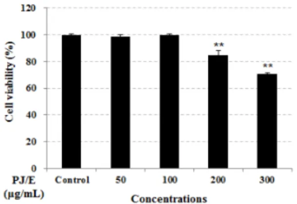
Cadmium으로 유도된 HaCaT 세포에서 머위 추출물 에 대한 세포보호효과를 확인해 보았다. 머위 추출물 에 의한 세포보호효과를 확인하기 위해 MTT assay를 시행하여 세포생존율을 확인하였다. 그 결과 머위 추 출물을 50, 100, 200, 300 µg/mL 처리하였을 때 각각 99.76 ± 1.54, 99.51 ± 1.21, 84.62 ± 3.74, 70.54 ± 1.06%
의 세포생존율을 나타내었으며(Figure 1), 200 µg/mL 이하의 농도에서는 세포독성을 나타내지 않아 이후 실 험에서는 50, 100 µg/mL 농도로 정하여 cadmium에 의 한 세포보호 효능을 관찰하였다.
Cadmium으로 세포사멸을 유도한 HaCaT 세포에서 47.99 ± 7.27%로 매우 낮은 세포생존율을 나타냈었다.
Figure 1. Effect of Petasites japonicus extract on Cd-induced cytotoxicity in HaCaT cell. The cells were treated with extract, and the viability of cells was determined by MTT assay. The values are the mean ± standard error of mean. **p < 0.001 compared the control group.
Figure 2. Cell protection effect of Petasites japonicus extract on Cd-induced HaCaT cell. The cells were treated with 0, 50, 100 µg/mL of extract, and the viability of cells was determined by MTT assay. The values are the mean ± standard error of mean. **p < 0.001 compared the control group.
반면 머위추출물을 50, 100 µg/mL의 농도로 처리한 군 에서는 각각 60.80 ± 1.99, 54.16 ± 3.77%로 cadmium을 단독으로 처리한 군에 비해 세포생존율이 증가하는 것 으로 나타났으며(Figure 2), 이는 세포보호효능으로 판 단되어 세포자멸사의 보호기전을 apoptosis regulatory factor들의 단백질 발현량을 조사함으로써 확인하였다.
3.2. Cadmium으로 유도된 HaCaT의 세포사멸에 대한 머위 추출물의 보호효과
3.2.1. Bcl-2의 발현에 미치는 영향
본 연구에서는 cadmium으로 유도된 HaCaT세포의 세포사멸에 대하여 cadmium 단독 처리군과 비교하였 을 때 머위 추출물 100 µg/mL 처리군에서 세포사멸 억 제인자인 Bcl-2의 발현이 53% 증가하는 것으로 확인되 었다(Figure 3). 이러한 결과는 cadmium이 일정부분 apoptosis를 유도하고 머위 추출물이 이를 유효하게 억 제함으로 세포보호효과가 있음을 의미한다.
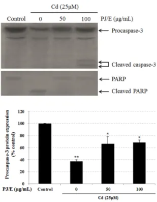
3.2.2. Procaspase-3의 발현에 미치는 영향
Cadmium으로 유도된 HaCaT세포의 세포사멸에 대 한 머위 추출물의 caspase 활성화에 미치는 영향을 확 인하기 위하여 procaspase-3의 발현을 조사하였다. 여 러 caspase family 중 caspase-3는 caspase-8과 caspase-9 의 신호를 증폭시켜 최종적으로 세포사멸을 유도하는 것으로 알려져 있다. 따라서 많은 연구자들이 이와 관 련하여 다양한 피부약리 소재들의 피부세포사멸억제 효능을 보고한 바 있다[13,14]. 그 결과 cadmium을 단 독으로 처리한 군에서는 음성대조군에 비하여 63% 감 소하는 것을 확인하였다. 반면 머위 추출물을 처리한 군에서는 cadmium 단독 처리군과 비교하였을 때 pro- caspase-3의 발현이 30% 증가함을 확인할 수 있었다 (Figure 4). 이러한 결과는 procaspase-3가 cadmium에 의 해 일정부분 apoptosis를 유도하고 머위 추출물이 이를 Figure 3. Effect of Petasites japonicus extract on the
expression of Bcl-2 in HaCaT keratinocyte. Cells were treated with 0, 50, 100 µg/mL of extract for 24 h. Protein were subjected to SDS-PAGE followed by immunoblot analysis.
Equal loading of protein was confirmed with β-actin antibody.
The values are the mean ± standard error of mean. **p <
0.001 compared the control group; *p < 0.05 compared the cadmium-alone group.
Figure 4. Effect of Petasites japonicus extract on the expression of Procaspase-3 and PARP and the cleavage of PARP in HaCaT keratinocytes. The cells were treated with specified concentrations of extract for 24 h. Procaspase-3 protein were subjected to SDS-PAGE followed by immunoblot analysis using anti-PARP antibody. The values are the mean ± standard error of mean. **p < 0.001 compared the control group; *p < 0.05 compared the cadmium-alone group.
유효하게 억제함으로 세포보호효과가 있음을 의미한다.
3.2.3. PARP의 발현에 미치는 영향
일반적으로 cadmium은, 노출정도 및 실험모델에 따 라서 necrosis와 apoptosis 모두를 유발할 수 있는 것으 로 알려져 있다. Apoptosis 시 세포의 핵 내에서는 poly-ADP Ribose Polymerase (PARP)의 분할이 일어나 고, 이러한 PARP의 분할은 DNA fragmentation과 chro- mosome condensation을 유도하면서, 세포사멸을 일으 키는 것으로 알려져 있다[15]. 이와 관련하여 본 연구 에서는 머위 추출물 50, 100 µg/mL 농도로 cadmium으 로 유도된 HaCaT세포에 처리하였으며 세포 내에서 세 포사멸과 관련된 PARP cleavage를 감소시킴을 확인할 수 있었다(Figure 4). 이러한 결과는 cadmium이 일정부 분 apoptosis를 유도하고 머위 추출물은 이를 유효하게 억제함으로 세포보호효과가 있음을 의미한다.
4. 결 론
본 연구에서는 머위 에탄올 추출물의 피부세포 보호 효과를 평가하기 위하여, HaCaT keratinocyte에 Cadmium 으로 세포사멸을 유도하여 피부세포 보호효능을 연구 하였다. Cadmium에 의해 발생되는 세포자멸사의 보호 기전을 Bcl-2와 procaspase-3 단백질 발현량을 조사함 으로써 중금속으로부터 피부보호 소재로서의 가능성 을 검증하였으며 다음과 같은 결론을 얻을 수 있었다.
1. 머위 추출물을 HaCaT 세포에 50, 100, 200, 300 µg/mL의 농도로 24 h 배양 후 MTT assay를 수행 한 결과 50, 100 µg/mL 농도에서 높은 생존율을 나타내었다.
2. Cadmium으로 세포사멸을 유도한 HaCaT 세포에 PJ/E를 50, 100 µg/mL의 농도로 처리한 결과, 단 독으로 cadmium을 처리한 군에서는 48.0%로 매 우 낮은 세포생존율을 나타냈으며 머위 추출물을 50 µg/mL 농도로 처리한 군에서는 60.8%로 cadmium에 의한 세포보호효능을 나타내었다.
3. 머위 추출물을 100 µg/mL 농도로 처리한 경우 apoptosis와 관련된 지표 단백질인 procaspase-3가 cadmium 단독 처리군 보다 40% 이상 증가하였다.
4. Cadmium으로 손상을 유도한 HaCaT 세포에 머위
추출물을 처리한 결과 세포내에서 세포사멸과 관 련된 PARP cleavage를 감소시킴을 확인할 수 있 었다.
이상의 결과는 머위 추출물이 중금속 독성에 대한 세포보호효과가 있음을 의미하는 것으로써 피부세포 사멸의 최종산출물인 caspase-3 활성을 감소시킨 효과 를 나타내었으므로 피부세포 보효 효능을 나타내는 천 연소재라 사료된다.
Reference
1. H. H. Kim, Y. W. Lim, J. Y. Yang, K. H. Moon, and D. C. Shin, Distribution of inorganic metals in blood of adults in urban area of Seoul, Korea. J. Environ.
Toxicol., 19. 327 (2004).
2. C. Jeon, J. Y. Park, and T. J. Yoo, Characteristics of metal removal using carboxylated alginic acid, Water Res., 36, 1814 (2002).
3. Y. H. Kim, Y. J. Yoo, and Y. J. Lee, Characterization of lead adsorption by Undaria pinnatifida. Biotechnol Lett., 17, 345 (1995).
4. S. H. Oh, Y. H. Yang, O. Y. Kwan, and M. R. Kim, Effects of diet with added butterbur (Petasites japoni- cus Maxim) on the plasma lipid profiles and anti- oxidant index of mice. J. East Asian Soc. Dietary Life, 16(4), 399 (2006).
5. B. S. Cho, J. J. Lee, J. O. Ha, and M. Y. Lee, Physicochemical composition of Petasites japonicus, Korean J. Food Preserv., 13(5), 661 (2006).
6. Y. Yaoita and M. Kikuchi, Petasiphenone a phenolic compound form rhizomes of Petasites Japonicus.
Phytochem., 37, 1773 (1994).
7. M. Kikuchi, Studies on the constituents of the flower stalk of Petasites japonicus Maxim VII. on the components of the volatile oil. Yakugaku Zasshi, 93, 123 (1973).
8. O. B. Choi, Anti-allergic effects of Petasites japoni- cum, Korean J. Food & Nutr., 15(4), 382(2002).
9. Y. H. Jee and C. S. Lee, Pathological changes on rats and mice fed with Petasites japonicus Maxim,
Korean J. Vet. Res., 36(2), 417 (1996).
10. C. H. Lee, M. C. Chung, H. J. Lee, and Y. H. Kho, An apoptosis regulator isolated from Petasites japoni- cus, Korean J. Food Sci. Technol., 32(2), 448 (2000).
11. H. S. Seo, B. H. Chung, and Y. G. Cho, The anti- oxidant and anticancer effects of butterbur (Petasites japonicus) extracts, Korean J. Plant Res., 21(4), 265 (2008).
12. J. H. Kim, Y. Na, G. S. Sim, B. C. Lee, and H. B.
Pyo, Antioxidative and anti-inflammatory effects of Petasites japonicus. J. Soc. Cosmet. Sci. Korea,
32(4), 263 (2006).
13. S. H. Seo, G. S. Bae, S. B. Choi, I. J. Jo, D. G. Kim,
J. Y. Shin, H. J. Song, S. J. Park, and M. O. Choi, The antioxidative and cytoprotective effect of Lonicerae japonicae FLOS water extracts on the ultraviolet (UV) B-induced human HaCaT keratino- cyte, Kor. J. Herbology, 29(6), 63 (2014).
14. A. Koh, S. Choi, Y. U. Kim, and G. H. Park, Protective effects of Pyrus pyrifolia NAKAI leaf extracts on UVB-induced toxicity in human dermal fibroblast, J. Soc. Cosmet. Sci. Korea, 42(1), 87 (2016).
15. C. Soldani and A. I. Scovassi, Poly (ADP-ribose) polymerase-1 cleavage during apoptosis: Apoptosis, 7, 321 (2002).