Bacillus amyloliquefaciens(SRCM 100730)로 발효된 청국장
추출물의 RAW 264.7 대식세포 면역증강 활성
추승빈
1․양 혜
1․정도연
2․정성엽
2․류명선
2․오광훈
3․유영춘
1 1 건양대학교 의학과 미생물학교실 2 (재)발효미생물산업진흥원 3 공주대학교 사범대학 체육교육과Immunomodulating Effect of Extract of Cheonggukjang Fermented with
Bacillus amyloliquefaciens (SRCM100730) on RAW 264.7 Macrophages
Seung Bin Choo1, Hui Yang1, Do-Yuon Jeong2, Seong-Yeop Jeong2,Myeong Seon Ryu2, Kwang-Hoon Oh3, and Yung Choon Yoo1 1
Department of Microbiology, College of Medicine, Konyang University
2
Microbial Institute for Fermentation Industry (MIFI)
3
Department of Physical Education, College of Education, Kongju National University
ABSTRACT Cheonggukjang is well known as a traditional fermented food in Korea and has various biological
activity. In this study, immune-enhancing activity of extract of cheonggukjang fermented with Bacillus amyloliquefaciens (SRCM100730) was examined in RAW 264.7 murine macrophages. Treatment with extract augmented production of nitric oxide (NO) and tumor necrosis factor-α (TNF-α) from RAW 264.7 macrophages in a dose-dependent manner. Similarly, increased mRNA expression of inducible nitric oxide synthase (iNOS) and TNF-α was observed. In addition, the extract synergistically enhanced production of NO and TNF-α from lipopolysaccharide (LPS)-stimulated macrophages. Analysis of intracellular pathways revealed that the immune-enhancing activity of cheonggukjang extract was related to activation of mitogen-activated protein kinases (MAPK) and nuclear factor kappa-light-chain-enhancer of activated B cells (NF-κB). These results suggest that cheonggukjang fermented with B. amyloliquefaciens (SRCM100730) is a beneficial food effective for activation of immune responses.
Key words: Bacillus amyloliquefaciens, immunostimulating activity, macrophages, MAPK, NF-κB
Received 5 September 2017; Accepted 3 November 2017 Corresponding author: Yung Choon Yoo, Department of Microbiol-ogy, College of Medicine, Konyang University, Daejeon 35365, Korea
E-mail: [email protected], Phone: +82-42-600-6495
서 론
최근 현대인들은 건강관리에 대한 관심이 커지면서 신체 건강에 유익한 천연소재와 식품에 대한 관심이 증가하고 있 는 추세이다(1). 이에 따라 생리활성을 갖는 식품소재를 탐 색하여 다양한 기능을 갖는 식품으로 개발하기 위해 많은 연구가 진행되고 있다(2). 특히 이러한 기능성 식품 연구 중 에서도 생체의 면역기능을 조절하는 식품소재 연구가 활발 히 수행되고 있어, 면역강화, 면역과민증의 억제, 생체 방어 작용 혹은 염증억제와 같은 활성을 지닌 소재 발굴이 목표가 되고 있다(3). 오래전부터 일부 발효식품이나 천연물 유래 물질은 숙주의 면역기능을 증강시켜, 암, 면역결핍증, 만성 감염 등에 효과를 나타내는 것으로 알려져 있다(4-6). 면역 계는 외부 항원 침입이나 조직 손상을 일으키는 물질이 체내 에 침입하였을 때 이로부터 신체를 보호하는 방어 수단으로 작용하고, 여기에는 림프구와 대식세포와 같은 면역세포들 이 관여한다(7-9). 대식세포는 항원의 침입에 대해 초기에 반응하여 항원의 탐식, 림프구에 항원 제시 및 병원체의 증 식 억제 등 선천면역을 담당하는 중요한 세포이다(10). 대식 세포가 활성화되면 증식과 운동성의 향상뿐 아니라 NO 및 각종 사이토카인(cytokine)의 생성을 동반하여 암세포와 각 종 유해균의 성장을 억제한다(10,11). 활성화된 대식세포가 분비하는 TNF-α와 같은 사이토카인은 감염초기에 반응하 여 병원균의 제거에 작용하며, NO는 숙주의 비특이적 방어 기작인 탐식작용, 병원균 및 암세포에 대한 증식 억제 등의 활성을 나타낸다(12). 우리나라의 대표적인 발효식품인 청 국장은 항암 효과, 항산화 효과, 면역증강 활성과 같은 다양 한 생리활성을 지니는 것으로 알려져 있다(4,13,14). 청국장 은 여러 종류의 발효균에 의해 제조되므로 균주의 종류와 특성에 따라 생리활성도 달라진다. 현재 청국장 발효에 관여 하는 주요 미생물로서 Bacillus subtilis, Bacilluslichen-iformis, Bacillus amyloliquefaciens, Bacillus pumilus 등 이 알려져 있다(15). 특히 본 연구에서 사용한 B. amyloli-quefaciens는 최근 연구 결과에 의하면 혈전을 용해하는 효 소를 분비하고(15), 또한 항산화 활성도 지니고 있는 것으로 보고되었다(16). 하지만 이 균주로 제조한 청국장에 대해 그 외의 생리활성에 대해서는 아직 보고된 바가 거의 없다. 본 연구에서는 B. amyloliquefaciens에 속하는 신규한 균주 인 B. amyloliquefaciens(SRCM 100730)로 발효한 청국장 의 면역조절 활성을 조사하기 위하여 80% 주정에 의해 추출 한 청국장 추출물을 이용하여 선천면역세포의 하나인 대식 세포에 대한 면역 활성화 효과를 측정하고, 이에 관련된 세 포 내 작용기전을 해석하였다.
재료 및 방법
시약 및 항체RAW 264.7 세포배양 배지로는 Dulbecco’s modified Eagle’s minimum essential medium(DMEM)(WelGENE, Seoul, Korea)을 사용하였으며, fetal bovine serum(FBS) 과 antibiotics(streptomycin/penicillin)는 Gibco BRL Life Technologies(Grand Island, NY, USA)의 제품을 사 용하였다. Lipopolysaccharide(LPS, Escherichia coli O111 :B4 유래)와 methyl thiazolyl tetrazolium(MTT)은 Sigma- Aldrich Co.(St. Louis, MO, USA) 제품을 사용하였다. Mitogen activated protein kinases(MAPK) 활성화 측정 을 위한 항체 phospho-p38(P-p38), p38, phospho-ERK (extracellular signal-regulated kinase; P-ERK), ERK, phospho-JNK(c-Jun-terminal kinase; P-JNK), JNK, 그 리고 NF-κB 활성화 측정을 위한 항체 phospho-IκB(in-hibitor of kappa B, P-IkB), IκB는 Cell Signaling Tech-nology(Beverly, MA, USA)에서 구매하였다. 그리고 house keeping 단백질인 anti-β-actin 항체는 Santa Cruz Bio-technology(Santa Cruz, CA, USA)에서 구매하여 사용하 였다. 균주 및 청국장 추출물 제조 청국장 제조에 사용된 대두는 전북 순창군에서 수확되는 백태를 구입하여 사용하였으며, 균주는(재)발효미생물산업 진흥원(Sunchang, Korea)이 보유하고 있는 전통 방법으로 제조된 고추장에서 분리한 B. amyloliquefaciens(SRCM 100730) 균주를 사용하였다. 청국장은 백태 3.5 kg을 24시 간 동안 침지하여 충분히 불린 후 체로 걸러 물기를 제거하 여 autoclave에서 121°C로 30분간 증자하였다. 증자된 백 태는 40°C 이하로 냉각시킨 후 tryptone soya broth(TSB, Oxoid Ltd., Basingstoke, UK) 배지에서 30°C, 24시간 배 양한 배양액을 백태 전 무게의 0.5%(v/w)로 접종 및 혼합하 였다. 발효 조건은 37°C에서 80% 습도로 24시간 동안으로 진행하였다. 제조된 청국장을 추출하기 위하여 발효가 끝난 청국장을 60°C에서 24시간 건조한 후 파쇄하여 청국장 분 말 시료로 사용하였다. 청국장 분말 시료의 10배수의 80% 발효주정을 이용하여 상온(25°C)에서 24시간 교반 추출하 여 저온감압농축(N-4000S, Sunileyela, Seongnam, Korea) 및 동결건조(FDT-12050, Hanil Science Medical, Dae-jeon, Korea) 후 분말화하여 주정 추출물 시료로 사용하였다. 세포배양 본 연구에 사용된 마우스 대식세포주인 RAW 264.7 세포 는 한국세포주은행(Seoul, Korea)에서 분양받았다. 세포는 DMEM 배지에 10% FBS와 1% antibiotics를 첨가해 만든 배지로 37°C, 5% CO2 조건에서 배양하였고, 실험과정에서 세포는 75~85% 정도의 밀도로 자랐을 때 계대 배양하였다. 세포독성 측정 청국장 추출물의 세포독성은 MTT assay를 통해 측정하 였다. RAW 264.7 세포(1×10⁴ cells/well)를 96-well plate 에 100 μL씩 분주하고 8시간 동안 안정화시킨 후 SRCM 100730 발효 청국장 추출물을 농도별(6.25, 12.5, 25, 50 μg/mL)로 처리하여 37°C, 5% CO2 incubator에서 24시간 배양하였다. 배양 후 5 mg/mL 농도의 MTT 시약을 10 μL 씩 첨가하여 2시간 추가배양하고 원심분리 후 상층액을 제 거하였다. 그 후 각 well에 dimethyl sulfoxide를 첨가하여 생성된 formazan을 녹여 이를 20분간 shake 후 Micro-plate reader(ELx 800-PC, BioTek, Winooski, VT, USA) 를 이용하여 540 nm에서 흡광도를 측정하였다. 생존율은 대조군에 대한 처리군의 흡광도 비를 백분율로 표시하여 계 산하였다.
NO 및 TNF-α측정
NO의 농도는 상층액 내의 nitrite 농도를 Griess 반응을 이용하여 측정하였다. RAW 264.7 세포는 5×10⁴ cells/well 농도로 조절 후 48-well plate에 분주하고 37°C, 5% CO2 incubator에서 8시간 안정화한 다음 세포에 발효 청국장 추 출물을 농도별(6.25, 12.5, 25, 50 μg/mL)로 처리하여 24시 간 배양하였다. 그 후 원심분리(Purispin 17R, Hanil Science Medical) 하여 상등액을 얻은 후 동량의 Griess 시약을 첨 가하여 실온에서 10분간 반응시키고 microplate reader를 이용하여 540 nm에서 흡광도를 측정하였으며, NO 생성량 은 농도별 표준곡선을 이용해 정량하였다. TNF-α는 NO를 측정하는 데 사용한 RAW 264.7 세포의 배양 상등액을 이용 하여 ELISA kit(BD Bioscience, San Jose, CA, USA)에 의해 정량하였다.
iNOS 및 TNF-α mRNA 분석
RAW 264.7 세포(5×10⁵ cells/well)를 6-well plate에 분주하고 세포독성실험과 동일한 조건에서 배양하였다. 발 효 청국장 추출물 혹은 positive control로 사용한 LPS(100
Table 1. Sequences of the primers for PCR
Primer Sequence
iNOS Anti-senseSense 5’-GTAGTGACAAGCACATTTGG-3’5’-GGCTCCACTTTTCACTCTGC-3’ TNF-α Anti-senseSense 5’-AGCCCCCAGTCTGTATCCTT-3’5’-CTCCCTTTGCAGAACTCAGG-3’ β-Actin Anti-senseSense 5’-AGCCATGTACGTAGCCATCC-3’5’-CTCTCAGCTGTGGTGGTGAA-3’
Fig. 1. Effect of the cheonggukjang fermented with Bacillus amyloliquefaciens (SRCM 100730) on cell viability in RAW
264.7 cells. Cells were treated with the indicated doses of the fermented cheonggukjang for 24 h. Cell viability was determined by MTT assay. Data were expressed as mean±standard errors. ng/mL)를 6시간 처리 후 원심분리기를 통해 상등액을 제거
하고 RNA를 TRIzol reagent(iNtRON Biotechnology, Lynnwood, WA, USA)를 이용하여 분리하였다. RNA로부 터 cDNA synthesis kits(Power cDNA synthesis kit, iNtRON Biotechnology, Kirkland, WA, USA)를 이용해 cDNA를 제작하였다. iNOS, TNF-α, β-actin에 대한 PCR primer는 Cosmo Genetech(Seoul, Korea)에서 구매하여 사용하였다(Table 1). 각 단백질 밴드는 Chemilumines-cence imaging system(BD Bioscience)을 이용하여 분석 하였다.
Western blot analysis
RAW 264.7 세포(1×10⁶ cells)를 cold PBS로 세척하고 lysis buffer[50 mM Tris-HCL(pH 7.4), 150 mM NaCl, 5 mM EDTA, 0.1% Triton X-100, protease 및 phospha-tase inhibitor cocktail]로 lysis 시킨 후 단백질 농도를 BCA protein assay kit(Sigma-Aldrich Co.)으로 정량한 후, 30 μg의 lysate를 10% SDS-PAGE에서 전개시켰다. 분리된 단백질은 polyvinylidene difluoride(PVDF) mem-brane(Bio-Rad Laboratories, Hercules, CA, USA)을 이 용하여 2시간 동안 전사시켰다. 이후 5% skim milk 용액으 로 상온에서 1시간 동안 blocking 하고, P-p38, p38, P- ERK, ERK, P-JNK, JNK, P-IκB 및 IκB에 대한 항체인 rabbit monoclonal antibodies(BD Bioscience)를 1:5,000 으로 희석하여 사용하였다. 2차 항체로는 horseradish peroxidase(HRP)가 결합된 anti-rabbit IgG를 1:5,000으 로 희석하여 상온에서 2시간 반응시키고, TBST(TBS con-taining 0.05% Tween-20)로 5분씩 총 8회 세정하고 ECL 기질과 반응시킨 후 각각의 단백질 밴드를 Chemilumines-cence imaging system을 이용하여 분석하였다.
LPS와의 시너지 효과 측정 발효 청국장 추출물에 의한 병용 효과를 조사하기 위하여 LPS(100 ng/mL)와 청국장 추출물을 함께 처리하고, 대식 세포로부터 NO와 TNF-α의 분비가 LPS 단독처리에 비해 상승하는가를 ELISA kit을 통해 측정하였다. 통계처리 모든 실험 결과는 평균±표준오차로 나타내었으며 통계 학적 유의성은 Student’s two-tailed t-test를 이용하여,
P<0.05 수준 이상에서 검증하였다.
결과 및 고찰
세포독성 평가 B. amyloliquefaciens(SRCM 100730) 균 발효 청국장 추출물의 RAW 264.7 대식세포에 대한 세포독성을 MTT assay에 의해 실시하여 독성을 나타내지 않는 농도를 결정 하였다. Fig. 1에서 나타난 결과와 같이 SRCM 100730 발 효 청국장은 50 μg/mL의 농도까지 세포증식에 영향을 주지 않는 것으로 확인되었다. 따라서 이후의 실험은 독성을 나타 내지 않는 안전한 농도인 50 μg/mL 이하에서 수행하였다. NO 및 TNF-α생성량 측정 NO와 TNF-α는 대식세포가 분비하는 대표적인 면역조 절물질로 외부에서 침입한 병원체의 제거와 증식 억제와 같 은 비특이 면역은 물론 림프구를 통한 항원 특이적인 면역반 응을 조절하는 중요한 물질이다(12). 발효 청국장 추출물의 면역세포 활성 효과를 확인하기 위해 청국장 추출물을 처리 한 RAW 264.7 세포의 세포배양액으로부터 NO와 TNF-α 를 측정하였다. Fig. 2A에서 나타난 결과와 같이 발효 청국 장 추출물 처리에 의해 NO의 생성량이 control과 비교하여 현저하게 증가하는 것을 확인할 수 있었다. 대식세포 활성화 지표 중 하나인 NO는 사이토카인이나 미생물의 영향을 받 아 대식세포에서 생성되는 반응질소 중간체로서 세포 내 감 염을 일으키는 미생물과 암세포를 제어하는 것으로 보고되 고 있고, NO가 과도하게 높은 농도로 생성될 경우에는 혈관 확장이나 염증반응에 의한 조직손상 등을 일으키는 것으로 알려져 있다(17,18). 이와 같이 NO는 양면적 특성을 나타내 는 면역조절물질이지만 세포독성을 유발하지 않는 농도에 서 NO가 생성될 경우 면역기능을 증가시키게 된다(19). 한 편 NO를 측정한 세포배양액을 이용하여 대식세포로부터 분 비되는 또 다른 면역조절 사이토카인인 TNF-α를 정량하였 다. NO 생성과 동일하게 발효 청국장 추출물의 처리에 의해 대식세포로부터 TNF-α가 농도 의존적으로 증가하는 것을Fig. 2. Effect of the cheonggukjang fermented with B. amyloli-quefaciens (SRCM 100730) on the production of NO and TNF-α
from RAW 264.7 cells. Culture supernatants of RAW 264.7 cells harvested 24 h after treatment of the fermented cheongukjang were used for determination of the level of NO and TNF-α. *P<0.05, **P<0.01, ***P<0.001; compared with the control (untreat-ed group).
Fig. 3. Effect of the cheonggukjang fermented with B. amyloliquefaciens (SRCM 100730) on the expression
of iNOS and TNF-α mRNA in RAW 264.7 cells. The cells were treated with the indicated doses of the fer-mented cheongukjang or LPS (100 ng/mL) for 6 h. The level of mRNA expression was iNOS, TNF-α, and β-actin was determined by PCR as described in Mate-rials and Methods. Densitometric analysis of the bands was performed by Chemiluminescence imaging system. *P<0.05, **P<0.01, ***P<0.001; compared with the con-trol (untreated group).
확인하였다(Fig. 2B). 면역세포로부터 분비되는 다양한 사 이토카인은 타 면역세포의 기능조절과 신호전달에 중요한 역할을 하는 단백질로 면역세포 간 network를 형성하여 면 역계 전체의 반응을 조율하는 것으로 알려져 있다(20). 또 한, TNF-α는 인체에 침입한 병원체에 대한 숙주의 방어에 관여하고 다른 사이토카인들의 분비를 유도함으로써 인체 면역반응을 조절한다(21). 이와 같이 SRCM 100730 발효 청국장 추출물 처리에 의해 RAW 264.7 세포로부터 TNF-α 의 생성이 증가한다는 것은 SRCM 100730 발효 청국장이 면역능을 조절하는 활성을 가진다는 것을 시사한다. iNOS와 TNF-αmRNA 발현 측정 발효 청국장 추출물 처리에 의해 RAW 264.7 세포로부터 NO와 TNF-α의 생성이 유도되었으므로, 이들 면역조절물 질의 합성에 관련된 iNOS(NO의 생합성 효소)와 TNF-α의 mRNA 발현 증가 여부를 조사하였다. RT-PCR법에 의해 이들 분자의 cDNA를 합성하고 PCR법에 의해 유전자의 발 현량을 증폭하여 분석한 결과, SRCM 100730 발효 청국장 추출물 처리에 의해 iNOS 및 TNF-α의 mRNA 발현이 증가 하는 것으로 확인되었다(Fig. 3). Fig. 2와 Fig. 3의 결과는
B. amyloliquefaciens(SRCM 100730) 발효 청국장 추출물 에 의한 NO 및 TNF-α 분비 유도 활성은 이들 면역조절물질 유전자 발현의 증폭을 통해 유도되는 것임을 시사한다. 청국 장의 생리활성 중 면역조절에 관한 연구 결과는 많이 보고되 어 있으나, 청국장의 종류에 따라 면역증강 효과를 나타내는 경우와 면역억제 혹은 염증제어 작용으로 구분된다. Kim 등(22)의 보고에 의하면 청국장 추출물이 염증세포에서 NO 의 생성을 억제한다는 결과를 제시하였다. 또한, 다른 연구 결과는 청국장 추출물이 ovalbumin(OVA)에 의해 유도된 알레르기를 억제하는 항알레르기 효과가 있음을 제시하였 다(23). 한편 또 다른 연구에서는 난소 적출 마우스에 청국 장을 경구 투여함으로써 T림프구의 기능이 향상되는 면역 증강 효과를 유도한다고 발표하였다(24). 본 연구에서는 B. amyloliquefaciens(SRCM 100730) 균주로 발효한 청국장 추출물이 대식세포를 자극하여 NO와 TNF-α와 같은 면역 조절물질을 분비하는 면역증강 활성이 있음을 제시하였다. 이러한 청국장 추출물에 의한 면역조절 활성의 차이는 아마 도 청국장의 원료인 콩의 종류는 물론 발효 균주의 성질에 따라 달라지는 것으로 생각된다. MAPK 및 NF-κB 활성화 측정 세포 내 신호 전달계 중 MAPK는 다양한 생물학적 기능을
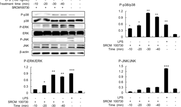
LPS (100 ng/mL) + Treatment time (min) 10 20 30 40 SRCM100730 + + + + -P-p38 p38 P-ERK ERK P-JNK JNK β-actin LPS + SRCM 100730 + + + + Time (min) 10 20 30 40 LPS + SRCM 100730 + + + + Time (min) 10 20 30 40 LPS + SRCM 100730 + + + + Time (min) 10 20 30 40
-Fig. 4. Analysis of MAPK activation by the cheonggukjang fermented with B. amyloliquefaciens (SRCM 100730) in RAW 264.7
cells. The phosphorylation of p38, ERK, and JNK MAP kinase in RAW 264.7 cells was determined by western blot. The cells were treated with the fermented cheonggukjang extract (12.5 mg/mL) or LPS (100 ng/mL) for the indicated times, and total cellular proteins obtained from the cells were used for western blot analysis. *P<0.05, **P<0.01, ***P<0.001; compared with the control
(untreated group).
조절하는데 일련의 MAPK의 활성화 과정은 AP-1의 연속적 인산화로 이루어진다고 보고되어 있다(25). AP-1은 Jun과 Fos family 단백질로 이루어진 전사인자로 c-Jun과 c-Fos 의 heterodimer 형태로 존재할 때 전사 활성이 가장 높다 (26). 대식세포와 같은 면역세포에 활성화 자극이 주어지면 자극에 의해 c-Jun과 c-Fos 단백질 발현이 증가하게 되며, c-Jun은 JNK와 p38의 인산화에 의해, c-Fos는 ERK의 인 산화에 의해 활성화된다(27). 그리고 NF-κB의 경우 세포질 에서 IκB와 결합되어 불활성화 상태로 핵 내 전사를 하지 못하지만, IκB가 LPS와 같은 자극에 의해 인산화되어 분해 되면 NF-κB가 핵 내로 전사되어 다양한 사이토카인 및 염 증매개물질의 합성을 촉진하게 된다(28). B. amylolique-faciens(SRCM 100730) 발효 청국장 추출물의 면역증강 활 성의 기전을 해석하기 위한 일환으로 MAPK와 NF-κB의 활성화 여부를 조사하였다. 그 결과 발효 청국장 추출물을 RAW 264.7 세포에 처리함으로써 MAPK 중 ERK와 p38의 인산화가 강력하게 유도되는 것으로 나타났으며, 이들 분자 의 인산화는 자극 후 10분 이내에 상승하여 20분 후부터는 유의하게 증가하는 것으로 확인되었다(Fig. 4). 하지만 MAPK 관련분자 중 JNK는 아무런 영향을 받지 않는 것으로 나타났 다. NF-κB의 활성화에 있어서도 MAPK와 동일하게 처리 후 10분 이내에 IκB의 인산화가 유도되기 시작하는 것으로 관찰되었다(Fig. 5). 한편 positive control로 사용한 LPS (100 ng/mL로 20분간 처리)가 B. amyloliquefaciens(SRCM 100730) 발효 청국장 추출물과는 달리 MAPK 분석에서 ERK, p38 및 JNK 등 모든 MAPK 관련분자를 인산화시킨 것으로 볼 때 B. amyloliquefaciens(SRCM 100730) 발효 청국장 추출물은 LPS의 활성화 경로인 toll-like receptor- 4(TLR-4)와는 다른 경로를 통해 대식세포를 활성화할 가능 성을 시사하였다. 일반적으로 세균을 포함하는 많은 천연물 의 면역증강 활성은 균주 자체가 TLR 분자와 같은 수용체를 통해 면역세포를 활성화하거나 혹은 발효균주에 의한 기질 의 분해 또는 형질전환을 통해 새롭게 생성된 물질에 의한 것으로 추정된다. 본 연구에서 사용한 청국장 추출물은 균주 로부터의 성분은 물론 기질에 포함된 성분이 함께 혼합되어 있는 상태이므로 면역증강 활성과 관련된 기전을 명확하게 해석하기 위해서는 B. amyloliquefaciens(SRCM 100730) 발효 청국장 추출물의 성분에 대한 분석과 면역학적 특성에 대한 해석이 필요하다. LPS와의 병용 효과 LPS는 대식세포를 활성화하는 대표적인 물질로서 본 연 구의 결과에서 보듯이 LPS 처리에 의해 대식세포는 MAPK 와 NF-κB의 활성화를 유도하고(Fig. 4, Fig. 5) iNOS 및 사이토카인의 mRNA의 발현을 촉진하여(Fig. 3) 이들 면역 조절물질의 분비를 증가시킨다. 발효 청국장 추출물에 의한 대식세포 활성화 실험 결과로부터 이 추출물은 LPS와는 다 른 경로를 통해 대식세포를 활성화시키는 것으로 관찰되었
LPS (100 ng/mL) + Treatment time (min) 10 20 30 40 SRCM100730 + + + + -P-IκB IκB β-actin LPS + SRCM 100730 + + + + Time (min) 10 20 30 40 LPS + SRCM 100730 + + + + Time (min) 10 20 30 40
-Fig. 5. Analysis of NF-κB activation by the cheonggukjang fermented with B. amyloliquefaciens (SRCM 100730) in RAW 264.7
cells. NF-κB activation was determined by phosphorylation of IκB in RAW 264.7 cells treated with the fermented cheonggukjang extract for the indicated times. Western blot analysis was carried out by the same method with that of MAPK activation. *P<0.05, **P<0.01, ***P<0.001; compared with the control (untreated group).
Fig. 6. Synergic effect of the cheonggukjang fermented with B. amyloliquefaciens (SRCM 100730) in LPS-priming RAW 264.7
cells. The cells were treated with various doses of the fermented cheonggukjang extract in the presence or absence of LPS (100 ng/mL) for 24 h. The level of NO and TNF-α in culture supernatants of the cells was determined using detection kits as described in Materials and Methods. *P<0.05, **P<0.01, ***P<0.001; compared with the control (untreated group).
으므로, 발효 청국장 추출물을 LPS와 함께 처리함으로써 대식세포 활성화의 시너지를 유도할 수 있는가를 조사하였 다. 그 결과 발효 청국장 추출물은 LPS와의 병용처리에 의 해 NO와 TNF-α 생성에 있어 시너지 효과를 나타내는 것으 로 확인되었다(Fig. 6). 이 결과는 SRCM 100730 발효 청국 장 추출물은 항원 노출에 의한 면역반응이나 혹은 면역세포 의 활성화에 기여하는 면역자극물질에 의한 면역세포 활성 화 과정에 함께 작용할 경우 상승적 효과를 나타낼 수 있음 을 시사한다. B. amyloliquefaciens는 혈전용해(15)와 항산화 활성 (16) 이 외에도 probiotic로서의 활성(29)과 장내세균총의 조절과 장관형태의 유지(30) 등과 같은 다양한 생리활성을 갖는 것으로 보고되었다. 하지만 B. amyloliquefaciens나 이 균에 속하는 균주 중에서 면역세포와의 상호작용을 통해 면역기능을 촉진한다는 연구보고는 거의 없는 상황이다. 최 근 B. amyloliquefaciens에 속하는 한 균주인 B. amyloli-quefaciens SQR9 균주가 선천면역세포의 하나인 수지상세 포의 성숙을 촉진하여 항원제시능을 높이며, 불활성화 조류 인플루엔자 백신(H9N2 type)에 대해 점막면역 백신의 ad-juvant 효과를 갖는다고 보고하였다(31). 본 연구에서도 B. amyloliquefaciens 균종에 속하는 신규한 균주인 B. amy-loliquefaciens(SRCM 100730)로 발효한 청국장 추출물이 대식세포를 활성화한다는 연구 결과를 제시하였다. 이러한 결과는 이 균주가 선천면역세포를 자극하여 면역기능을 향 상시키는 청국장 제조에 유용한 발효균주임을 시사한다.
요 약
청국장은 한국의 전통발효식품으로서 다양한 생리활성이 보고되어 있다. 본 연구에서는 B. amyloliquefaciens 균종에속하는 신규한 균주인 B. amyloliquefaciens(SRCM 100730) 에 의한 발효 청국장이 선천면역 세포인 RAW 264.7 대식세 포를 활성화하는 효과를 가지는가를 검토하였다. 청국장 추 출물을 RAW 264.7 세포에 처리한 결과 처리농도에 의존하 여 TNF-α와 NO의 생성이 증가하였으며, 이러한 면역조절 물질의 증가는 iNOS와 TNF-α mRNA 발현에서도 확인되 었다. 또한, 청국장 추출물에 의한 대식세포 활성화와 관련 한 세포 내 작용기전을 해석한 결과 청국장 처리에 의해 p38 과 ERK와 같은 MAPK의 인산화와 NF-κB 활성화가 유도 되는 것으로 밝혀졌다. 하지만 MAPK에 속하는 JNK에 대해 서는 아무런 영향을 주지 않는 것으로 나타났다. 한편 청국 장 추출물을 LPS와 함께 처리한 실험에 있어서 청국장 추출 물은 LPS에 의한 대식세포 활성화를 촉진하여 TNF-α와 NO의 생성을 증가시키는 시너지 효과를 나타내었다. 이들 결과로부터 B. amyloliquefaciens(SRCM 100730) 균주로 발효한 청국장 추출물은 대식세포를 활성화하여 선천면역 을 증가시키는 효과가 있는 것으로 확인되었다.
감사의 글
이 논문은 2017년도 과학기술정보통신부(과제번호 2016M 3C1B5907053)의 재원으로 한국연구재단-전통문화융합 연구사업과 한국연구재단 기본연구지원사업(과제번호 2015 R1D1A1A010 60440) 및 2016년도 농림수산식품부 기술 사업화지원사업과제(과제번호 855001-3)의 지원에 의해 수행되었음.REFERENCES
1. Lyu HN, Park MH, Hong SG. 2007. Development of bio-logically active compounds from edible plant sources-ⅩⅩⅤ. Immunostimulating effect of edible plant extracts. Korean
J Food Sci Technol 39: 708-714.
2. Park SY, Kim JE, Choi CY, Lee DW, Kim KM, Yoon G, Yoon IS, Moon HS, Cho SS. 2015. Analytical validation of rosmarinic acid in water extract of Perilla frutescens Britton var. acuta Kudo as functional health ingredient. J
Korean Soc Food Sci Nutr 44: 85-88.
3. Ryu HS, Kim JH, Kim HS. 2008. Effects of plant water extract mixture Ixeris sonchifolia Hance, Oenanthev
jav-anica, Fagopyrum esculentum Moench, Hisikia fusiforme, Zingiber officinele Roscoe on mouse immune cell activation
ex vivo. Korean J Nutr 41: 141-146.
4. Min HK, Kim HJ, Chang HC. 2008. Growth-inhibitory ef-fect of the extract of porphyran-chungkookjang on cancer cell. J Korean Soc Food Sci Nutr 37: 826-833.
5. Hara C, Kumazawa Y, Inagaki K, Kaneko M, Kiho T, Ukai S. 1991. Mitogenic and colony-stimulating factor-inducing activities of polysaccharide fractions from the fruit bodies of Dictyophora indusiata FISCH. Chem Pharm Bull 39: 1615-1616.
6. Lee JS, Lee SH, Jang YM, Lee JD, Lee BH, Jung JY. 2011. Macrophage and anticancer activities of feed additives on β-glucan from Schizophyllum commune in breast cancer cells. J Korean Soc Food Sci Nutr 40: 949-955.
7. Belardelli F. 1995. Role of interferons and other cytokines in the regulation of the immune response. APMIS 103: 161-179.
8. Liu YJ. 2001. Dendritic cell subsets and lineages, and their functions in innate and adaptive immunity. Cell 106: 259- 262.
9. Salazar-Mather TP, Hokeness KL. 2003. Calling in the troops: Regulation of inflammatory cell trafficking through innate cytokine/chemokine networks. Viral Immunol 16: 291-306. 10. Iontcheva I, Amar S, Zawawi KH, Kantarci A, Van Dyke
TE. 2004. Role for moesin in lipopolysaccharide-stimulated signal transduction. Infect Immun 72: 2312-2320.
11. Kim HS, Kang JS. 2008. Preparation and characteristics of bread by medicinal herb composites with immunostimulat-ing activity. J Korean Soc Food Sci Nutr 37: 109-116. 12. Janeway CA Jr, Medzhitov R. 2002. Innate immune
recog-nition. Annu Rev Immunol 20: 197-216.
13. Shon MY, Seo KI, Lee SW, Choi SH, Sung NJ. 2000. Bio-logical activities of chungkugjang prepared with black bean and changes in phytoestrogen content during fermentation.
Korean J Food Sci Technol 32: 936-941.
14. Hwang JS, Kim SJ, Kim HB. 2009. Antioxidant and blood- pressure reduction effects of fermented soybean, chungkook- jang. Korean J Microbiol 45: 54-57.
15. Shin SH, Hong SW, Chung KS. 2013. Purification and char-acterization of a fibrinolytic enzyme produced by Bacillus
amyloliquefaciens HC188. Kor J Microbiol Biotechnol 41:
33-43.
16. Lee JY, Shin JM, Liu X, Yao Z, Lee KW, Cho KM, Kim GM, Shin JS, Kim JS, Kim JH. 2016. Inhibition of Bacillus
cereus in cheonggukjang fermented with Bacillus starters
with antimicrobial activities. J Korean Soc Food Sci Nutr 45: 736-745.
17. Flurkey WH. 1991. Identification of tyrosinase in mush-rooms by isoelectric focusing. J Food Sci 56: 93-95. 18. Jung DW, Park SI. 2005. Effect of green tea power on the
growth inhibition of oral bacteria in yoghurt. Korean J Food
Sci Ani Resour 25: 500-506.
19. Byun MW. 2013. Immunomodulatory activities of apple seed extracts on macrophage. J Korean Soc Food Sci Nutr 42: 1513-1517.
20. Starr R, Willson TA, Viney EM, Murray LJ, Rayner JR, Jenkins BJ, Gonda TJ, Alexander WS, Metcalf D, Nicola NA, Hilton DJ. 1997. A family of cytokine-inducible in-hibitors of signalling. Nature 387: 917-921.
21. Jeong HJ, Chung HS, An HJ, Seo SW, Kim TG, Won JH, Shin JY, Ahn KS, Kim HM. 2004. The immune-enhancing effect of the herbal combination Bouum-Myunyuk-Dan.
Biol Pharm Bull 27: 29-33.
22. Kim HG, Lee MJ, Kim HJ, Kim KC, Bose S. 2012. Effects of fermented soybean upon anti-inflammation and intestinal mucous membrane permeability. J Soc Korean Med Obesity
Res 12: 33-47.
23. Bae MJ, Shin HS, See HJ, Chai OH, Shon DH. 2014. Cheong-gukjang ethanol extracts inhibit a murine allergic asthma via suppression of mast cell-dependent anaphylactic reactions.
J Med Food 17: 142-149.
24. Park H, Yoon L, Kim HS. 2013. Effects of fermented soy-bean paste Chungkukjang on the immunoreactivity in ovar-iectomized mice. J Korean Soc Food Sci Nutr 42: 1930- 1939.
25. Pawson T, Scott JD. 1997. Signaling through scaffold, an-choring, and adaptor proteins. Science 278: 2075-2080. 26. Chen CC, Mo FE, Lau LF. 2001. The angiogenic factor
Cyr61 activates a genetic program for wound healing in hu-man skin fibroblasts. J Biol Chem 276: 47329-47337. 27. Fisher GJ, Talwar HS, Lin J, Lin P, McPhillips F, Wang
Z, Li X, Wan Y, Kang S, Voorhees JJ. 1998. Retinoic acid inhibits induction of c-Jun protein by ultraviolet radiation that occurs subsequent to activation of mitogen-activated protein kinase pathways in human skin in vivo. J Clin Invest 101: 1432-1440.
28. Nam NH. 2006. Naturally occurring NF-κB inhibitors. Mini
Rev Med Chem 6: 945-951.
29. Zurmiati, Wizna, Hafil Abbas M, Endo Mahata M, Fauzano R. 2017. Effect of Bacillus amyloliquefaciens as a probiotic
on growth performance parameters of pitalah ducks. Int J
Poultry Sci 16: 147-153.
30. Lei X, Piao X, Ru Y, Zhang H, Peron A, Zhang H. 2015. Effect of Bacillus amyloliquefaciens-based direct-fed micro-bial on performance, nutrient utilization, intestinal morphol-ogy and cecal microflora in broiler chickens.
Asian-Austra-las J Anim Sci 28: 239-246.
31. Huang L, Qin T, Yin YY, Gao X, Lin J, Yang Q, Yu Q. 2016. Bacillus amyloliquefaciens SQR9 induces dendritic cell maturation and enhances the immune response against inactivated avian influenza virus. Sci Rep 6: 21363.