64
Inhibitory Effect of Epidermal Growth Factor on the Proli- feration of Lung Cancer Cell Lines
Purpose: Epidermal growth factor (EGF) is known to cause cellular proliferation, differentiation and survival. However, there are a few articles that have reported on the cell killing effect of EGF. We evaluated the effects of EGF on the survival of some lung cancer cell lines and we tried to determine the mechanism of action. Materials and Methods: We examined various lung cancer cell lines. The cultured cells were exposed to radiation alone (0, 2, 5, and 10 Gy), EGF alone (0∼1,000 nM) or a combination of radiation and EGF (10 nM). Survival was measured using a clonogenic assay and the expressions of epidermal growth factor receptor (EGFR), Ki-67 and cleaved- PARP were detected by western blot analysis. K-ras mutations were detected using polymerase chain reaction and sequencing. Results: Treatment of EGF induced cell death in the lung cancer cell lines. The addition of EGF (10 nM) to radiation (0, 2, 5, and 10 Gy) resulted in an increased cell killing effect of radiation. The EGFR expression decreased with the addition of EGF. EGF increased the expression of cleaved-PARP, but it did not increase the expression of Ki-67. The effects of EGF were not correlated with EGFR mutation or K-ras mutation. Conclusion: In our study, EGF inhibited the proliferation of lung cancer cell lines and it induced apoptosis. EGF enhanced the radiosensitivity of lung cancer cells when EGF was combined with radiation. It is suggested that EGF seem to be one of the cytotoxic agents for lung cancer cell lines. (J Lung Cancer 2010;9(2):64 71)
Key Words: Epidermal growth factor, Radiation, Apoptosis, Lung cancer cell lines
Seung-Hee Lee, M.S. and Hong-Gyun Wu, M.D., Ph.D.
Department of Radiation Oncology &
Cancer Research Institute, Seoul National University College of Medicine and Institute of Radiation Medicine, Medical Research Center, Seoul National University, Seoul, Korea Received: July 29, 2010 Revised: August 9, 2010 Accepted: August 19, 2010 Address for correspondence Hong-Gyun Wu, M.D., Ph.D.
Department of Radiation Oncology, Seoul National University College of Medicine, 28, Yeongeon-dong, Jongno- gu, Seoul 110-744, Korea
Tel: 82-2-2072-3177 Fax: 82-2-765-3317 E-mail: [email protected]
This work supported by research grant from Korean Association for the Study of Lung Cancer in 2008 and grant No. 2009-0083886 from the Korea Science and Engineering Foundation grant funded by the Korea government (MEST).
서 론
상피세포성장인자(epidermal growth factor, EGF)는 미국 의 스탠리 코헨이 바로 태어난 쥐 등에서 눈꺼풀이 열리는 효과를 갖는 단백질로 처음 발견하였고, 53개의 아미노산 과 3개의 분자 내 이황화 결합을 가진 분자량 6,045-Da이다 (1-3). EGF는 세포막에 있는 EGF수용체(epidermal growth factor receptor, EGFR)와 결합함으로써 tyrosine kinase를 활 성화시켜 신호전달을 통해 세포의 분열을 유도하고 증식을 촉진하는 것으로 알려져 있다. 따라서 상처가 발생하게 되 면 혈액으로부터 공급되어 상피세포를 증식시키고 신생혈 관을 형성하여 상처의 회복을 향상시키기 때문에(4,5) 당뇨
성 족부궤양이나 방사선치료 시의 부작용으로 나타나는 구 강점막염과 손상된 각막 등의 치료에 좋은 영향을 미친다 고 보고되고 있다(6-8).
EGFR의 과발현은 여러 상피성 암종에서 관찰되어 암의 발생, 진행, 표준 치료법에 대한 내성, 불량한 예후 등과 관 련이 있는 것으로 알려져 있다(9,10). 최근에는 다양한 EGFR 억제제가 개발되어 암의 치료에 이용되고 있다 (11-13).
그러나 일반적으로 EGF가 세포를 증식시킨다는 개념과 반대로 EGFR이 과발현하는 암세포주에서는 세포의 성장 을 억제시킨다는 보고가 있으며(14-16), EGF의 세포분화 작 용으로 인해 항암제나 방사선 치료와 EGF를 병행한 연구도 보고되고 있다. 여러 종류의 암세포주에 항암제와 함께
EGF를 병행했을 때 항암 효과가 높게 나타났고(17), 방사선 조사 시 EGF와 병행함으로써 효과가 상승하였다(18,19). 본 연구실에서도 EGF가 정상 섬유모세포의 증식을 촉진시키 지만 두경부암 세포주인 AMC-HN3에서 현저하게 세포사 를 일으키고 다른 암세포주에서도 방사선과 병용 시 방사 선 효과를 저해하지 않으며 오히려 방사선 효과를 증가시 키는 것을 확인하였다(20).
따라서 본 연구에서는 EGFR을 발현하는 폐암세포주에 서 EGF의 작용을 평가하고 작용기전을 알아보고자 하였다.
또한 방사선과 병행 시의 효과도 평가하고자 하였다.
대상 및 방법 1) 세포배양
EGF의 효과를 알아보기 위하여 여러 가지 폐암 세포주를 이용하였다. A549는 American Type Culture Collection (ATCC) 에서 구입하였고, HCC-827, HCC-1438, NCI-H1437, NCI- H157, NCI-H1650은 분당서울대학교병원의 이춘택 교수로 부터 공급 받았다. 10% fetal bovine serum (Sigma, St. Louis, MO, USA)을 첨가한 RPMI 1640 (Gibco BRL, Gaithersburg, MD, USA) 배지에서 37oC, 5% 이산화탄소 하에서 배양하였 다.
2) EGF 처리 및 방사선 조사
EGF는 대웅제약(Seoul, Korea)에서 무상 제공 받았으며 세포 배양액에 원하는 농도로 희석하여 투여하였다. 세포 증식을 보기 위한 실험에서는 0∼1,000 nM의 농도로 처리 하여 배양액을 교환하지 않은 상태로 세포집락이 형성될 때까지 배양하였고, 방사선과 병용 처치 시에는 예비 실험 에서 구한 최적의 농도 10 nM을 사용하여 EGF 처리 후 바 로 방사선을 조사하였다. Western blot을 위한 실험에서도 10 nM로 사용하였다. EGFR과 Ki-67, cleaved-PARP 발현을 평가하기 위하여 4시간, 24시간, 48시간 동안 EGF를 처리한 후 단백질을 추출하였다. 방사선은 선형가속기(Clinac 4/100;
Varian, Palo Alto, CA, USA)를 이용하여 0, 2, 5, 10 Gy 선량 으로 조사하였다.
3) Clonogenic assay
6 well 배양접시에 102∼103개의 세포를 심고 1일 후에 EGF를 처치 또는 방사선을 조사하고 10∼15일간 세포집락 이 형성되도록 배양하여 차가운 100% methanol로 고정시킨 후 0.5% crystal violet 용액으로 10분 염색하여 광학현미경 하에서 집락의 수를 계측하였다. 집락을 형성한 세포 수가
50개 이상일 때 하나의 집락으로 인정하였다.
4) 단백질 추출 및 western blotting
세포를 모은 후 cell lysis buffer (iNtRON Biotechnology, Seoul, Korea)에 초음파 파쇄기로 용해한 뒤 13,000 rpm으로 20분간 원심분리하여 상층액을 취해 단백질을 정량하였다.
적절한 양의 단백질을 6∼8% SDS-PAGE로 전기영동한 다 음, PVDF (Millipore Co., Bedford, MA, USA)로 단백을 이동 시켰다. 이를 5% 탈지유와 0.1% Tween-20을 포함하는 Tris-buffered saline (TBST)에 1시간 동안 처리하고 1차 항체 를 4oC에서 하룻밤 처리하였다. TBST로 세척 후 상온에서 2차 항체를 1시간 동안 반응시킨 다음 WEST-ONE TM chemiluminescent substrate (iNtRON Biotechnology)로 발색시 킨 후 LAS-3000 (FujiFilm, Tokyo, Japan)으로 band를 관찰하 였다. 사용한 1차 항체는 EGFR (Cell Signaling Technology, Beverly, MA, USA), Ki-67 (Abcam, Cambridge, UK), cleaved- PARP (Cell Signaling Technology)이었다.
5) Mutation 평가
세포에 트립신 처리 후 QIAamp DNA Mini Kit (QIAGEN, Hilden, Germany)을 이용한 proteinase K 숙성방법을 사용하 여 DNA를 추출하였다. K-ras 변이를 검색하기 위한 중합효 소 연쇄반응(PCR)의 시발체는 엑손 1번의 코돈 12, 13과 엑 손 2번의 코돈 61을 포함하도록 제작하였다(codon12,13- forward: 5’-GAGATGTTCTAATATAGT-3’, reverse: 5’-CTG- TATCAAAGAATGGTCCT-3’; codon61-forward: 5’-GACTG- TGTTTCTCCCTTCT-3’, reverse: 5’-ACTATAATTACTCCT- TAATGTC-3’). 제작된 시발체는 10 pmole/μL의 농도로 Premix Taq (TaKaRa, Tokyo, Japan)과 혼합하여 94oC에서 5 분간 전변성 시킨 후 94oC에서 30초, 50oC에서 45초, 73oC에 서 45초로 35 cycle을 증폭하고 73oC에서 5분간 반응시켰다.
증폭된 PCR 산물은 2% agarose gel에서 전기영동으로 확인 하고 마크로젠(Macrogen Co., Seoul, Korea)에 DNA 정제 및 염기서열 분석을 의뢰하였다.
결 과 1) EGF에 의한 세포증식 억제효과
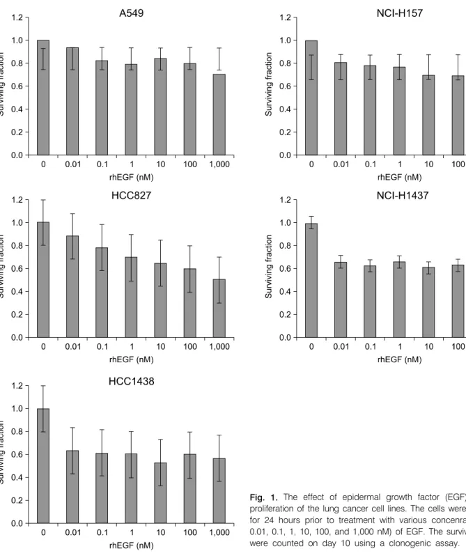
Clonogenic assay를 이용하여 0∼1,000 nM의 EGF 농도에 따른 폐암 세포주의 세포 집락수를 비교하였다. HCC-827에 서는 EGF를 처리하였을 때 농도의 증가와 비례하는 세포증 식 억제효과가 나타났으며, 다른 세포주에서도 농도와 비 례하는 양상은 관찰되지 않았으나 EGF를 처리하지 않은 대
조군에 비하여 처치군에서 세포집락수가 감소했다(Fig. 1).
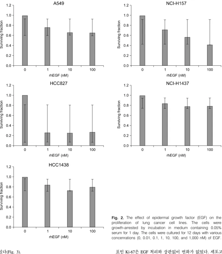
혈청에는 다양한 성장인자와 성장과 관련된 다양한 호르 몬이 존재하기 때문에 EGF에 의한 효과를 확인하기 위하여 0.05% 혈청을 첨가한 배지에서 24시간 동안 serum starvation시킨 후 같은 실험을 반복했으나 혈청 유무에 관 계없이 같은 결과를 보였다(Fig. 2).
2) EGF가 방사선 효과에 미치는 영향
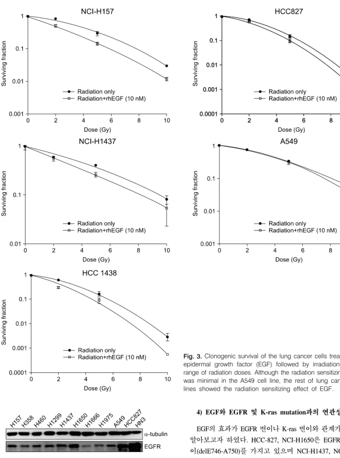
EGF와 방사선을 병행했을 때 효과를 알아보기 위하여 10 nM의 EGF와 0, 2, 5, 10 Gy의 방사선을 함께 처리하였다.
A549 세포주에서는 효과가 적었으나, 다른 모든 폐암 세포 주에서 방사선 단독 처리 시 보다 EGF를 병용 처리하였을 때 방사선의 세포성장 억제효과가 증가하는 결과를 보여주
Fig. 1. The effect of epidermal growth factor (EGF) on the proliferation of the lung cancer cell lines. The cells were cultured for 24 hours prior to treatment with various concenrations (0, 0.01, 0.1, 1, 10, 100, and 1,000 nM) of EGF. The surviving cells were counted on day 10 using a clonogenic assay.
었다(Fig. 3).
3) EGFR, Ki-67, cleaved-PARP 발현의 변화
단백면역분석으로 여러 종류의 세포주에서 EGFR이 발 현되는 것을 확인하였다(Fig. 4). 모든 세포주에서 EGF 처치 후 4시간, 24시간, 48시간으로 시간이 경과함에 따라 EGFR 의 발현이 뚜렷하게 감소하였다. 세포 증식을 나타내는 지
표인 Ki-67은 EGF 처리와 상관없이 변화가 없었다. 세포고 사와 관련이 있는 cleaved-PARP는 HCC-1438, NCI-H1437, NCI-H1650에서 EGF 처리 24시간 후 현저히 증가하였고, HCC-827과 NCI-H157에서는 48시간 후에 증가하였다(Fig.
5).
Fig. 2. The effect of epidermal growth factor (EGF) on the proliferation of lung cancer cell lines. The cells were growth-arrested by incubation in medium containing 0.05%
serum for 1 day. The cells were cultured for 12 days with various concenrations (0, 0.01, 0.1, 1, 10, 100, and 1,000 nM) of EGF.
Fig. 4. Expression of epidermal growth factor receptor (EGFR) in various cancer cell lines.
4) EGF와 EGFR 및 K-ras mutation과의 연관성
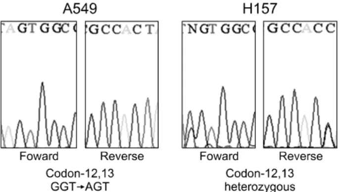
EGF의 효과가 EGFR 변이나 K-ras 변이와 관계가 있는지 알아보고자 하였다. HCC-827, NCI-H1650은 EGFR결실 변 이(delE746-A750)를 가지고 있으며 NCI-H1437, NCI-H157, A549는 EGFR wild type (wt)인 것으로 알려져 있고(18,19), A549와 H157에서는 코돈 12, 13의 K-ras 변이가 발견되었다 Fig. 3. Clonogenic survival of the lung cancer cells treated with epidermal growth factor (EGF) followed by irradiation with a range of radiation doses. Although the radiation sensitizing effect was minimal in the A549 cell line, the rest of lung cancer cell lines showed the radiation sensitizing effect of EGF.
Fig. 5. The epidermal growth factor receptor (EGFR), Ki-67 and cleaved PARP expressions in response to epidermal growth factor (EGF) treatment on the lung cancer cell lines. Treatment with EGF decreased the expression of EGFR and it increased the expression of cleaved PARP.
Fig. 6. Mutation analysis of K-ras in the lung cancer cell lines.
(Fig. 6). 그러나 EGFR 변이나 K-ras 변이가 있는 세포주들 과 변이가 없는 H1437을 비교했을 때 EGF의 성장억제 효과 에서 차이점을 보이지 않았다(Figs. 1, 3).
고안 및 결론
EGFR은 erb-B2 (HER2/neu), erb-B3, erb-B4와 함께 인체 내 상피성 수용체인 tyrosine kinase에 속하는 세포막 당단백 으로, 자체 tyrosine kinase 활성을 가지고 있는데, EGF와 결
합하여 활성화되면 유전자 발현, 세포 증식, 주변 조직에 대 한 침범, 세포고사 억제, 혈관 신생 등의 효과를 유발하여 암을 일으키거나 더욱 악화시킬 수 있다(21,22). EGFR은 비 소세포폐암을 포함한 많은 상피세포종양에서 비정상적으 로 과발현 되는데(23), 최근 EGFR 억제제인 gefitinib, erlotinib, cetuximab 등이 표적치료제로 개발되어 일부 암의 치료에 사용되고 있다(11-13).
그러나 EGF는 세포의 성장을 촉진하는 기능 외에 높은 농도로 암세포에 처리하였을 때 암세포의 성장을 억제하고 세포고사를 촉진한다는 연구 결과들이 보고되었다(24,25).
특히 EGFR의 발현이 적은 세포에서는 EGF 처리 시 성장이 촉진되지만 EGFR이 과발현하는 세포에서는 EGF가 성장을 저해하고 결국 세포고사를 유도하는 것으로 나타났다 (26-28). 세포고사의 기전은 caspase라고 하는 단백질 분해 효소에 의해 세포 내 단백질이 분해되면서 신호가 전달되 는 과정으로 여러 종류의 caspase들이 세포사멸에 관여하며 caspase의 활성을 통해 세포고사의 정도를 파악할 수 있다 (29). 최종적으로 활성화된 caspase-3는 PARP를 활성화시키 고 활성화된 PARP는 DNA 분절화 현상을 유도하면서 세포 고사를 일으키는 것으로 보고되고 있다(30). 또한 방사선 조사와 EGF를 병행하였을 때 그 효과가 상승된다는 보고가
있다(18,19).
본 연구에서도 여러 가지 폐암 세포주를 대상으로 EGF를 처리하였을 때 세포사가 일어나는 것을 확인하였고, 시간 에 따른 EGFR 발현의 감소와 cleaved-PARP의 발현 증가로 그 기전이 세포고사의 증가에 의한 것임을 알 수 있었다.
방사선 조사와 EGF를 병행하여 처리했을 때는 A549에서 그 효과가 미약하기는 하였지만 다른 폐암 세포주에서는 방사선 조사 단독 처리 결과보다 효과가 있는 것으로 나타 났다. Ki-67은 세포증식을 나타내는 지표로서 세포주기의 G0기와 초기 G1기를 제외한 모든 세포의 핵 내에 존재하 고, 활발하게 분열하고 있는 세포들의 분포를 반영하며 종 양의 재발 및 악성도와 연관이 있다(31). 본 연구결과에서 Ki-67은 모든 세포주에서 변화를 보이지 않았다. 이는 EGF 가 폐암 세포주를 증식시키지 않는다는 것을 보여주었다.
본 연구자들은 이전 실험에서 EGF가 정상섬유아세포의 증 식을 촉진시키고 방사선 조사에 의한 세포사를 억제시키는 반면 두경부암 세포주인 AMC-HN3에서는 세포사를 증가 시키는 것을 확인하였다(20). 또한 EGFR 변이와 K-ras 변이 는 상호배타적이지만 폐암에 있어서 기능적으로 같은 역할 을 한다(32,33). EGFR 변이는 선암, 비흡연자, 동양인, 여성 에게서 많이 발견되는데 EGFR 표적치료제의 임상결과에 효과적이며 K-ras 변이는 약물 저항성의 예측인자라고 할 수 있다. 항암제의 효과가 EGFR 변이 또는 K-ras 변이와 밀 접한 관련이 있는 것으로 알려져 있기 때문에 EGF의 암세 포 증식억제 효능이 변이에 의한 것인지 확인하였다. 결과 적으로 변이의 유무에 따른 차이는 나타나지 않았으므로 EGF는 변이와 무관하게 효과가 있는 것으로 생각된다.
본 연구에서 EGF의 세포고사를 통한 폐암 세포주 증식 억제효과를 확인하였다. 또한 방사선과 병용 시 방사선 단 독 처리보다 그 효과가 증가되었으므로 방사선 민감제로서 의 역할도 기대할 수 있을 것으로 생각된다. 향후 추가적인 실험을 통하여 세포고사를 일으키는 기전과 EGF가 정상조 직에 대한 최소한의 부작용을 보이면서 암 조직의 성장을 억제시킬 수 있는 가능성에 대한 확인이 필요하다.
REFERENCES
1. Carpenter G, Cohen S. Epidermal growth factor. J Biol Chem 1990;265:7709-7712.
2. Carpenter G, Cohen S. Epidermal growth factor. Annu Rev Biochem 1979;48:193-216.
3. Cohen S. Isolation of a mouse submaxillary gland protein accelerating incisor eruption and eyelid opening in the new- born animal. J Biol Chem 1962;237:1555-1562.
4. Servold SA. Growth factor impact on wound healing. Clin
Podiatr Med Surg 1991;8:937-953.
5. Brown GL, Curtsinger L 3rd, Brightwell JR, et al. Enhance- ment of epidermal regeneration by biosynthetic epidermal growth factor. J Exp Med 1986;163:1319-1324.
6. Hong JP, Jung HD, Kim YW. Recombinant human epidermal growth factor (EGF) to enhance healing for diabetic foot ulcers. Ann Plast Surg 2006;56:394-398.
7. Lee SW, Jung KI, Kim YW, Jung HD, Kim HS, Hong JP.
Effect of epidermal growth factor against radiotherapy-induced oral mucositis in rats. Int J Radiat Oncol Biol Phys 2007;67:
1172-1178.
8. Mishima H, Nakamura M, Murakami J, Nishida T, Otori T.
Transforming growth factor-beta modulates effects of epider- mal growth factor on corneal epithelial cells. Curr Eye Res 1992;11:691-696.
9. Bianco R, Gelardi T, Damiano V, Ciardiello F, Tortora G.
Rational bases for the development of EGFR inhibitors for cancer treatment. Int J Biochem Cell Biol 2007;39:1416-1431.
10. Arteaga CL. Epidermal growth factor receptor dependence in human tumors: more than just expression? Oncologist 2002;7 (Suppl 4):31-39.
11. Paez JG, Jänne PA, Lee JC, et al. EGFR mutations in lung cancer: correlation with clinical response to gefitinib therapy.
Science 2004;304:1497-1500.
12. Lynch TJ, Bell DW, Sordella R, et al. Activating mutations in the epidermal growth factor receptor underlying responsive- ness of non-small-cell lung cancer to gefitinib. N Engl J Med 2004;350:2129-2139.
13. Pao W, Miller V, Zakowski M, et al. EGF receptor gene mutations are common in lung cancers from "never smokers"
and are associated with sensitivity of tumors to gefitinib and erlotinib. Proc Natl Acad Sci U S A 2004;101:13306-13311.
14. Gill GN, Lazar CS. Increased phosphotyrosine content and inhibition of proliferation in EGF-treated A431 cells. Nature 1981;293:305-307.
15. Kamata N, Chida K, Rikimaru K, Horikoshi M, Enomoto S, Kuroki T. Growth-inhibitory effects of epidermal growth factor and overexpression of its receptors on human squamous cell carcinomas in culture. Cancer Res 1986;46:1648-1653.
16. MacLeod CL, Luk A, Castagnola J, Cronin M, Mendelsohn J. EGF induces cell cycle arrest of A431 human epidermoid carcinoma cells. J Cell Physiol 1986;127:175-182.
17. Kröning R, Jones JA, Hom DK, et al. Enhancement of drug sensitivity of human malignancies by epidermal growth factor.
Br J Cancer 1995;72:615-619.
18. Christen RD, Hom DK, Eastman A, Howell SB. Epidermal growth factor regulates the ability of human ovarian carcinoma cells to repair DNA damage. Proc Am Assoc Cancer Res 1991;32:430.
19. Kwok TT, Sutherland RM. Enhancement of sensitivity of human squamous carcinoma cells to radiation by epidermal growth factor. J Natl Cancer Inst 1989;81:1020-1024.
20. Kwon EK, Lee SH, Kim KB, Wu HG, Lee SW. Differential effect of recombinant human EGF on proliferation and radiation survival of normal fibroblast and cancer cell lines.
Open Transl Med J 2009;1:9-15.
21. Reynolds NA, Wagstaff AJ. Cetuximab: in the treatment of metastatic colorectal cancer. Drugs 2004;64:109-118.
22. Simmonds PC. Palliative chemotherapy for advanced colorectal cancer: systematic review and meta-analysis. Colorectal Cancer Collaborative Group. BMJ 2000;321:531-535.
23. Suzuki S, Dobashi Y, Sakurai H, Nishikawa K, Hanawa M, Ooi A. Protein overexpression and gene amplification of epidermal growth factor receptor in nonsmall cell lung carcinomas. An immunohistochemical and fluorescence in situ hybridization study. Cancer 2005;103:1265-1273.
24. Grudinkin PS, Zenin VV, Kropotov AV, Dorosh VN, Nikolsky NN. EGF-induced apoptosis in A431 cells is dependent on STAT1, but not on STAT3. Eur J Cell Biol 2007;86:591-603.
25. Filmus J, Trent JM, Pollak MN, Buick RN. Epidermal growth factor receptor gene-amplified MDA-468 breast cancer cell line and its nonamplified variants. Mol Cell Biol 1987;7:251- 257.
26. Gulli LF, Palmer KC, Chen YQ, Reddy KB. Epidermal growth factor-induced apoptosis in A431 cells can be reversed by reducing the tyrosine kinase activity. Cell Growth Differ 1996;7:173-178.
27. Armstrong DK, Kaufmann SH, Ottaviano YL, et al. Epidermal
growth factor-mediated apoptosis of MDA-MB-468 human breast cancer cells. Cancer Res 1994;54:5280-5283.
28. Brabyn CJ, Franks DJ, Kleine LP. Long-term exposure to epidermal growth factor results in apoptosis in T51B cells.
Biochem Cell Biol 1994;72:429-438.
29. Alnemri ES. Mammalian cell death proteases: a family of highly conserved aspartate specific cysteine proteases. J Cell Biochem 1997;64:33-42.
30. Soldani C, Scovassi AI. Poly(ADP-ribose) polymerase-1 cleavage during apoptosis: an update. Apoptosis 2002;7:
321-328.
31. Scopa CD, Tsamandas AC, Zolota V, Kalofonos HP, Batistatou A, Vagianos C. Potential role of bcl-2 and ki-67 expression and apoptosis in colorectal carcinoma: a clinicopathologic study. Dig Dis Sci 2003;48:1990-1997.
32. Shigematsu H, Lin L, Takahashi T, et al. Clinical and biolo- gical features associated with epidermal growth factor receptor gene mutations in lung cancers. J Natl Cancer Inst 2005;97:
339-346.
33. Kosaka T, Yatabe Y, Endoh H, Kuwano H, Takahashi T, Mitsudomi T. Mutations of the epidermal growth factor receptor gene in lung cancer: biological and clinical implica- tions. Cancer Res 2004;64:8919-8923.