††주 저자 (e-mail: [email protected]) call: 031-280-5865
††교신저자 Su Na Kim (e-mail: [email protected]) Hyoung-June Kim (e-mail: [email protected])
call: 031-280-5827
Vol. 47, No. 3, September 2021, 255-263 http://dx.doi.org/10.15230/SCSK.2021.47.3.255
모낭기관배양과 임상에서 제주푸른콩발효추출물의 육모 효능에 미치는 영향
이 용 희*,†ㆍ신 승 현*ㆍ김 세 현*ㆍ정 규 상*ㆍ홍 민 정*ㆍ박 혁 곤**ㆍ김 승 훈**ㆍ 김 은 주**ㆍ강 영 규*ㆍ박 병 철***ㆍ박 원 석*ㆍ김 수 나*,††ㆍ김 형 준*,††
*아모레퍼시픽, R&D센터, 기반혁신연구디비전
**아모레퍼시픽, R&D센터, 스킨케어연구디비전
***단국대학교병원 피부과, 교수
(2021년 9월 23일 접수, 2021년 9월 28일 수정, 2021년 9월 29일 채택)
Fermented Jeju Soybean Extract Promotes Hair Growth in Human Hair Follicle Organ Culture and Clinical Trial
Yonghee Lee
1,†, Seung Hyun Shin
1, Sehyun Kim
1, Gyusang Jeong
1, Min Jung Hong
1, Hyeokgon Park
2, Seung Hun Kim
2, Eun Joo Kim
2, Young-Gyu Kang
1, Byung Cheol Park
3, Won Seok Park
1, Su Na Kim
1,††, and Hyoung-June Kim
1,††1Basic Research & Innovation Division, AMOREPACIFIC Corporation, R&D Center, 1920 Yonggu-daero, Giheung-gu, Yongin-si, Gyeonggi-do 17074, Korea
2Skincare Research Division, AMOREPACIFIC Corporation, R&D Center
3Department of Dermatology, Dankook Medical College
(Received September 23, 2021; Revised September 28, 2021; Accepted September 29, 2021)
1)
요 약: 콩 추출물은 노화, 암, 비만 및 심혈관 질환과 같은 질병의 예방 및 치료에 중요한 역할을 하는 것으로 알려져 있다. 최근 연구에 따르면 콩 추출물은 in vitro, in vivo 및 임상 연구에서 모발 성장에 강력한 영향을 미치는 것으로 나타났다. 최근에는 발효추출물이 일반추출물에 비해 다양하고 높은 효능을 나타내는 것으로 보고되고 있다. 그러나 발효된 콩 추출물의 모발 성장을 유도하는 근본적인 매커니즘은 잘 알려져 있지 않다.
본 연구는 미녹시딜과 유사한 기본 매커니즘에 초점을 두고 제주푸른콩발효(FJS)추출물이 모발 성장에 미치는 영향을 확인하였다. 우리는 in vitro, ex vivo와 임상 연구를 수행하였다. 우리의 연구결과, FJS 추출물은 모낭모유 두세포증식, VEGF 수준 및 포타슘 채널 활성을 향상시킴을 확인하였다. 이러한 효과는 FJS 추출물 이 함유된 토닉을 사용하였을 때, 24주 후 모발 밀도 개선을 확인한 임상결과에서도 종합적으로 입증되었다. 결론적으로, 본 연구의 결과는 FJS 추출물이 모낭의 미녹시딜과 유사한 매커니즘을 통해 모발 성장을 촉진하고 탈모를 억제한 다는 것을 보여준다.
Abstract: Soybean extract is known to play an important role in preventing and treating diseases associated with aging,
cancer, obesity, and cardiovascular disease. A recent has revealed that soybean extract has a potent effect on hair growth
in in vitro, in vivo, and clinical studies. Recently, it has been reported that their fermented extracts exhibit numerous
and high efficacy, as compared to general extracts. However, the underlying mechanisms that induce hair growth after
using fermented soybean extract are not well understood. The present study aimed to determine the effects of fermented
Jeju soybean (FJS) extract on hair growth, with a focus on the underlying mechanisms similar to those of minoxidil.
We conducted in vitro and ex vivo investigations and clinical studies. FJS extract enhanced dermal papilla cell proliferation, VEGF levels, and potassium channel opening. Moreover, it promoted human hair follicle elongation. These effects were comprehensively demonstrated in the clinical results, in which FJS extract-containing shampoo improved hair density after 24 weeks of utilization. Collectively, the results of this study demonstrate that FJS extract promotes hair growth and inhibits hair loss through a mechanism similar to that of minoxidil in hair follicles.
Keywords: fermented Jeju soybean extract, hair growth, human dermal papilla cells, hair follicle, minoxidil
1. Introduction
Hair loss is a common disease, defined as an abnormal loss of hair on the head or body. Hair loss is not a disability, but it is considered a serious social and psychological problem because it affects appearance[1]. As the cause of hair loss is found to be not only genetic but also complex factors, such as mental stress, irregular lifestyle, and excessive use of chemical hair products, research on various treatments and prevention is being conducted.
Finasteride and minoxidil (MNX) are approved by the United States Food & Drug Administration (FDA) for hair loss treatment[2]. However, these drugs have therapeutic limits because of their unpredictable response rate and adverse effects; therefore, more effective and safer treatment options are needed for hair loss patients. Finasteride can cause birth defects when women of childbearing potential are exposed to it[3], and MNX can cause side effects such as pruritus, dermatitis, and irritation[4]. Therefore, recently, research on natural products with hair growth potential and materials containing active ingredients is increasing.
Jeju soybeans are native resources found only in the southern part of Jeju Island. They are taller than ordinary beans; hence, they are vulnerable to wind and difficult to grow because of the long cultivation period. Recently, we found that Jeju soybean contains more isoflavones than ordinary soybeans, and when fermenting Jeju green soybeans, various complex bacteria, fungi, and enzymes generated from them act more effectively than while fermenting ordinary soybeans. This extract of fermentation is known to be more effective than other extracts. In addition, Jeju green beans contain high amounts of poly γ-glutamic acid (γ-PGA) and 7,8,4′-trihydroxyisoflavone, major metabolites of daidzin and daidzein, which are representative isoflavones (data not shown).
Fermentation is one of the new drug development methods.
It is a process of decomposing natural products through catalytic decomposition by microorganisms at constant relative humidity and temperature after specific treatments. This process is carried out under stringent temperature and humidity conditions using various methods, depending on the type of natural product. Glycosides that are not absorbed through fermentation can be bioconverted to non-glycosides, which increases the absorption and bioavailability of pharmaceutical ingredients in the body[5]. In this study, we investigated the effect of fermented Jeju soybean (FJS) extract on hair growth as well as the underlying mechanisms.
2. Experimental
2.1. Preparation of FJS Extract
First, in 10 g of Jeju soybean, Bacillus subtilis was inoculated with 1.0 × 10
8colony forming unit (CFU)/mL (the strain obtained by culturing in ATCC Medium 3 was suspended in phosphate-buffered saline (PBS) and then fermented at a temperature of approximately 37 °C for 72 h to obtain the fermented product of Jeju green beans. Purified water was added to the fermented product to prepare a reactant such that the weight ratio of the fermented product to purified water was 1 : 1. Subsequently, the reaction product was obtained by incubating at a temperature of approximately 90 °C for 3 h.
Then, it was filtered through a 0.3
µm filter, and the filtrate
obtained was dried to obtain filtered dry matter 1 (dry matter
1). Next, 10 g of Jeju green soybean and 95% ethanol
aqueous solution (ethanol : purified water volume ratio = 95 :
5) were prepared so that the weight ratio of the reactants was
1 : 1, and then left at a temperature of approximately 22 °C
for approximately 72 h. The reaction product was filtered with
a 0.3
µm filter, and the resulting filtrate was dried to obtain
dry matter 2 (dry matter 2). The fermented Jeju green soybean was derived by mixing dry matter 1 and dry matter 2 in a 1 : 1 ratio by weight. An extract of fermented Jeju soybean extract was prepared. Fermented of soybean, and black soybean extract used in the experiment were collected from Yeongwolsan Mountain (Korea) and fermentation was carried out in the same condition as FJS extract.
2.2. Huma Hair Follicles (HFs) and Cell Culture Non-balding scalp specimens were obtained from patients undergoing hair transplantation surgery. The Medical Ethical Committee of the Dankook Medical College (Department of Dermatology, Korea) approved all the described studies, and informed written consent was obtained from all patients ( IRB:
DKUH 2013-08-012-001). HFs were isolated and cultured using a previously described method with minor modifications [6,7]. Briefly, HFs were cultured in William’s E medium (Sigma-aldrich, USA) supplemented with 2 mM L-glutamine (Sigma-aldrich, USA), 10
µg/ mL insulin (Sigma-aldrich, USA), 10 ng/mL hydrocortisone (Sigma-aldrich, USA), 0.1% fungizone (Gibco, USA), and 1% antibiotic-antimycotic (Gibco, USA).
Human dermal papilla cells (hDPCs) were isolated from HFs and NIH-3T3 cells were purchased from the Korean Cell Line Bank (Korea). hDPCs and NIH-3T3 cells were cultured in Dulbecco’s modified Eagle’s medium (DMEM; Hyclone Laboratories, USA) supplemented with 10% fetal bovine serum (FBS, Gibco, USA) and 1% antibiotic–antimycotic and incubated at 37 °C and 5% CO
2.
2.3. Human Dermal Papilla Cells Proliferation Human dermal papilla cells (hDPCs) were plated in 96 well plates at a density of 2,000 cells/well. After cell attachment, FJS extract and another soybean ferment extract concentration of 20 parts per million (ppm) were added to FBS-free medium. As a positive control, 5% FBS was used.
After 72 h, cell proliferation was assessed using a commercially available kit (Cell Counting Kit-8 [CCK-8], Dojindo Molecular Technologies, Japan).
2.4. Potassium Channel Assay
The potassium channel assay is a method used to identify the effect of potassium channel opening according to the
viability of NIH-3T3 cells co-treated with potassium channel blockers (tolbutamide [TBT]; Sigma-aldrich, USA). This experiment was performed according to a previously described method[8]. Briefly, NIH-3T3 cells were plated at a density of 8,000 cells/well in 96 well plates and incubated for 24 h at 37 °C and 5% CO
2. After each well was washed with PBS, TBT (2.5 mM) was dissolved in DMEM (without phenol red) with 10% FBS and penicillin (100 U/mL). After 10 min of pre-treatment with 2.5 mM TBT medium, each well was treated with 20 ppm FJS extractr for 48 h. MNX treatment served as the positive control. The efficacy of potassium channel opening was determined using CCK-8 assay.
2.5. VEGF Expression Analysis
VEGF secretion was quantified using a VEGF enzyme- linked immunosorbent assay (ELISA) kit (R&D Systems, USA) according to the manufacturer’s protocol. For the measurement of secreted VEGF content from hDPCs treated with FJS extract and MNX, hDPCs passage 3 were plated overnight at a density of 1.5 × 10
5cells per 35 mm of the culture dish. The cells were washed three times with PBS and incubated in serum-free medium for 24 h. Then, 20 ppm FJS extract and 50
µM of MNX in serum-free medium were added, and the concentration of VEGF in the medium were measured after 24 h. Briefly, 100
µL of culture medium was loaded per well using titer plates coated with VEGF capture antibody and incubated for 2 h. Each well was then washed three times with washing buffer and incubated with 100
µL of VEGF detection antibody for 2 h. Then, each well was washed three times and incubated with 100
µL of streptavidin-HRP in the dark. Each well was then washed again, and then 100
µL of substrate solution was added and the sample was incubated for 20 min in the dark. Optical density was measured using an ELISA reader at 450 nm.
Serial dilutions of recombinant human VEGF were used to establish a standard curve.
2.6. Quantitative Real-time Polymerase Chain Reaction (qRT-PCR)
To analyze VEGF gene expression, 1.5 × 10
5hDPCs were
cultured in 6 well plates for 24 h, followed by exchange with
FBS-free media containing FJS extract at a concentration of
20 ppm. After an additional 48 h of incubation, the cells and supernatant were isolated for qRT-PCR. To analyze VEGF gene expression, messenger RNA (mRNA) isolation was performed using a commercially available kit (RNeasy Mini Kit, Qiagen, United Kingdom) and then complementary was performed DNA synthesis using the PrimeScript™ 1st strand cDNA Synthesis Kit (Takara, Japan). Gene amplification and quantification were performed using a thermocycler (7500 Fast Real-Time PCR Instrument System, Applied Biosystems, USA) and TaqMan™ Universal Master Mix II (Applied Biosystems, USA).
2.7. HF Organ Culture and Assessment of Hair Elongation Anagen HFs were obtained from human scalp skin specimens. Six HFs per well in 24 well plates were cultured in William’s E medium at 37 °C in a humidified atmosphere with 5% CO
2in 500
µL of basal medium supplemented with 10
µg/mL insulin, 10 ng/mL hydrocortisone, 2 mM L-glutamine, 0.1% Fungizone, 10
µg/mL streptomycin, and 100 U/mL penicillin according to the method described by Philpott[9]. Each experimental group contained at least 30 anagen HFs derived from three different human donors/volunteers. MNX and vehicle served as the positive and negative controls, respectively. Both FJS extract and MNX were added at concentrations of 20 ppm and 50
µM, respectively. The incubation medium was renewed every two days. HF elongation was measured directly after 6 d of culture using a stereo microscope (Olympus, Japan)[10].
2.8. Clinical Study 2.8.1. Participants
Forty-four Korean men and women aged 28 to 54 years (mean ± SD, 46.614 ± 6.288 years) were recruited as subjects for the clinical study. Subjects diagnosed with androgenetic alopecia were selected according to basic and specific (BASP) and Ludwig’s classification. In detail, the Basic type is men and women with androgenic alopecia diagnosed with M1 or higher, C1 or higher, or U1 or higher, and the specific type is men and women with androgenic alopecia diagnosed with V1 or higher or F1 or higher. In the case of women subjects, those diagnosed with 1 or more by Ludwig classification are included.
Those who had undergone procedures, such as hair transplantation, or those who were treated with oral hair growth agents within the previous 6 months were excluded.
This clinical study was performed according to the ethical principles of the Helsinki Declaration and the Korea Food and Drug Administration (KFDA) guidelines (IRB approval number KC-IRB-014). All participants provided informed written consent to participate. The topical solution, in which 5% of PG extract dissolved in 15% ethanol solution, was supplied to the test group and 15% ethanol vehicle was offered to control group. All test materials were applied to the scalp once a day for 24 weeks.
2.8.2. Protocol
This clinical study was conducted in a double-blind manner.
Hair density was assessed at baseline and at 8, 16, and 24 weeks of treatment. To measure hair density, the center of the measurement area was labeled with a dye to evaluate the same location. Then, the hair around the labeled spot was cut to be shorter than 2 mm. Hair density was determined by analyzing the total number of hairs in a 1 cm
2area of the photographed image using a phototrichogram instrument (Folliscope 2.8, LeadM, Korea)[11]. To observe the overall changes in hair density, global photographs of the vertex and hairline areas were captured.
2.9. Statistical Analyses
SPSS version 23.0 (IBM Corporation, USA) was used for the statistical analyses. Data were compared using the student’s t-test. They are expressed as mean ± standard deviation (SD); differences with p < 0.05 were considered statistically significant.
For the analysis of clinical study, Repeated-measure ANOVA and Friedman test were used to analyze the results of the present study. Normality test was performed with the shapiro-wilk normality test, and as a result, parameter statistics were performed.
3. Results and Discussion
3.1. Cell Proliferation Effect of FJS Extract on hDPCs
To investigate the potential role of FJS extract on the
proliferation of hDPCs, we performed and compared the results of the CCK-8 assay 3 d after treatment with FJS extract and another type of soybean extract, such as fermented of soybean and black soybean. The results showed that FJS extract enhanced the proliferation of hDPCs compared to that of untreated negative controls and other soybean extracts.
Compared to that in the untreated negative controls, cell proliferation in the FJS extract-treated (20 ppm) group increased by 41% (Figure 1).
3.2. The Function of FJS Extract as a Potassium Channel Activator
To evaluate the effect of the FJS extract on potassium
channel activation, we performed a potassium channel assay with NIH-3T3 fibroblasts, according to a previously described method[12]. MNX is a well-known potassium channel opener that restores NIH-3T3 fibroblast proliferation, which is inhibited by TBT[13]. TBT decreased cell viability by approximately 60%. By contrast, cell viability in the FJS extract-treated group was approximately 89%, which was the same as that of the MNX-treated group (Figure 2). We demonstrated that FJS extract induced potassium channel opening against the action of TBT, which, in turn, improved cell survival. These results suggest that FJS extract exerts a vasodilation effect on hair HFs, with a mechanism of action similar to that of MNX[14–16].
Figure 1. The effect of fermented jeju soybean (FJS) extract on human dermal papilla cells (hDPCs) proliferation. FJS extract enhanced the cell proliferation. (
*p < 0.05,
**p < 0.01 vs. control,
##p < 0.01 vs. compared group)
µM
Figure 2. FJS extract as a potassium channel opener against TBT. FJS restored cell growth inhibition by TBT to levels similar to
those by minoxidil. (
*p < 0.05,
***p < 0.001 vs. compared group)
3.3. Modulation of VEGF Expression by FJS Extract in hDPCs
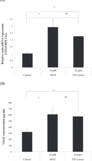
VEGF is a typical growth factor that enhances hair growth, and the effect of MNX on VEGF expression has already been investigated[17]. To further investigate the relevance of the hair growth effects of FJS extract, we examined changes in the expression of VEGF using real-time PCR and VEGF ELISA kits.
The mRNA level of VEGF was enhanced by a factor of 2.92 in the positive control group (50 µM MNX) and by a factor of 2.26 in the FJS extract-treated group (Figure 3A).
Moreover, MNX and FJS extracts significantly stimulated the expression of secreted VEGF in hDPCs ( Figure 3B). These results suggest that FJS extract could promote hair growth via VEGF stimulation, similar to the action of MNX.
3.4. Hair Shaft Elongation was Promoted by FJS Extract in Human HF Organ Culture
To examine the effect of FJS extract at the organ level, we performed an ex vivo culture of whole human scalp HFs.
During a 5-d culture with 20 ppm FJS extract, HFs grew to an average length of 1.88 mm, which was significantly greater than that in the control group, which grew to an average of only 1.64 mm ( Figure 4). These results suggested that FJS extract was as efficacious as MNX in promoting HF growth.
3.5. Effect of FJS Extract on Hair Loss in the Clinical Study
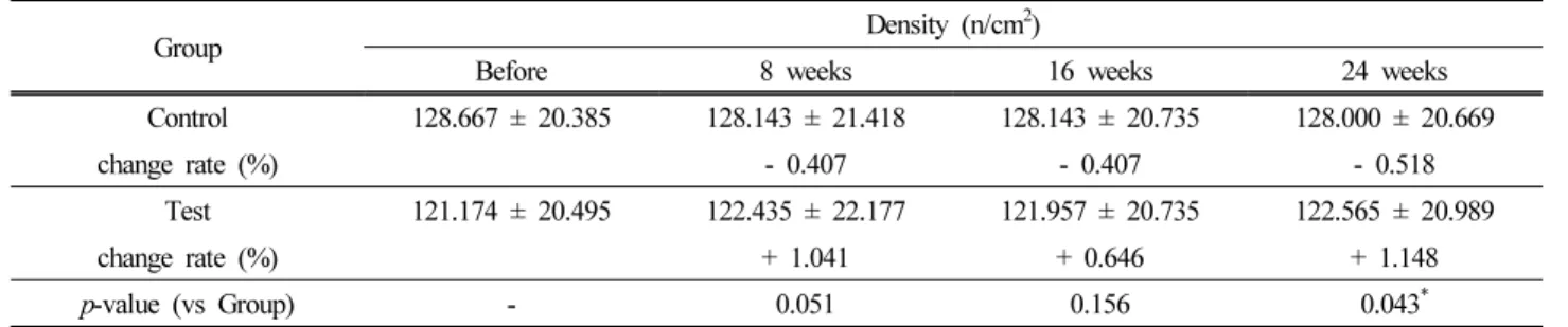
The results of the phototrichogram showed that the hair density of the test group (5% FJS extract) increased during treatment. However, both the control and test groups did not exhibit statistically significant changes in hair density during the 24 week trial (Table 1). However, comparison of hair density changes between the control and test groups revealed significant differences after 24 weeks of product use (Figure 5A). During the 24 week test period, hair density increased by 1.148% in the test group but decreased by 0.518% in the control group. Efficacy results were visually confirmed using clinical photographs from global vertex and hairline images (Figure 5B). These results suggest that FJS extract alleviates hair loss progression and induces hair growth in patients experiencing hair loss.
μM
(A)
μM
(B)