Effects of α-lipoic acid on cell proliferation and apoptosis in MDA-MB-231 human breast cells*
Mi Hee Na, Eun Young Seo and Woo Kyoung Kim§
Department Food Science and Nutrition, Dankook University, 126 Jukjeon-dong, Suji-gu, Yongin-si, Gyunggi 448-701, Korea
Abstract
The role that antioxidants play in the process of carcinogenesis has recently gained considerable attention. α-Lipoic acid, a naturally occurring disulfide molecule, is a powerful antioxidant that reportedly exerts beneficial effects in patients with advanced cancer by reducing the level of reactive oxygen species and increasing glutathione peroxidase activity. In this study, we examined changes in the protein and mRNA expression associated with cell proliferation and apoptosis in MDA-MB-231 breast cancer cultured in the presence of various concentrations (0, 250, 500, and 1000 μmol/L) of α-lipoic acid. The results revealed that α-lipoic acid inhibited the growth of breast cancer cells in a dose-independent manner (P < 0.05). Additionally, ErbB2 and ErbB3 protein and mRNA expressions were significantly decreased in a dose-dependent manner in response to α-lipoic acid (P < 0.05).
Furthermore, the protein expression of phosphorylated Akt (p-Akt) levels and total Akt, and the mRNA expression of Akt were decreased dose-dependently in cells that were treated with α-lipoic acid (P < 0.05). Bcl-2 protein and mRNA expressions were also decreased in cells that were treated with α-lipoic acid (P < 0.05). However, Bax protein and mRNA expressions were increased in cells treated with α-lipoic acid (P < 0.05). Finally, caspase-3 activity was significantly increased in a dose-dependent manner in cells treated with α-lipoic acid (P < 0.05). In conclusion, we demonstrated that α-lipoic acid inhibits cell proliferation and induces apoptosis in MDA-MB-231 breast cancer cell lines.
Key Words: α-Lipoic acid, proliferation, apoptosis, breast cancer, MDA-MB-231 cells
Introduction2)
Breast cancer is a major health problem in women (Hortobagyi et al., 2005). In Korea, the incidence of breast cancer among women is second only to thyroid cancer, with a frequency of 31.0 per 100,000 being observed in 2005 (Ministry of Health and Welfare and Family, 2008). Genetic factors are one of the main causes of breast cancer (Palacios et al., 2008). It is also known that there is a close relationship between the incidence of cancer and diet, and dietary factors can act as either risk or protective factors (Zhai et al., 2003).
α-Lipoic acid was first separated from bovine livers in 1950 and has since been found to occur naturally in extremely small amounts in many plants and animals (Reed et al., 1951). α-Lipoic acid acts as an important coenzyme for enzymes inside the mitochondria (Cameron et al., 1998), such as pyruvate dehydrogenase and α-ketoglutarate dehydrogenase; therefore, it has been treated as a vitamin-like substance (Gurer et al., 1999).
α-Lipoic acid is easily absorbed and converted into the reduced form of dihydrolipoic acid in a variety of cellular tissues (Packer, 1998). α-Lipoic acid and dihydrolipoic acid mutually form a redox couple, which then acts as an antioxidant in water-soluble and
fat-soluble environments. The anticancer effects of α-lipoic acid were found in various cancer types such as ovarian epithelial cancer cells (Vig-Varga et al., 2006) and B16F10 murine melanoma cells (Packer, 1998). And Wenzel et al. (2005) reported that α-lipoic acid increases the level of active glutathione peroxidase in the body and reduces the oxidative stress, and therefore has a carcinostatic effect in cancer patients. But the anticancer effects of α-lipoic acid on breast cancer have not been fully understood. Accordingly, we investigated the effect of α -lipoic acid on the proliferation and apoptosis of MDA-MB-231 human breast cancer cells.
Materials and Methods
Materials and Reagents
α-Lipoic acid (Sigma, St. Louis, MO, USA) was dissolved in ethanol and diluted in cell culture medium. MDA-MB-231 cells were purchased from American Type Culture Collection (ATCC, Rockville, MD, USA). The following reagents and chemicals were obtained from the respective suppliers: Dulbecco’s modified
* This work was supported by the Korea Research Foundation Grant funded by the Korean Government (KRF-2006-531-C00058).
This research was supported by the Graduate Research Assistantship of Dankook University in 2009.
§Corresponding Author: Woo Kyoung Kim, Tel. 82-31-8005-3172, Fax. 82-31-8005-3170, Email. [email protected] Received: October 7, 2009, Revised: November 9, 2009, Accepted: November 11, 2009
ⓒ2009 The Korean Nutrition Society and the Korean Society of Community Nutrition
This is an Open Access article distributed under the terms of the Creative Commons Attribution Non-Commercial License (http://creativecommons.org/licenses/by-nc/3.0/) which permits unrestricted non-commercial use, distribution, and reproduction in any medium, provided the original work is properly cited.
maintained in DMEM/F12 containing 100 mL/L fetal bovine serum (FBS) with 100,000 U/L penicillin and 100 mg/L streptomycin as described previously (Kim et al., 2005). Before MDA-MB-231 human breast cancer cells were treated with α -lipoic acid, the cell monolayers were rinsed and starved of serum for 24 h by culture in DMEM/F12 supplemented with 5 mg/L transferrin, 1 g/L BSA, and 5 μg/L selenium (serum-free medium, SFM). After serum starvation, the medium was replaced with fresh SFM with the indicated concentrations of α-lipoic acid (0, 25, 500, and 1,000 μmol/L). Cell proliferation was then estimated 12, 24, and 48 h after the cells were exposed to α-lipoic acid using the 3-[4,5-dimethylthiazol-2-yl]-2,5-diphenyltetrazolium bromide (MTT) assay, as described previously (Kim et al., 2005).
Three independent experiments were done.
Western blotting analysis
Cell lysates were prepared as previously described (Lee et al., 2004). The total cell lysates were resolved on a sodium dodecylsulfate 40-200 g/L polyacrylamide gel and then transferred to a polyvinylidene fluoride membrane (Millipore, Bedford, MA).
Next, the blot was blocked for 1 h in 10 g/L BSA in TBS-T (20 mmol/L Tris-Cl, pH 7.5, 150 mmol/L NaCl, 1 g/L Tween 20) or 50 g/L milk TBS-T, after which it was incubated for 1 h with either anti-ErbB2, anti-ErbB3, anti-Akt, anti-p-Akt, anti-phospho-Tyr, anti-Bcl-2, or anti-Bax antibody. The blot was then incubated with antimouse or antirabbit HRP-conjugated antibody. Signals were detected by the enhanced chemiluminescence method using Super-Signal West Dura Extended Duration Substrate (Pierce, Rockford, IL). Finally, the relative abundance of each protein band was analyzed by scanning the exposed films densitometrically using the Image J Launcher (provided by NCBI).
RNA expression (RT-PCR)
Total RNA was isolated using Tri-reagent (Sigma), and cDNA was synthesized using 2 μg of total RNA with SuperScriptTM II reverse transcriptase (Invitrogen). For amplification of cDNA, primers for ErbB2 (upstream primer, 5’-GTTTCCCAGATGA
GGAGGGCGCATGCC-3’; downstream primer, 5’-TTCTCCC CATCAGGGATCCAGATGCCC-3’ , annealing at 62℃ for 1 min with 37 cycles), primers for ErbB3 (upstream primer, 5’-GG
TCAGCCCATCTTCTTCCAGA-3’, annealing at 55℃ for 1 min with 32 cycles), and primers for β-actin (upstream primer, 5’-GTTTGAGACCTTCAACACCCC-3’; downstream primer, 5’-GTGGCCATCTCCTGCTCGAAGTC-3’, annealing at 60℃
for 1 min with 35 cycles) were used. The expression of human β-actin transcripts was examined as an internal control. The PCR products were separated on a 1% agarose gel and stained with ethidium bromide. Bands corresponding to each specific PCR product were quantified by densitometric scanning of the exposed film using the Bio-profile Bio-IL application (Vilber-Lourmat).
Activity of caspase-3
The activity of caspase-3 was evaluated as previously described using a commercially available caspase-3 assay kit from Clontech Laboratories, Inc (Seo & Kim, 2006). Briefly, cells that were treated with α-lipoic acid were collected and suspended in 50 μl of ice cold lysis buffer at a concentration of 2×10⁶ cells. The cell lysates were then incubated on ice for 10 min, after which they were centrifuged at 1,500 rpm for 5 min. Next, the supernatant was placed in 96-well plates and incubated with 50 μl 2× reaction buffer/DTT and caspase-3 colorimetric substrate DEVD-pNA for 1 h at 37℃. Finally, the absorbance at 405 nm was measured using a plate-reading micro plate reader.
Statistical analysis
Statistical analysis was conducted using the Statistical Analysis System (SAS Institute, Cary, NC). All data were expressed as the means ± standard deviations. Differences among groups were evaluated by analysis of variance (ANOVA), followed by Duncan's multiple range test when necessary. All data were tested at α=0.05.
Results
Inhibition of breast cancer cell proliferation by α-lipoic acid treatment
To evaluate the effects of α-lipoic acid on breast cancer cell proliferation, an MTT assay was conducted after cultivating the cells for 0, 12, 24, or 48 h in the presence of 0, 250, 500, and 1,000 μmol/L α-lipoic acid. α-Lipoic acid had no effect on cell
Fig. 1. Effect of α-lipoic acid on cell proliferation in MDA-MB-231 cells.
MDA-MB-231 cells were plated at a density of 2.5×10⁴ cells/ml in a 24 well plate with DMEM/F12 supplemented with 10% FBS. The monolayers were then serum-starved with DMEM/F12 supplemented with 5 μg/ml transferrin, 5 ng/ml selenium, and 1 mg/ml bovine serum albumin for 24 h. After serum starvation, the monolayers were incubated in serum - free medium with 0, 250, 500, or 1,000 μ mol/L α-lipoic acid for 0, 12, 24, or 48 h Each bar represents the mean ± S.D.
of three independent experiments. Different letters indicate significant differences among groups at α=0.05 as determined by Duncan's multiple range test.
Fig. 3. Effect of α-lipoic acid on Py-20 expression in MDA-MB-231 cells.
MDA-MB-231 cells were plated in a 100 mm dish at a density 1×10⁶ cells/dish with DMEM/F12 supplemented with 10% FDS for 48 h. The cells were then incubated in serum free medium for 24 h, after which they were incubated in the presence of α-lipoic acid at concentrations of 0, 250, 500 or 1,000 μmol/L for three days. Equal amounts of cell lysates (30 μg) were resolved by SDS-PAGE, transferred to a membrane and probed with py-20.
Fig. 4. Effect of α-lipoic acid on ErbB2 and ErbB3 mRNA expression in MDA-MB-231 cells. MDA-MB-231 cells were treated with α-lipoic acid. Total RNA was isolated and RT-PCR was performed to investigate the mRNA expression of ErB2 (A) and ErB3 (B). (a) Photographs of ethidium bromide-stained gel, which were representative of three independent experiments, are shown. (b) Quantitative analysis of RT-PCR. Relative abundance of each band was estimated by densitomertric analysis. Each bar represents the mean ± S.D. calculated from three independent experiments. Comparisons among different concentrations of the α-lipoic acid that yielded statistically significant differences among groups at α=0.05 as determined by Duncan's multiple range test.
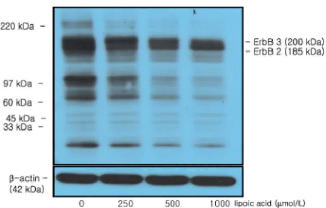
Fig. 2. Effect of α-lipoic acid on ErbB2 and ErbB3 protein expression in MDA-MB-231 cells. MDA-MB-231 cells were plated in a 100 mm dish at a density 1×10⁶ cells/dish with DMEM/F12 supplemented with 10% FDS for 48 h. The cells were then incubated in serum free medium for 24 h, after which they were then incubated in the presence of α-lipoic acid at concentrations of 0, 250, 500, or 1,000 μmol/L for three days. Equal amounts of cell lysates (30 μg) were then resolved by SDS-PAGE, transferred to a membrane and probed with ErbB2 (A) and ErbB3
(B). a) Photographs of chemiluminiscent detection of the blots, which were representative of three independent experiments. b) Quantitative analysis of the western blots. Each bar represents the mean ± S.D. of three independent experiments. Different letters indicate significant differences among groups at α
=0.05 as determined by Duncan's multiple range test.
Fig. 5. Effect of α-lipoic acid on Akt, p-Akt protein expression and mRNA expression in MDA-MB-231 cells. For Proteins expression, MDA-MB-231 cells were plated in a 100 mm dish at a density 1×10⁶ cells/dish with DMEM/F12 supplemented with 10% FDS for 48 h. The cells were then incubated in serum free medium for 24 h, after which they were incubated in the presence of α-lipoic acid at concentrations of 0, 250, 500 or 1,000 μmol/L for three days. Equal amounts of cell lysates (30 μg) were then resolved by SDS-PAGE, transferred to a membrane and probed with Akt (A) and p-Akt (B). a) Photographs of chemiluminiscent detection of the blots, which were representative of three independent experiments. b) Quantitative analysis of the western blots. For RT-PCR, total RNA was isolated and RT-PCR was performed to investigate the mRNA expression Akt (C). (a) Photographs of ethidium bromide-stained gel, which were representative of three independent experiments, are shown. (b) Quantitative analysis of RT-PCR. Relative abundance of each band was estimated by densitomertric analysis. Each bar represents the mean ± S.D. calculated from three independent experiments. Comparisons among different concentrations of the α-lipoic acid that yielded statistically significant differences among groups at α=0.05 as determined by Duncan's multiple range test.
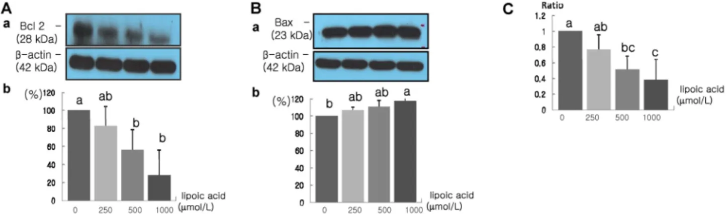
Fig. 6. Effect of α-lipoic acid on Bcl-2 and Bax protein expression in MDA-MB-231 cells. MDA-MB-231 cells were plated in a 100 mm dish at a density 1×10⁶ cells/dish with DMEM/F12 supplemented with 10% FDS for 48 h. The cells were then incubated in serum free medium for 24 h, after which they were incubated in the presence of α-lipoic acid at concentrations of 0, 250, 500 or 1,000 μmol/L for three days. Equal amounts of cell lysates (30 μg) were then resolved by SDS-PAGE, transferred to a membrane and probed with Bcl-2 (A) and Bax (B). a) Photographs of chemiluminiscent detection of the blots, which were representative of three independent experiments.
b) Quantitative analysis of western blots. The Bcl-2/Bax ratio was calculated (C). Each bar represents the mean ± S.D. of three independent experiments. Different letters indicate a significant difference among groups at α=0.05 as determined by Duncan's multiple range test.
proliferation after 12 and 24 h of incubation. However, incubation with α-lipoic acid at a concentration of 250 μmol/L or higher for 48 h led to a significant decrease in cell proliferation (P
< 0.05) (Fig. 1).
Inhibition of protein and mRNA expression associated with cancer cell proliferation by α-lipoic acid treatment
The protein expression of ErbB2 and ErbB3, which are epidermal growth factor receptors (EGFR) that control the cell growth signal of cancer cells, was decreased significantly in response to treatment with 250 μmol/L α-lipoic acid or higher (P < 0.05) (Fig. 2). In addition, when protein expression was observed using antibody Py20 to examine the tyrosine phosphorylation of these proteins, phosphorylated ErbB2 and ErbB3 were confirmed to be inhibited
as the concentration of the α-lipoic acid treatment increased (P
< 0.05) (Fig. 3). And the mRNA expression of ErbB2 and ErbB3
was decreased significantly in response to treatment with 500 μmol/L α-lipoic acid or higher (ErbB2) and 250 μmol/L α-lipoic acid or higher (ErbB3) (P < 0.05) (Fig. 4).
The expression of Akt protein, which receives the signal of the epidermal growth factor receptors to control cell proliferation, was found to be reduced significantly in response to treatment with 500 μmol/L α-lipoic acid or higher, whereas the protein expression of the active form of Akt (p-Akt) was reduced significantly in response to treatment with concentrations of 250 μmol/L and above (Fig. 5). The mRNA expression of Akt also was reduced significantly in response to treatment with 250 μ mol/L α-lipoic acid or higher (Fig. 5).
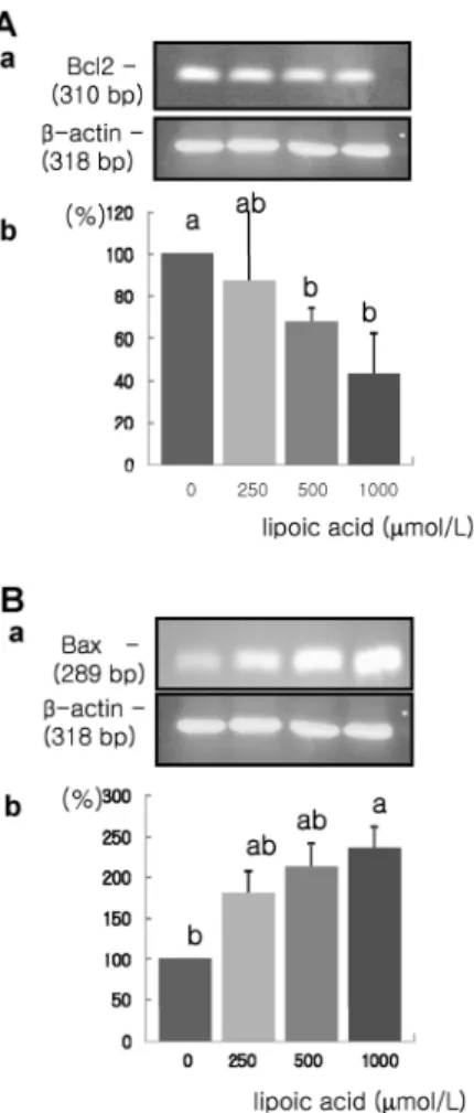
Fig. 7. Effect of α-lipoic acid on Bcl-2 and Bax mRNA expression in MDA-MB-231 cells. MDA-MB-231 cells were treated with α-lipoic acid. Total RNA was isolated and RT-PCR was performed to investigate the mRNA expression of Bcl-2 (A) and Bax (B). (a) Photographs of ethidium bromide-stained gel, which were representative of three independent experiments, are shown. (b) Quantitative analysis of RT-PCR. Relative abundance of each band was estimated by densitomertric analysis. Each bar represents the mean ± S.D. calculated from three independent experiments. Comparisons among different concentrations of the α-lipoic acid that yielded statistically significant differences among groups at α=0.05 as determined by Duncan's multiple range test.
Fig. 8. Effect of α-lipoic acid on caspase-3 activities in MDA-MB-231 cells. Each bar represents the mean ± S.D. of three independent experiments. Different letters indicate significant differences among groups at α=0.05 as determined by Duncan's multiple range test.
Induction of cancer cell apoptosis by α-lipoic acid treatment When treated with α-lipoic acid at concentrations of 500 μ mol/L and higher, the protein expression of Bcl-2 was decreased significantly, whereas the protein expression of Bax was increased significantly at concentration of 1,000 μmol/L α-lipoic acid (P < 0.05) (Fig. 6). The Bcl-2/Bax ratio, which was used as the index of cell apoptosis, was also significantly reduced in response to treatment with α-lipoic acid at concentrations of 500 μmol/L and above (P < 0.05).
And mRNA expression of Bcl-2 was decreased significantly, whereas the mRNA expression of Bax was increased significantly in response to treatment with 1,000 μmol/L α-lipoic acid (P <
0.05) (Fig. 7).
Increase of caspase-3 activity
The activity of caspase-3 was significantly higher in cells that were treated with 1,000 μmol/L α-lipoic acid (P < 0.05) (Fig. 8).
Discussion
In the present study, we investigated the effects of α-lipoic acid on the proliferation and apoptosis of MDA-MB-231 human breast cancer cells.
α-Lipoic acid was found to significantly inhibit the proliferation of breast cancer cells (Fig. 2) in this study. Vig-Varga et al.
(2006) reported that the proliferation of ovarian epithelial cancer cells separated from 8- week-old mice was significantly decreased by 75%, 85% and 100% in response to treatment with 10, 100, and 1000 μmol/L α-lipoic acid, respectively. Additionally, Pack et al. (2002) reported that treatment of Jurkat and CCRF-CEM human T lymphoma leukaemic cells with 0.01 mmol/L~4 mmol/L α-lipoic acid led to the dose-dependent inhibition of DNA replication and cell proliferation. Moreover, Packer (1998) reported that treatment of WM35 melanoma cells with 10~30 μmol/L α-lipoic acid inhibited cell growth, while Jeoung (2006) reported that the survival rate of B16F10 murine melanoma cells treated with 0.1~7.5 mmol/L α-lipoic acid was decreased in a dose-dependent manner in response to treatment with α-lipoic acid at concentrations of 2.5 mmol/L and above. Taken together, these findings indicate that α-lipoic acid inhibits the proliferation of a wide variety of cancer cells.
The epidermal growth factor receptors (EGFR) play an important role in cancer cell proliferation and differentiation.
EGFRs are composed ofErbB1/HER1, ErbB2/HER2, ErbB3/HER3, and ErbB4/HER4 (Bianco et al., 2007). The ErbB family receptors combine with an extracellular domain in homodimer and heterodimer forms (Perez-Nadales & Lloyd, 2004). Because ErbB3 cannot participate in intracellular reactions (Guy et al., 1994), it forms a dimer with ErbB2 for signal transduction (Xue et al., 2006). When phosphorylation of this receptor dimer occurs,
was found to be significantly reduced in response to treatment with α-lipoic acid treatment (Fig. 2, 4), which indicates that α -lipoic acid inhibits cancer cell proliferation via reduction in the excessive expression of EGFR s.
Akt is a serine/threonine kinase catalyzed by PI3K that inhibits cell apoptosis and promotes cell survival. Simbula et al., (2007) reported that treatment of FaO mice liver cancer cells with 500 μmol/L α-lipoic acid resulted in a reduction in the protein expression of the active form of Akt, p-Akt. Additionally, Laghero et al., (2007) found that treatment of HUVE cells with α-lipoic acid led to a reduction in the phosphorylation of the signal transduction substances Erk and Akt over time. In this experiment, we were also able to verify the inhibition of Akt expression by α-lipoic acid. Taken together, these results indicate that the inhibition of Akt protein and mRNA expression by α -lipoic acid resulted in the reduction of cell proliferation observed in this study.
There are several mechanisms involved in cancer cell apoptosis, including Bcl-2 family proteins and caspases (Alnemri, 1997;
Marsh et al., 2005). Simbula et al. (2007) reported that α-lipoic acid increased the protein expression of p27 and p21, and that this signal in turn promoted the expression of Bax and induced the release of cytochrome C into the cytoplasm, which led to a subsequent increase in caspase-3 activity, thereby inducing cell apoptosis. Their results are consistent with the results of the present study. Sen et al. (1999) reported that treatment of blood cancer cells with α-lipoic acid led to an increase in caspase-3 activity.
Moungjaroen et al. (2006) reported that α-lipoic acid treatment induced cell apoptosis of H460 lung cancer cells, and that this occurred via inhibition of the production of reactive oxygen species and increased expression of antioxidant enzymes such as superoxide dismutase by α-lipoic acid.
The results of our study indicate that α-lipoic acid exerts an inhibitory effect on cell proliferation via EGFRs and Akt signal transduction and induces cancer cell apoptosis in MDA-MB-231 human breast cancer cells. Overall, these results indicate that α -lipoic acid has the potential for use in the prevention of cancer.
References
Alnemri ES (1997). Mammalian cell death proteases: a family of highly conserved aspartate specific cysteine proteases. J Cell
(1994). Insect cell-expressed p180 possesses an impaired tyrosine kinase activity. Proc Natl Acad Sci U S A 91:8132-8136.
Hortbagyi GN, de la Garza Salazar J, Pritchard K, Amodori D, Haidinger R, Hudis CA, Khaled H, Liu MC, Martin M, Namer M, O’Shaughnessy JA, Shen ZZ & Albain KS (2005). The global breast cancer burden:variation in epidemiology and survival. Clin Breast Cancer 6:391-401.
Huang WY, Newman B, Milliken RC, Conway K, Hulka BS, Schell MJ & Liu ET (2000). Risk of breast cancer according to the status of HER-2/neu oncogene amplification. Cancer Epidemiol Biomarkers Prev 9:65-71.
Jeoung SY (2006). The effect of lipoic acid on antioxidant enzyme system in murine melanoma cells. Master thesis, SookMyung University, Seoul. Republic of Korea
Kang HJ, Kim SW, Yun YK, Oh SK, Choe KJ & Noh DY (2001).
Expression of p53, c-erbB2, bcl-2, Cathepsin D in lnfiltrating Ductal Cancer of the Breast. Jornal of the Korean Surgical Society 60:592-599.
Kim WK, Bang MH, Kim ES, Kang NE, Jung KC, Cho HJ & Park JHY (2005). Quercetin decreases the expression of ErB2 and ErB3
proteins in HT-29 human colon cancer cells. J Nutr Biochem 16:155-162.
Larghero P, Vene R, Minghelli S, Travaini G, Morini M, Ferrari N, Pfeffer U, Noonan DM, Albini A & Benelli R (2007). Biological assays and genomic analysis reveal lipoic acid modulation of endothelial cell behavior and gene expression. Carcinogenesis 28:1008-1020.
Lee HS, Seo EY & Kim WK (2004). Resveratrol induces apoptosis in SW480 human colon cancer cell lines. Food Sci Biotechnol 13:80-84.
Marsh SA, Laursen PB, Pat BK, Gobe GC & Coombes JS (2005).
Bcl-2 in endothelial cells is increased by vitamin E and alpha-lipoic acid supplementation but not exercise training. J Mol Cell Cardiol 38:445-451.
Ministry of Health and Welfare and Family (2008). Annual report of National Cancer Registration. https://u-lib.nanet.go.kr/dl/Simple View.php. Accessed on 8/14/2009.
Moungjaroen J, Nimmannit U, Callery PS, Wang L, Azad N, Lipipun V, Chanvorachote P & Rojanasakul Y (2006). Reactive oxygen species mediate caspase activation and apoptosis induced by lipoic acid on human lung epithelial cancer cell through Bcl-2 down regulation. J Pharmacol Exp Ther 319:1062-1069.
Olayioye MA, Neve RM, Lane HA & Hynes NE (2000). The ErbB signaling network: receptor heterodimerization in development and cancer. EMBO J 19:3159-3167.
Pack RA, Hardy K, Madigan MC & Hunt NH (2002). Differential effects of the antioxidant alpha-lipoic acid on the proliferation of mitogenstimulated peripheral blood lymphocytes and leukemic T cell. Mol Immunol 38:733-745.
Packer L (1998). alpha-Lipoic acid: a metabolic antioxidant which regulates NF-kappa B signal transduction and protects against oxidative injury. Drug Metab Rev 30:245-275.
Palacios J, Robles-Frias MJ, Castilla MA, Lopez-Garcia MA &
Benitez J (2008). The molecular pathology of hereditary breast cancer. Pathobiology 75:85-94.
Perez-Nadales E & Lloyd AC (2004). Essential function for ErbB3 in breast cancer proliferation. Breast Cancer Res 6:137-139.
Reed LJ, Debusk BG, Cunsalus IC & Hornberger CS Jr (1951).
Crystalline alpha-lipoic acid; a catalytic agent associated with pyruvate dehydrogenase. Science 27:93-94.
Riese DJ & Stern DF (1998). Specificity within the EGF family/ErbB receptor family signaling network. Bioessays 20:41-48.
Sen CK, Sashwati R & Packer L (1999). Fas mediated apoptosis of human Jurkat T-cells: intracellular events and potentiation by redox-active alpha-lipoic acid. Cell Death Differ 6:481-491.
Seo EY & Kim WK (2006). Effect of [6]-gingerol on bcle-2 and Bax expression in MDA-MB-231 human breast cancer cell line.
Journal of the Korean Society of Food Science and Nutrition
35:671-676.
Simbula G, Columbano A, Ledda-Columbano GM, Sanna L, Deidda M, Diana A & Pibiri M (2007). Increased ROS generation and p53 activation in alpha-lipoic acid -induced apoptosis of hepatoma cells. Apoptosis 12:113-123.
Vig-Varga E, Benson EA, Limbil TL, Allison BM, Geobl MG &
Harrington MA (2006). Alpha-lipoic acid modulate ovarian surface epithelial cell growth. Gynecol Oncol 103:45-52.
Wenzel U, Nickel A & Daniel H (2005). alpha-Lipoic acid induces apoptosis in human colon cancer cells by increasing mitochondrial respiration with a concomitant O2-*- generation. Apoptosis 10:359-368.
Xue C, Liang F, Mahmood R, Vuolo M, Wyckoff J, Qian H, Tsai KL, Kim MM, Locker J, Zhang ZY & Segall JE (2006).
ErbB3-Dependent Motility and Intravasation in Breast Cancer Metastasis. Cancer Res 66:1418-1426.
Zhai H, Chen X & Hu Z (2003). Study on the relationship between intake of trace elements and breast cancer mortality with chemometric methods. Comput Biol Chem 27:581-586.