Anthocyanin의 Delphinidin이 MDA-MB-231 유방암세포에 미치는 영향
장혜연1․이송희1․안인정1․이해님1․김혜리1․박영석1․박병권1․김병수1 김상기1․조성대2․남정석3․최창순4․정지윤1†
1공주대학교 특수동물학과, 2전북대학교 구강병리학교실
3가천의과학대학교 이길여암당뇨연구원, 4중앙대학교 식품공학부
Effects of Delphinidin in Anthocyanin on MDA-MB-231 Breast Cancer Cells
Hye-Yeon Jang1, Song-Hee Lee1, In-Jung An1, Hae-Nim Lee1, Hye-Ri Kim1, Young-Seok Park1, Byung-Kwon Park1, Byeong-Soo Kim1, Sang-Ki Kim1, Sung-Dae Cho2,
Jeong-Seok Nam3, Chang-Sun Choi4, and Ji-Youn Jung1†
1Dept. of Companion and Laboratory Animal Science, Kongju National University, Chungnam 340-702, Korea
2School of dentistry, Institute of Oral Bioscience, Chonbuk National University, Jeonbuk 561-756, Korea
3Lee Gil Ya Cancer and Diabetes Institute, Gachon University of Medicine and Science, Incheon 406-840, Korea
4School of Food Science and Technology, Chung-Ang University, Gyeonggi 456-756, Korea
ABSTRACT This research showed that delphinidin, an anthocyanin present in blueberry, induced apoptosis in MDA-MB-231 breast cancer cells as well as mediated anti-cancer effects in vivo. As a result, growth of cancer cells as assessed by MTT assay decreased in a concentration-dependent manner. Chromatin condensation by DAPI staining significantly increased in a concentration-dependent manner, indicating apoptosis. The level of p53-protein increased while those of anti-apoptotic molecules (Bcl-2, p-GSK3β) decreased in the western blot. Tumor size decreased in cells treated with 10 mg/kg of delphinidin compared with the control group in vivo. Cell apoptosis assessed by TUNEL assay significantly increased, and tumor inhibitory effect was confirmed. Therefore, delphinidin can be developed for cancer preventive medicine due to its growth inhibitory effects and induction of apoptosis in human breast cancer cells.
Key words: delphinidin, anthocyanin, anticancer, breast cancer, p53
Received 27 August 2013; Accepted 14 January 2014
†Corresponding author.
E-mail: [email protected], Phone: +82-41-330-1526
서 론
우리나라는 최근 10년간 암에 의한 사망률이 점차 증가하 는 추세이며, 그중에서도 유방암은 여성에게 발병률이 높은 암으로 보고되고 있다(1). 국내 유방암 발생빈도는 2011년 발표된 중앙 암 등록본부 자료에 의하면 2009년 전체 암 발생의 7%로 6위, 여성의 암 중에서 2위를 차지하였다(2).
유방암은 원발부위로부터 림프절이나 혈관을 타고 다른 곳으로 쉽게 이동하여 2차성 암을 형성하는 전이능력을 가 지며 특히 선택적으로 뼈나 폐로 전이가 잘된다고 알려졌는 데 이러한 전이성 유방암 환자들은 수술과 여러 치료요법으 로도 개선되기 힘들다(3,4).
최근까지 개발된 외과적 기술, 방사선 치료와 약물치료, 호르몬 요법 등과 같은 유방암 치료 방법은 암 환자의 생존 율을 다소 향상시키기는 하지만 일반적으로 알려진 항암 치 료는 강한 독성으로 인해 부작용이 크고 재발 위험도 큰 것
으로 보고되고 있다. 그에 따라 항암제를 통해 치료하는 기 존의 화학요법과 달리 부작용이 적으면서 인체에 미치는 고 통을 최소화하는 암 예방 및 치료 요법을 개발하는 데 사람 들의 관심이 증가되었다. 더불어 자연으로부터 쉽게 얻을 수 있고 부작용은 적으면서 암을 효율적으로 치료할 수 있 는, 식물 유래의 천연 물질들에 많은 관심을 두게 되었다.
대표적으로 블루베리의 항암효과가 대중매체를 통해 주목 받고 있다.
블루베리에 포함된 안토시아닌(anthocyanin)은 식물계 에 널리 분포되어 있는 페놀 화합물 중의 하나로 과실, 줄기, 잎, 뿌리 등 식물체 각 부위에 폭넓게 분포되어 있는 적색, 자색, 청색 등의 매력적인 색을 띠는 수용성 색소이다(5).
기능성인 측면에서 안토시아닌은 항산화 활성(6,7), 신경계 질환(8), 심장독성(9), 항염증(10) 등에 효과가 있으며 암 발생 억제(7,11), 항바이러스 작용(12,13), 지질대사 조절 (14,15), 시력(16) 및 난소 기능 증강효과(17) 등 생리활성 연구가 보고되어 있다.
특히 채소 및 과실류에서 안토시아닌 중 cyanidin이 50%
정도로 가장 많으며 delphinidin, pelargonidin, peonidin이 각각 12% 정도, petunidin과 malvidin은 7%로 분포하는데,
Fig. 1. Chemical structure of delphinidin.
그중 본 연구에 쓰인 delphinidin(Fig. 1)은 담자색, 적자색 및 청색을 나타내고 항산화 작용을 비롯한 콜레스테롤 저하 와 항암 및 항염증(18), DNA 손상의 보호(19,20) 등에 효과 가 있는 것으로 알려졌다. 또한 시력과 기억력이 좋아지고 비만을 억제하여 심장, 뇌혈관 질환과 당뇨병 및 관절염에 효과가 있다고 알려졌다(21). 채소과실류에 가장 많이 분포 하고 있는 cyanidin에 관한 연구는 활발하게 진행되고 있는 반면에 delphinidin의 생리적 기능성 효과가 petunidin과 malvidin에 비하여 더 우수한 것으로 알려졌음에도 불구하 고 현재 이에 대한 연구는 미비한 실정이다.
그리하여 본 연구에서는 블루베리에서 항암효과를 나타 낼 것으로 기대되는 delphinidin이 인간 유래의 MDA-MB- 231 유방암세포의 증식을 억제하는지 확인하고, in vivo 실 험에서도 항암효과가 나타나는지 알아보고자 하였다.
재료 및 방법
시험물질
Delphinidin chloride는 Sigma-Aldrich(St. Louis, MO, USA)에서 구입하여 in vitro 실험에서는 DMSO에 희석하여 사용하였고, in vivo 실험에서는 D・W증류수에 희석하여 사 용하였다.
MDA-MB-231 유방암세포배양
실험에 사용된 MDA-MB-231 유방암세포는 American Type Culture Collection(Rockville, MD, USA)에서 구입 하였다.
MDA-MB-231 유방암세포 배양을 위해 RPMI-1640배 지, 5% fetal bovine serum(FBS; Life Technologies Inc., Gaithersburg, MD, USA)과 항생제(1% penicillin-strep- tomycin 0.525 mL)를 첨가하여 37°C, 5% CO2가 유지되는 배양기(Thermo Electron Corp., Marietta, OH, USA) 내 에서 세포를 배양하였다. 세포가 80~90% 정도 flask를 덮 으면 phosphate buffered saline solution(PBS)으로 2번 씻어내고 trypsin-EDTA를 처리하여 세포를 계대 배양하고 배지는 2~3일마다 교환하였다.
MTT assay
MDA-MB-231 유방암세포를 96 well plate의 각 well당 세포수가 2.5×10⁴ cells/mL가 되도록 넣고 plates에 붙을 수 있도록 RPMI 1640 성장배지에서 약 24시간 동안 배양했 다. 그러고 나서 그 성장배지는 제거하고 delphinidin을 0, 12.5, 25, 50 µM 농도 조건으로 처리하였다. 37°C, 5% CO2
가 유지되는 배양기 내에서 약 24시간 동안 배양하였다. 그 후 각 well당 40 µL의 PBS에 2 mg/mL의 MTT를 녹인 용액 을 넣고 호일로 감싼 후 37°C 배양기에 2시간 동안 방치하 였다. 다음으로 MTT 용액을 제거하고 well당 100 µL의 DMSO를 첨가하여 형성된 formazan을 녹인다. 완전히 용 해된 formazan의 흡광도를 ELISA-reader(Bio-Rad Lab- oratories Inc., Hercules, CA, USA)로 595 nm에서 측정하 였다.
DAPI stain
체외 배양중인 MDA-MB-231 유방암세포의 블루베리 처리 시 세포의 핵 변화를 시각화하기 위하여 DNA염기 서 열에 특이적으로 반응하는 DAPI 핵 염색을 수행하여 핵의 변화를 분석하였다. 96 well plates에 MDA-MB-231 유방 암세포(2×10⁵ cells/mL)를 넣은 후 37°C 배양기에서 배양 하였다. 처리 시간이 끝나면 배양기에서 배양접시를 상온으 로 꺼낸 후 PBS로 조심스럽게 세척한 후에 4% paraf- ormaldehyde solution으로 15분간 고정하였으며, PBS로 다시 세척 후 PBS로 10배 희석한 DAPI stain 용액을 500 µL/well 처치하였다. 처치 후 암실에서 형광현미경(Zeiss fluorescence microscope, Thornwood, NY, USA)으로 관찰하였다.
Western blot
175T flask에 MDA-MB-231 유방암세포를 24시간 배 양한 후 delphinidin을 0, 25 µM 농도로 24시간 동안 처리하 였다. 그 후 trypsin-EDTA를 처리하여 세포를 부유상태로 만들어 원심분리(1,000 rpm, 5 min, 4°C)하였다. 차가운 PBS로 세포를 세척하고 원심분리하여 cell pellet에 cell lysis buffer(Invitrogen, Carlsbad, CA, USA)를 첨가하여 4°C에서 30분간 반응시켰다. Lysate는 13,000 rpm에서 5 분 동안 원심분리하여 상등액을 취해 cell lysate로 사용하 였다. 추출한 단백질의 농도는 Bradford protein assay를 이용해 측정하였다. 단백질을 12% sodium dodecyl sul- fate polyacrylamide gel electrophoresis(SDS-PAGE)로 크기별로 분리한 후 nitrocellulose membranes(Bio-Rad Laboratories Inc.)에 이동시켰다. Membrane은 5% skim milk로 1시간 동안 blocking한 후 anti-Bcl-2, anti-p53, anti-p-GSK3β, anti-β-actin의 1차 항체를 각각 첨가하여 4°C에서 overnight 하였다. 그 후 anti-rabbit IgG를 첨가 하여 1시간 반응시켰다. 각 protein band는 ECL detection reagents(Pierce, Rockford, IL, USA)를 이용하여 실험결
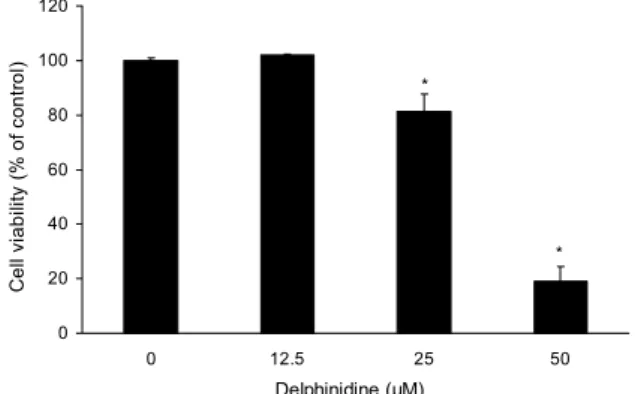
0 20 40 60 80 100 120
0 12.5 25 50
Delphinidine (µM)
Cell viability (% of control) .
*
*
Fig. 2. The effects of delphinidin on the cell viability in MDA- MB-231 cells. MDA-MB-231 cells were treated with delphini- din for 24 hr, and the cell viability was determined by MTT assay as described in Materials and Methods. Datas are mean±
standard deviation (SD) for three samples. The significance was determined by Student's t-test (*P<0.05 compared with untreated control).
과를 보았다.
Xenograft
37°C, 5% CO2가 유지되는 배양기에서 5% FBS를 첨가 한 RPMI-1640 배지에 MDA-MB-231 유방암세포를 배양 하였다. 75T flask에 80~90%의 세포가 배양되었을 때 trypsin-EDTA를 첨가하여 세포를 부유시켜 원심분리 (1,200 rpm, 5 min, 4°C)하였다. 그 후 PBS로 세척 후 다시 한 번 원심분리(1,200 rpm, 3 min, 4°C)하여 cell pellet을 얻어 1×10⁷ cells/mL가 되도록 하여 RPMI-1640 배지에 분주하였다. (주)오리엔트(Seoul, Korea)에서 5주령의 nude mice 암컷 10마리를 구입하여 1주일의 순화기간을 보낸 nude mice 등에 200 µL(2×10⁷ cells/mL)씩 MDA-MB- 231 유방암세포를 주입하여 종양을 유발시켰다. 종양조직 이 어느 정도 자라면 nude mice를 마취시켜 종양을 적출한 후 1 mm3 크기로 절편하여 다시 nude mice에 주입하였다.
그 다음 종양 사이즈가 군별로 일정하게 나오도록 군 분류를 한 후 실험군 3마리는 delphinidin을 선행연구논문(22)에서 생체실험에서 효과가 있는 것으로 판단되는 최저농도를 선 정하여 D・W증류수 200 µL에 희석시켜 매주 3번 10 mg/kg 을 같은 시간에 4주간 경구투여 하였고, 대조군은 D・W증류 수 200 µL를 매주 3번 같은 시간에 4주간 경구투여 하였다.
투여기간 동안 마우스의 일반적인 상태를 살피고 종양사이 즈는 주 2회 측정하였다.
TUNEL assay
Dead End™ Colorimetric TUNEL System(Promega, Madison, WI, USA)를 이용한 TUNEL assay를 통해 apoptosis 세포를 확인하였다. 먼저 xylene을 이용해 탈파 라핀화를 거친 후 ethanol을 100, 95, 85, 70, 50% 순서로 이용하여 함수시켰다. 그 후 PBS로 세척 후 Proteinase K를 각 슬라이드에 떨어트려 실온에서 15분간 반응시키고, equilibration buffer와 Biotinylated Nucl. Mix, rTDT를 섞어 각 슬라이드에 처리하여 37°C에서 1시간 반응시켰다.
그 후 0.3% hydrogen peroxide를 PBS와 섞어 5분간 반응 후 Streptavidin HRP를 각 슬라이드에 처리하였고, 각 슬라 이드에 3,3'-diaminobenzidine tetrahydrochloride(DAB) solution을 10분 반응시켰다. 마지막으로 methyl green으 로 염색 후 광학현미경으로 관찰하였다(×200).
통계처리
모든 실험결과는 평균치와 표준오차를 사용하여 나타내 고 각 군간 비교는 one-way ANOVA에 이은 t-test분석을 실시하였다. 대조군과 비교하여 P 값이 0.05 미만일 때를 통계학적으로 유의성이 있다고 판정하였다.
결과 및 고찰
Delphinidin이 MDA-MB-231 유방암세포 생존에 미치는 영향
Delphinidin이 MDA-MB-231 유방암세포 생존에 미치 는 영향을 확인하기 위하여 delphinidin을 농도(0, 12.5, 25, 50 µM)별로 처리하여 MTT assay을 통해 관찰하였다.
Delphinidin을 각각 0, 12.5, 25, 50 µM로 24시간 처리하였 을 때 각각 농도별 세포 생존율은 100, 100, 80, 20%로 대조군에 비해 25 µM의 농도에서부터 농도 의존적으로 세 포 생존에 대하여 유의적인 감소를 보였다(Fig. 2). 따라서 delphinidin이 MDA-MB-231 유방암세포에 25 µM부터 농 도 의존적으로 세포사멸을 유도하는 것으로 판단되었다.
Yun 등(23)의 연구에 의하면 HCT116 cell에 delphinidin 을 30, 60, 120 µM로 처리하였을 때 농도 의존적으로 감소 한다고 나타났으며, Kim 등(6)에서는 Jurkat T cell에서 100 µg/mL, MCF-7 cell에서 125 µg/mL 감소하는 것으로 나타났다. Pal 등(24)의 연구에서도 A549, NHBE, NCI- H441, SK-MES-1 cell에 delphinidin을 농도별로 처리한 결과 NHBE cell을 제외하고 나머지 cell에서 20 µM의 농도 에서부터 농도 의존적으로 감소를 보였다. 이러한 결과는 본 연구에서 delphinidin이 MBA-MD-231 유방암세포에 농도 의존적으로 세포생존율을 감소시킨 것과 같은 결과로 나타나 delphinidin이 암세포에 억제효과를 보이는 것으로 사료된다.
Delphinidin에 의한 MDA-MB-231 유방암세포의 apoptosis 효과
MTT assay을 통해 관찰된 세포사멸이 apoptosis에 의 해 나타난 결과인지 확인하기 위해 DAPI stain을 실시하였 다. MDA-MB-231 유방암세포에 delphinidin을 농도(0, 12.5, 25, 50 µM)별로 24시간 처리 후 DAPI 염색을 이용하
Fig. 3. Effect of delphinidin on the chromatin condensation in MDA-MB-231 cells. MDA-MB-231 cells were treated with delphinidin for 24 hr and stained with DAPI.
0 25 (μM) p-GSK3β
p53
Bcl-2
β-actin
Fig. 4. Effect of delphinidin on apoptosis protein. Cells were treated with dephinidin (0, 25 µM and 24 hr). Cell lysates were prepared as described in the Materials and Methods and ana- lyzed by 12% SDS-PAGE followed by western blotting. The membranes were incubated with anti-p-GSK3β, anti-Bcl-2 and anti-p53 antibodies. The blots were also probed with anti-β-actin antibodies to confirm equal sample loading.
여 형광현미경으로 관찰하였다(Fig. 3). 그 결과 delphinidin 을 25 µM 처치한 투여군에서부터 apoptosis 발현세포의 증가가 관찰되었으며, MTT assay 결과에서 농도 의존적으 로 세포의 성장이 감소한 것과 동일하게 DAPI 염색에서도 대조군과 비교하였을 때 delphinidin을 처리한 군에서 세포 수가 줄어들었고 세포질의 위축, 염색사의 응축, apoptotic body와 같은 apoptosis형태가 관찰되었다. Hafeez 등(25) 의 연구에서도 PC3 cell에 대한 delphinidin의 apoptosis를 현미경으로 관찰한 결과 60 µmol/L에서 apoptosis의 발현 을 관찰하였고 120, 180 µmol/L로 농도가 증가할수록 발현 이 증가한 것으로 보고하였다. 이러한 결과로부터 delphi- nidin이 MDA-MB-231 유방암세포에 대한 세포사멸이 apoptosis 유도에 의해 이루어지는 것으로 여겨진다.
Delphinidin이 apoptosis의 경로 단백질 발현에 미치는 영향
이전 실험에서 MDA-MB-231 유방암세포에 delphini- din을 25 µM 처치한 투여군에서부터 apoptosis 양성세포의 유의적인 증가가 관찰되었으므로 Western blot을 통해 apoptosis를 조절하는 p53, Bcl-2, p-GSK3β 단백질의 유 전자 발현 정도를 관찰하여 delphinidin이 apoptosis 발현 유전자의 역할에 어떠한 영향을 미치는지 확인하였다. p53 은 종양억제 유전자의 산물로서 세포 증식을 억제하며 유전 자가 손상되었을 경우 복구하거나 괴사를 유도하고, apop- tosis를 유발하는 종양억제 유전자로 알려졌다(26). West- ern blot을 실시하여 delphinidin 처리에 따른 p53 단백질의 발현 여부를 관찰한 결과, p53 단백질은 대조군에 비해 del- phinidin의 처리농도 25 µM에서 증가하였고, anti-apop- totic 인자로서 apoptosis 유발을 억제하는 Bcl-2, p-GSK3 β 단백질의 비율은 25 µM에서 유의적으로 감소하였다(Fig.
4). Yun 등(23)의 연구에서는 HCT116 cell에 delphinidin 을 처리하였을 때 30 µM에서부터 농도 의존적으로 p53 단 백질이 증가하였고 Bcl-2 단백질은 감소한 것으로 나타났 으며, Hafeez 등(25)의 연구에서는 PC3 cell에 delphinidin 을 60, 120, 180 µmol/L로 처리하였을 때 p21, p27은 농도 의존적으로 증가하였고 pNF-κB/p50과 pNF-κB/p65가 농 도 의존적으로 감소하였다. 결과적으로 delphinidin이 MDA-
MB-231 유방암세포의 apoptosis 유도에 매우 중요한 역할 을 하고 있음을 알 수 있다.
Delphinidin이 xenograft를 이용해 이식한 종양에 미치는 영향
Xenograft를 실시한 마우스의 MDA-MB-231 유방암세 포 증식에 delphinidin이 어떠한 영향을 미치는지 관찰하였 다. 종양사이즈는 주 2회 측정하였고, delphinidin(10 mg/
kg)은 D・W증류수 200 µL에 희석시켜 주 3회 같은 시간에 4주 경구투여하였고, 대조군은 D・W증류수 200 µL를 주 3 회 같은 시간에 4주 경구투여하였다. 그 결과 경구투여 11일 째부터 대조군과 비교하였을 때, 종양의 사이즈가 감소하는 경향을 보였다. 25일째 대조군에 비해 delphinidin을 처리 한 군에서 43% 정도의 억제율을 보였다(Fig. 5A). 최종 종 양의 크기는 대조군은 평균 3,953 mm3, delphinidin 투여군 은 1,566 mm3이었으며, 종양의 무게는 대조군 1.575 g, delphinidin 투여군은 1.008 g으로 delphinidin 투여군의 종 양이 감소하는 경향을 보였다(Fig. 5B). Hafeez 등(25)의 연구에서는 PC3 cell로 xenograft를 실시한 마우스에 del-
0
1000 2000 3000 4000 5000 6000 7000
1 4 8 11 15 18 22 25
Day after tumor implantation
Tumor volume (mm³) .
control 10mg/kg
Control 10 mg/kg
0 0.5 1 1.5 2 2.5 3
Control 10 mg/kg
Concentration
Tumor weight (g) .
(A) (B)
(C)
Control 10 mg/kg
Fig. 5. Inhibition of MDA-MB-231 breast tumor growth and increase of apoptosis in MDA-MB-231 breast tumors by delphinidin.
Xenograft experiment was performed as described in the Material and Methods. (A) To identify the effect of delphinidin in MDA- MB-231 breast tumor growth, nude mice were treated with delphinidin (0, 10 mg/kg) for 25 days (n=5, *P<0.05). (B) The graph expresses final tumor weight. The data are presented as the mean±standard deviation (SD) of five mice, *P<0.05. (C) Nude mice were treated delphinidin (0, 10 mg/kg) of 25 days. Tumor tissues were observed using microscope and photographed (×200). Paraffin- embedded tumors were sectioned into 5 μm. Scale bar size: 10 μm.
phinidin을 주 3회, 2 mg을 복강투여하여 8주 동안 관찰한 결과 최종 종양의 크기는 대조군은 1,200 mm3, delphinidin 투여군은 361 mm3로 높은 억제율을 나타내었고, delphini- din 투여군의 종양크기가 12주가 되었을 때 대조군의 1,200 mm3와 비슷해졌다. 이러한 결과는 본 연구에서 나타난 결과 와 일치하여 delphinidin이 MDA-MB-231 유방암세포에 대한 억제효과를 보이는 것으로 판단되어진다.
Delphinidin이 미치는 조직수준에서의 apoptosis 관찰 Xenograft 부검을 통해 채취한 종양조직에 대하여 apoptosis 유도에 의한 항암효능을 확인하고자 Tunel as- say를 실시하였다. 그 결과 대조군보다 10 mg/kg delphi- nidin을 투여한 군에서 apoptosis가 일어난 세포의 유의적 증가를 관찰할 수 있었다(Fig. 5C). Anthocyanin 성분들의 암에 대한 연구 중 가장 분포가 많은 cyanidin에 대한 연구 로, Chu 등(27)의 연구에서는 cyanidin이 MDA-MB-231 유방암세포의 이동성과 침윤성에 미치는 영향을 조사했는 데 cyanidin의 첨가 농도가 세포 증식에 영향을 주지 않는 0, 5, 10, 20 μM에서 이동성 및 침윤성이 유의적으로 감소되 었고, Zhang 등(28)의 연구에서는 AGS cell, HCT-116 cell, MCF-7 cell, NCI-H460 cell, SF-268 cell에 cyani- din을 100 μM 이상으로 처리했을 때 유방암세포인 MCF-7
에서 세포 증식이 유의적으로 감소되었다고 보고하였다.
Cyanidin, malvidin에 관한 Choi(29)의 연구에서는 cyani- din과 malvidin이 산화적 스트레스에 의한 WI-38 cell의 지질 과산화 생성을 유의적으로 감소시켰으며 이를 통해 cyanidin, malvidin의 산화적 스트레스 개선을 통한 항노화 효과가 뛰어남을 확인할 수 있었다. 그리고 Kim 등(6)의 연 구 결과에 따르면 anthocyanin 함량 중 특히 cyanidin 함량 은 타 계통보다 2배 이상 높았고, 암세포에 대한 독성효과로 JurKat T 세포와 MCF-7 세포에 cyanidin, delphinidin, petunidin을 농도별로 처리한 결과 petunidin은 cyanidin에 비해 조금 떨어지지만, delphinidin은 cyanidin에 뒤지지 않 는 암세포 독성효과가 있는 것으로 나타났다. 이러한 결과는 본 연구에서 delphinidin이 MDA-MB-231 유방암세포에 농도 의존적으로 세포생존율을 감소시킨 것과 같은 결과로 나타나 delphinidin이 암세포에 억제효과를 보이는 것으로 사료된다.
그동안 delphinidin, petunidin에 비해 cyanidin의 효과 가 부각된 것은 대부분 식물이 함유하고 있는 anthocyanin 이 cyanidin인 까닭에 그동안의 연구 결과는 cyanidin 중심 으로 이루어진 것으로 생각된다. 따라서 cyanidin을 포함한 anthocyanin의 개별 성분에 대한 더 깊은 연구가 필요할 것으로 판단된다. 본 연구는 anthocyanin의 성분 중 항암
가능성이 있을 것으로 여겨지는 delphinidin이 MDA-MB- 231 유방암세포에 미치는 영향을 알아보았고, delphinidin 이 MDA-MB-231 유방암세포에 대해 apoptosis를 유발시 켜 암세포 증식을 억제한다는 것을 확인하여 delphinidin이 암 예방제나 치료제로 개발될 수 있을 것으로 사료된다.
요 약
본 연구에서는 블루베리에 포함된 anthocyanin 중 delphi- nidin이 인간 유래의 MDA-MB-231 유방암세포의 성장을 억제시키고 apoptosis를 유발하는지 살펴보고 in vivo 실험 에서도 항암효과가 나타나는지 확인하였다. 그 결과 cell viability를 보기 위한 MTT assay에서는 농도 의존적으로 암세포의 성장을 억제시켰으며, apoptosis의 확인을 위한 DAPI stain에서 농도 의존적으로 chromatin condensation 이 유의적으로 증가하는 것을 확인하였다. 또한 western blot에서 암 억제 유전자인 p53 단백질이 증가하였고, an- ti-apoptotic 분자인 Bcl-2 단백질과 p-GSK3β 단백질은 감소하였다. In vivo 실험에서는 대조군과 비교해 10 mg/kg delphinidin을 투여한 군에서 종양의 크기가 감소하였으며, TUNEL assay를 통해 apoptosis 세포 또한 통계학적으로 유의적인 증가가 관찰되어 종양 억제 효과를 확인하였다.
이상의 결과들로 볼 때, MDA-MB-231 유방암세포에서 delphinidin은 암세포의 증식을 억제시키고, apoptosis를 유발시키는 효과를 보이므로 암 예방제나 치료제로 개발될 수 있을 것으로 사료된다.
감사의 글
본 연구는 농촌진흥청의 연구비 지원으로 수행된 연구 결과 의 일부이며(Project NO. PJ007884), 이에 감사드립니다.
REFERENCES
1. Nam MK. 2011. The effect of red cabbage extract (Brassica Oleracea L. Var. Capitata F. Rubra) on the apoptosis in hu- man breast cancer MDA-MB-231 cells. PhD Dissertation.
Duksung Women's University, Seoul, Korea.
2. National cancer center. Cancer Incidence Table. http://www.
cancer.go.kr/mbs/cancer/subview.jsp?id=cancer_040102000 000 (accessed May 2012).
3. Block KI, Gyllenhaal C, Tripathy D, Freels S, Mead MN, Block PB, Steinmann WC, Newman RA, Shoham J. 2009.
Survival impact of integrative cancer care in advanced meta- static breast cancer. Breast J 15: 357-366.
4. Gupta GP, Minn AJ, Kang Y, Siegel PM, Serganova I, Cordon-Cardo C, Olshen, AB, Gerald WL, Massague J.
2005. Identifying site-specific metastasis genes and functions.
Cold Spring Harb Symp Quant Biol 70: 149-158.
5. Koo BS, Song DS. 2010. Extraction of natural red color pig- ment concentrate and manufacturing characteristics of pig- ment powder from purple-fleshed sweet potato. Korean J Food & Nutr 23: 23-29.
6. Kim YH, Kim DS, Woo SS, Kim HH, Lee YS, Kim HS, Ko KO, Lee SK. 2008. Antioxidant activity and cytotoxicity on human cancer cells of anthocyanin extracted from black soybean. Korean J Crop Sci 53: 407-412.
7. Park EH. 2012. Anthocyanins prevent helicobacter pylori-in- duced inflammation on gastric epithelial Cells. PhD Disser- tation. Gyeongsang National University, Jinju, Korea.
8. Choung MG, Lim JD. 2012. Antioxidant, anticancer and immune activation of anthocyanin fraction from Rubus cor- eanus Miquel fruits (Bokbunja). Korean J Medicinal Crop Sci 20: 259-269.
9. Kim YK. 2012. Anthocyanin extracts from black soybean (Glycine Max L.) protect human glial cells against oxygen- glucose deprivation by promoting autophagy. PhD Disser- tation. The Catholic University, Seoul, Korea.
10. Choi EY. 2009. Protective effect of anthocyanin-rich bil- berry (Vaccinium myrtillus L.) extract against doxorubicin- induced cardiotoxicity. PhD Dissertation. Ewha Womans University, Seoul, Korea.
11. Kang HH. 2009. Determination of biological activities of Korean berries and their anthocyanin identification. PhD Dissertation. Gyeongsang University, Jinju, Korea.
12. Lacombe A, Tadepalli S, Hwang CA, Wu VC. 2013. Phyto- chemicals in lowbush wild blueberry inactivate Escherichia coli O157:H7 by damaging its cell membrane. Foodborne Pathog Dis 10: 944-950.
13. Chung TH, Kim JC, Lee CY, Moon MK, Chae SC, Lee IS, Kim SH, Hahn KS, Lee IP. 1997. Potential antiviral ef- fects of Terminalis chebula, Sanguisorba officinalis, Rubus coreanus and Rheum palmatum against duck hepatitis B vi- rus (DHBV). Phytotherapy Res 11: 179-182.
14. Wu T, Tang Q, Gao Z, Yu Z, Song H, Zheng X, Chen W. 2013. Blueberry and mulberry juice prevent obesity de- velopment in C57BL/6 mice. PLoS One 8: e77585.
15. Tsuda T, Horio F, Uchida K, Aoki H, Osawa T. 2003.
Dietary cyanidin 3-O-beta-D-glucoside-rich purple corn col- or prevents obesity and ameliorates hyperglycemia in mice.
J Nutr 133: 2125-2130.
16. Liu Y, Song X, Zhang D, Zhou F, Wang D, Wei Y, Gao F, Xie L, Jia G, Wu W, Ji B. 2012. Blueberry anthocyanins:
protection against ageing and light-induced damage in reti- nal pigment epithelial cells. Br J Nutr 108: 16-27.
17. Kim BS, Park YK, Kang BS. 2001. The effect of RUBI FRUCTUS on the ovulation and ovary in rats. PhD Disser- tation. Dongguk University, Seoul, Korea.
18. Hyeong KE. 2013. Suppression of induced regulatory T cell by metabolites of delphinidin chloride. PhD Dissertation.
Chungang University, Seoul, Korea.
19. Liu W, Lu X, He G, Gao X, Xu M, Zhang J, Li M, Wang L, Li Z, Wang L, Luo C. 2013. Protective roles of Gadd45 and MDM2 in blueberry anthocyanins mediated DNA repair of fragmented and non-fragmented DNA damage in UV-ir- radiated HepG2 cells. Int J Mol Sci 14: 21447-21462.
20. Liu W, Lu X, He G, Gao X, Li M, Wu J, Li Z, Wu J, Wang J, Luo C. 2013. Cytosolic protection against ultraviolet in- duced DNA damage by blueberry anthocyanins and antho- cyanidins in hepatocarcinoma HepG2 cells. Biotechnol Lett 35: 491-498.
21. Ko EK. 2009. Delphinidin chloride effects on the expression of cell adhesion molecules. PhD Dissertation. Chonnam University, Gwangju, Korea.
22. Jin X, Yi L, Chen ML, Chen CY, Chang H, Zhang T, Wang L, Zhu JD, Zhang QY, Mi MT. 2013. Delphinidin-3-gluco- side protects against oxidized low-density lipoprotein-induced
mitochondrial dysfunction in vascular endothelial cells via the sodium-dependent glucose transporter SGLT1. PLoS One 8: e68617.
23. Yun JM, Afaq F, Khan N, Mukhtar H. 2009. Delphinidin, an anthocyanidin in pigmented fruits and vegetables, in- duces apoptosis and cell cycle arrest in human colon cancer HCT116 cells. Mol Carcinog 48: 260-270.
24. Pal HC, Sharma S, Strickland LR, Agarwal J, Athar M, Elmets CA, Afaq F. 2013. Delphinidin reduces cell pro- liferation and induces apoptosis of non-small-cell lung can- cer cells by targeting EGFR/VEGFR2 signaling pathways.
PLoS One 8: e77270.
25. Hafeez BB, Siddiqui IA, Asim M, Malik A, Afaq F, Adhami VM, Saleem M, Din M, Mukhtar H. 2008. A dietary antho- cyanidin delphinidin induces apoptosis of human prostate
cancer PC3 cells in vitro and in vivo: involvement of nuclear factor-kappaB signaling. Cancer Res 68: 8564-8572.
26. Ryu MJ, Chung HS. 2011. Effects on hot water extract of Schizandra chinensis on colon cancer cell line. Food Engineering Progress 15: 64-69.
27. Chu SK, Seo EY, Kim WK, Kang NE. 2008. Effect of cya- nidin on cell motility and invasion in MDA-MB-231 human breast cancer cells. Korean J Nutr 41: 711-717.
28. Zhang Y, Vareed SK, Nair MG. 2005. Human tumor cell growth inhibition by nontoxic anthocyanidins, the pigments in fruits and vegetables. Life Sci 76: 1465-1472.
29. Choi MJ. 2008. Study on anti-oxidant and anti-aging effect of balck rice with protective mechanism. MS Thesis. Pusan University, Busan, Korea.