©2008 The Korean Nutrition Society
Anthocyanin계 성분인 Cyanidin이 인체 유방암세포 MDA -MB-231의 이동성과 침윤성에 미치는 영향
*추수경1)·서은영2)·김우경2)·강남이3)§
을지대학교 간호학과,1) 단국대학교 식품영양학과,2) 을지대학 식품영양학과3)
Effect of Cyanidin on Cell Motility and Invasion in MDA -MB-231 Human Breast Cancer Cells
*Chu, Su Kyoung1)·Seo, Eun Young2)·Kim, Woo Kyoung2)·Kang, Nam E3)§ Department of Food and Nutrition,1) Daejeon University, Daejeon 701-214, Korea
Department of Nursing,1) Eulji University, Seongnam 461-713, Korea Department of Food Science & Nutrition,2) Dankook University, Yongin 448-701, Korea
Department of Food & Nutrition,3) Eulji University, Seongnam 461-713, Korea
ABSTRACT
Anthocyanidins, the aglycones of anthocyanins, are natural colorants belonging to the flavonoid family. Cyanidin is one of the anthocyanidins, used for their antioxidant properties. Furthermore, previous studies have shown anthocyani- din-rich material extracts or aglycone form inhibit growth and induce apoptosis of cancer cells. But, Tumor metastasis is the most important cause of cancer death, and various treatment strategies have targeted on preventing the occurrence of metastasis. This study investigated the effects of cyanidin on metastasis processes, including motility, invasion and activity of MMP-2 and MMP-9 in MDA-MB-231 human breast cancer cell lines. We cultured MDA-MB-231 cells in presence of various concentrations 0, 5, 10 and 20 μM of cyanidin. The cell motility was significantly decreased dose- dependently in cells treated with cyanidin (p < 0.05) and cyanidin treatment caused the significant suppression of the invasion (p < 0.05). MMP-2 and MMP-9 activities, and MMP-9 mRNA express were not affected by anthocyanin treat- ment. In conclusion, cyanidin inhibits cell motility, invasion in MDA-MB-231 human breast cancer cell lines. (Korean J Nutr 2008; 41(8): 711 ~717)
KEY WORDS: cyanidin, metastasis, motility, invasion, breast cancer.
서 론
암의 발병 원인 중에서 80% 정도가 환경적인 요인에 의 해 발생하며, 이 중에서도 식생활에 의한 암 발생이 35%
정도를 차지하고 있다고 한다.1,2) 여러 암 중에서도 유방암, 자궁암, 전립선암 등의 호르몬 관련 암과 위암과 대장암 등 의 소화기관 관련 암 등에서 식사 요인이 암 발병의 중요한 위험인자로 지목되고 있어 이러한 암들과 식품 섭취와의 관 련성에 대한 연구가 활발히 이루어지고 있다.3) 특히 유방암 의 경우 우리나라에서 유방암으로의 사망률이 1995년에 비
해 2005년에는 무려 1.75배 정도 증가하였다.4)
암은 원발 조직에서 빠르게 성장, 증식하여 혈액을 통해 다른 조직으로 전이하게 되는데 암 환자에게 있어 전이는 치료의 어려움과 사망의 위험을 증가시키는 원인이 된다.5) 암세포의 침윤 (invasion)과 전이 (metastasis)는 원발 병 소로부터 다른 장기로 이동하면서 일어나는 세포 진행의 한 형태로써, 복합적이고 연속적인 분자들의 기전에 의해 일어 난다.
특히 암세포로부터 분비되는 단백질 분해 효소에 의해 빠른 시간 내에 세포외 기질이 파괴되는 것이 특징이다.6) 암 전이의 첫 번째 단계는 침윤 과정으로 초기 종양으로부 터 전이성 암세포가 떨어져 나와 주위의 세포외 기질 (ex- tracellular matrix, ECM)에 부착하게 된다. 유착한 종양세 포가 가수분해 효소를 분비하여 용해된 matrix 간격을 통해 이동한 후 성장인자를 자극하여 빠르게 성장하고 신생혈관 을 형성하게 된다.7) 암의 전이는 이러한 과정이 연속적으로 접수일:2008년 09월 2일 / 수정일:2008년 9월 20일
채택일:2008년 11월 8일
*This work was supported by the Eulji University Research Fund, 2007
§To whom correspondence should be addressed.
E-mail:[email protected]
진행되어 이루어지게 된다.8)
최근 여러 역학 연구에서 천연 과일과 채소에 많이 함유 되어 있는 색소 성분들이 항산화 능력을 가지고 있어, 이러 한 식물성 식품을 섭취하면 여러 암의 위험이 감소될 수 있 는 것으로 보고되고 있다.9) 특히 anthocyanin은 식물계에 광범위하게 분포되어 있는 천연 색소10,11)로써 최근 많은 연 구가 진행되고 있다. Anthocyanin은 flavonoid 중의 하나 로 베리류, 적포도, 적색 감자, 적색 양배추와 같은 색이 짙 은 과일과 채소에 함유되어 있으며 효능으로는 산화적 스 트레스와 관련된 질병을 예방하고,12) 항염증,13) 항산화,14-16) 항돌연변이,17) 항암작용을 하는 것으로 보고되고 있다.18,19) Anthocyanin을 가수분해하면 당 부분 (단당류, 소당류)과 비당 부분인 anthocyanidin으로 분해된다.20) 비당 부분인 anthocyanidin은 cyanidin, delphinidin, malvidin, pelago- nidin, peonidin 등 약 20종으로 나눌 수 있으며, 이 중 cya- nidin이 항산화력이 크다고 보고되고 있다.21) 그러나 지금까 지의 cyanidin 관련 연구는 대부분이 aglycon 형태를 사용 하였으며 유방암에서의 효과 연구는 많이 되어있지 않다.
본 연구에서는 cyanidin이 전이성이 강한 인체 유방암 세포 인 MDA-MB-231의 이동성과 침윤성, 침윤 단계에서 분비 되는 단백분해효소인 matrix matalloproteinase-2 (MMP-2), matrix matalloproteinase-9 (MMP-9)의 활성에 미치는 영 향을 알아보고자 하였다.
재료 및 방법
세포 배양
실험에 사용된 인체 유방암 세포 MDA-MB-231 cell은 American Type Culture Collection (ATCC, Rockville, MD, USA)에서 구입하였다. 세포는 습윤한 5% CO2, 37℃
incubator에서 Dulbecco’s modified Eagle’s medium/
Nutrient Mixture Ham’s F12 (DMEM/F12, Gibco/BRL.
Gaithersburg, MD, USA), 10% FBS, 100 units/mL pen- icillin, 100 μg/mL streptomycin (Gibco/BRL)이 포함되 도록 medium을 만들어 배양하였다.22) 세포가 80~90% 정 도 dish를 덮으면 phosphatate buffered saline solution (PBS)으로 2번 씻어내고 trypsin-EDTA (Gibco/BRL)를 처리하여 세포를 모은 후 계대 배양하고 medium은 2~3일 마다 교환하였다.23) Cyanidin (Roth, Karlsruhe, Germa- ny)은 dimethyl sulphoxide (DMSO, Fisher scientific)에 100 mM로 stock을 만들어 냉동 보관하여 사용하였고 대 조군을 포함하여 모든 well에는 DMSO의 농도가 동일하게 포함되도록 하였다.
세포 증식
Cyanidin의 첨가 농도 증가에 따른 세포의 성장을 알아 보기 위해 24 well plate에 2.5 × 10⁴ cells/mL의 농도로 plating 하고, 48시간 후에 fetal bovine serum (FBS)을 첨가하지 않은 serum free medium (SFM, 0.1% BSA, 5 μg/mL transferrin, 5 ng/mL selenium, 1,000 units/mL penicillin, 1,000 μg/mL streptomycin, Gibco)으로 me- dium을 교환하여 24시간 serum starvation 시키고, SFM 에 cyanidin을 0, 5, 10, 20 μM로 처리한 군과 0, 25, 50, 100 μM로 처리한 군으로 medium을 교환하였다.24) Treat- ment 후 0, 12, 24시간이 경과한 후 Trypan blue (Sigma) 로 염색하여 hemocytometer로 세포의 수를 측정하였다.25)
세포 이동성 (Boyden chamber motility assay)
세포의 motility를 측정하기 위해 boyden chamber as- say를 실시하였다. Low chamber에는 attractant로 10%FBS medium을 26 μL씩 분주하고 미리 gelatin 용액으로 코팅된 filter를 올린 후 upper chamber와 단단히 고정하 였다. 세포는 trypsin-EDTA로 세포를 모은 후 2 × 10⁶ cells/mL로 만들고 cyanidin을 농도별로 처리하여 upper chamber에 55 μL씩 분주하여 37℃ CO2 incubator에서 12시간 배양하였다. 배양이 끝난 후 filter의 윗부분을 종이 타올로 닦아내고 Diff-quick solution으로 염색한 후 filter 아래로 이동한 세포면이 아래로 가도록 하여 유리판에 올려 놓고 건조시켰다. 전자 현미경을 이용하여 임의로 5곳을 지 정하여 세포 수를 측정하였다.26)
세포 침윤성 (Matrigel invasion assay)
냉동 보관된 matrigel chamber를 실온이 되도록 한 후 SFM을 well과 insert에 500 μL씩 넣어 2시간 재수화 시 켰다. 24 well plate에 세포 농도를 1 × 10⁶ cells/mL로 0.5 mL씩 넣었다. 48시간 후에 SFM으로 교체한 후 다시 24 시간 후에 cyanidin을 0, 25, 50, 100 μM로 처리하였다. 재 수화된 well에는 attractant로 10% FBS medium을 750 μL 첨가하고 insert를 얹은 후 insert에는 treatment된 cell 을 500 μL씩 분주하여 37℃ incubator에서 12시간 배양 시켰다. 배양이 끝난 후 upper chamber의 matrigel을 통과 하지 못한 insert의 cell을 면봉으로 닦아내었다.27) 통과된 cell을 Diff-quick stain solution을 이용하여 염색하고 현미 경을 이용하여 임의의 5곳을 지정하여 세포수를 측정하였다.
MMPs의 활성
Cell을 1 × 10³ cells/mL로 6 well plate에 분주하고 48 시간 후 PBS로 1번 세척한 후 SFM로 2번 세척하고 농도
별 treatment 용액을 well당 1.1 mL씩 분주하였다. 12시간 동안 배양시킨 후 medium을 15 mL tube에 모으고, 3,000 rpm에서 10분 원심분리하고 2 mL centricon에 나누어 넣 어 3,000 rpm에서 30분간 농축하였다. 농축된 medium은 -70℃에 보관하면서 실험에 이용하였다. 1% gelatin이 들 어있는 polyacrylamide에 sample을 각 well에 넣고 전기 영동하였다. 전기 영동된 gel은 renaturing buffer로 30분 씩 2번 세척하고 developing buffer로 30분간 세척한 후 37℃ shaker에서 48시간 배양시켰다. 0.25% commassie blue 용액으로 12시간 염색하고 destain solution (meth- anol: acetic acid: water = 50:10:40)으로 10분간 de- staining 한 후 증류수로 microwave에서 5분씩 2번을 세 척하여 band를 확인하였다.28)
mRNA 발현
Cyanidin이 mRNA 수준에서 MMPs를 변화시키는지 알 아보기 위해 실험하였다. 100 mm dish에 1 × 10⁵ cells/
mL 농도로 cell을 분주하고 48시간 배양 후 SFM으로 med- ium을 교환하였다. 24시간 후에 cyanidin 0, 5, 10, 20 μM 로 처리한 SFM으로 48시간 배양하여 medium을 제거하 고 Trireagent 1 mL를 첨가하여 scraper로 cell를 분리하 였다. cDNA는 Superscript Ⅱ reverse transcriptase (Invitrogen) 0.5 μL를 첨가하여 합성하였다. cDNA 증폭 (amplifica tion)은 2.5 λ mix buffer (10 × PCR buffer, 50 mM MgC12, 10 mM NTP mix, autoclave water)와 sense, antisense primer taq polymerase를 혼합하여23) forward (5’-TGG GCTACGTGACCTATGACCAT-3’),
reverse (5’-GCCCA GCCCACCTCCACTCCTG-3’) 55 ℃ 에서 1분간 35사이클을 수행하였다. Ethidium bromide로 염색한 1% argarose gel로 전기영동을 실시하여 결과를 확인하였다.29)
통계 처리
본 연구의 실험으로 얻어진 결과는 SAS 프로그램을 이용 하여 각 실험군의 평균과표준 편차로 계산되었고, 각 군 간 의 차이는 ANOVA 분석 후 α = 0.05 수준에서 Duncan’s multiple range test를 실시하여 유의성을 검증하였다.30)
결 과
세포 증식
인체 유방암 세포인 MDA-MB-231에 cyanidin을 0, 25, 50, 100 μM로 처리하고 0, 12, 24시간 후에 살아있는 세 포를 염색하여 세포의 수를 측정한 결과, 12시간 배양하였 을때 첨가 농도가 증가함에 따라 유의적으로 세포의 성장 이 억제되었다 (p < 0.05)(Fig. 1A). 본 실험에서는 cya- nidin이 세포 증식을 억제하여 이동성과 침윤성에 영향을 미치지 않는 실험 조건을 정하기 위하여 cyanidin의 처리 농 도를 25 μM 이하인 0, 5, 10, 20 μM 농도로 처리하고 배 양시간을 12, 24시간 후로 다시 실험하였다. 결과는 cya- nidin을 0, 5, 10, 20 μM 농도로 처리하고 배양시간을 12, 24시간 후에는 세포의 증식에 아무런 영향을 미치지 않았다 (p < 0.05)(Fig. 1B). 그러므로, 본 실험에서는 cya- nidin이 증식 억제작용과는 독립적으로 이동성과 침윤성에
140
120
100
80
60
40
20
0
0 12 24 Time (hr)
Cell number (103 cell/mL)
NS
a
a a
b a
a ab b 00 25
50 100 (μM)
A
140
120
100
80
60
40
20
0
0 12 24 Time (hr)
Cell number (103 cell/mL)
NS 00 25 50 20 (μM)
NS
NS
B
Fig. 1. Effect of cyanidin on cell growth in MDA-MB-231 cells. MDA-MB-231 cells were plated at a density of 2.5 × 10⁴ cells/mL in 24 well plate with DMEM/F12 supplemented with 10% FBS for 48 hour, the monolayers were serum-starved with DMEM/F12 supple- mented with 5 μg/mL transferrin, 5 μg/mL selenium, and 1 mg/mL bovine serum albumin for 24 hour. After serum starvation, the mo- nolayer were incubated in serum free medium with 0, 5, 10, 20 μM cyanidin (A) and 0, 25, 50, 100 μM (B). The cell numbers were counted for hematocytometer. Each bar represents the mean ± S.D. from three independent experiments. Comparison among dif- ferent concentrations of cyanidin that yielded significant differences (p < 0.05) are indicated by different letters above each bar.
미치는 영향을 알아보기 위해서 cyanidin이 서로 증식에 영 향을 미치지 않도록 처리 농도를 25 μM 이내로 하였고, 배 양시간은 24시간 이내로 결정하여 실험을 수행하였다.
세포 이동성
세포의 이동성을 알아보기 위해 boyden chamber assay 를 실시하였다. 0.01% gelatin으로 코팅되어 8 μm의 구멍 을 가지고 있는 filter를 암세포가 통과하여 이동하는 정도 를 알아본 boyden chamber assay 결과는 cyanidin을 0, 5, 10, 20 μM로 12시간 처리하였을 때는 42%, 52.4%, 64.1%
로, 유의적으로 성장력이 억제되었다 (p < 0.05)(Fig. 2).
세포 침윤성
침윤성 실험은 생체의 기저막 성분을 화학적으로 조합한 matrigel을 이동하는 세포를 측정하는 것으로 gelatin이 코 팅된 filter를 통과하는 boyden chamber motility assay와 차이점이 있다. 세포를 12시간 배양시킨 후에 matrigel로
코팅된 filter를 통과한 세포는 cyanidin의 첨가 농도가 0, 5, 10, 20 μM에서 35.8%, 50%, 64.3%로 침윤성이 유의 적으로 감소되었다 (p < 0.05)(Fig. 3).
MMPs의 활성과 mRNA 수준에서의 발현
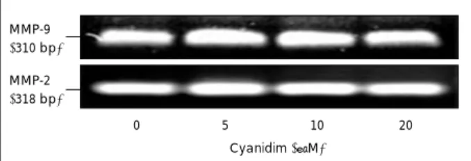
Gelatin zymography는 gelatin이 함유한 polyacryla- mide gel상에서 시료에 포함된 단백분해효소에 의해 gel- atin이 분해되는 gel의 정도로 MMP의 활성 정도를 알아보 는 방법이다. 본 실험에서는 MMP-2, MMP-9의 활성을 알 아본 결과 MMP-2, MMP-9 모두 cyanidin의 처리 농도에 따라 활성의 차이가 나타나지 않았으며 (Fig. 4), MMP-9의 mRNA 발현도 cyanidin에 의해 영향을 받지 않았다 (Fig. 5).
고 찰
암은 초기 발병을 막는 것도 중요하지만, 암 발병 이후 다
(%) 120
100
80
60
40
20
0
0 5 10 20 Cyanidin (μM) a
b
c d
B A
Fig. 3. Effects of cyanidin on invasion in MDA-MB-231 cells. The cells were cultured in the presence of various concentrations of cyanidin for 12 hour with invasion chamber. A: Microphotography of cells treated with cyanidin on invasion chamber. B: Quan- titative analysis of invasion assay. Each bar represents the mean ± S.D. from three independent experiments. Comparisons among different concentration of cyanidin that yielded significant difference (p < 0.05) are indicated by different letters above each bar.
Fig. 2. Effects of cyanidin on cell motility in MDA-MB-231 cells. The cells were cultured in the presence of various concentrations of cyanidin for 12 hour with boyden chamber. A: Microphotography of cells treated with cyanidin on cell migration assay. B: Quan- titative analysis of cell motility assay. Each bar represents the mean ± S.D. from three independent experiments. Comparisons among different concentration of cyanidin that yielded significant difference (p < 0.05) are indicated by different letters above each bar.
(%) 120
100
80
60
40
20
0
0 5 10 20 Cyanidin (μM) a
b b
c
B A
른 장기로의 전이를 차단시키는 것이 암으로 인한 사망률 을 줄일 수 있는 데에 매우 중요한 의의를 갖는다. 그러므로 본 연구에서는 cyanidin이 인체 유방암 세포인 MDA-MB- 231의 전이 과정 중 이동성 (motility), 침윤성 (invasion), 침윤 시 암세포에서 분비되는 단백 분해 효소인 MMP-2, MMP-9의 활성과 mRNA 수준에서의 발현에 미치는 영향 을 관찰하였다.
Yanjun 등31)의 연구에서는 AGS (stomach cancer cell), HCT-116 (colon cancer cell), MCF-7 (breast cancer cell), NCI-H460 (lung cancer cell), SF-268 (central nervous cancer cell) cell에 cyanidin을 0~200 μM로 처리하여 48시간 배양 후에 MTT assay를 실시한 결과, cyanidin 을 100 μM 이상으로 처리했을 때 유방암 세포인 MCF- 7에서 세포 증식이 유의적으로 감소되었다고 보고하였다.
Ye 등32)의 보고에서는 포도 씨에서 추출한 proanthocya- nidin을 MCF-7 cell에 50 μM로 처리하여 24, 48, 72시 간 배양한 후에 MTT assay를 실시한 결과 각각 11, 35, 47% 세포 증식을 억제하였다고 하였다. 본 연구 결과에서 도 cyanidin을 0, 5, 10, 20 μM로 처리하고 12, 24시간 배 양 시에는 cyanidin이 세포의 증식에 영향을 미치지 않았으 나, cyanidin의 농도를 높여 0, 25, 50, 100 μM로 처리하 였을 때는 배양 시간 12시간 이후부터 세포 증식을 유의적 으로 억제하는 것으로 관찰되었다. 이러한 결과로 본 연구 의 실험 조건은 cyanidin을 20 μM 이내로 처리하였고, 배 양 시간은 24시간 이내로 정하여 세포의 이동성과 침윤성 에 미치는 영향을 알아보았다.
세포의 이동성에 anthocyanin이 미치는 영향을 알아본 연구로는 Sylvie 등33)이 glioblastoma U-87 cell에 7개의 anthocyanidin (cyanidin, delphinidin, malvidin, pelargo- nidin, peonidin, petunidin)과 1개의 anthocyanin (del- phinidin 3-O-beta-glucopyranoside)을 25 μM로 처리하 여 4시간 후에 이동성을 측정한 결과 petunidin, cyanidin,
delphinidin 처리군에서 유의적으로 이동성이 감소하였으며 이 3가지 처리 물질의 농도를 0~50 μM로 달리했을 때 cya- nidin은 처리 농도 20 μM 이상에서 유의적으로 세포의 이 동을 억제하였다. Chen 등34)은 Huh-7, HeLa, SCC-4 cell 에 흑미 (O. sativa L. indica)에서 추출한 peonidin-3-glu- coside와 cyanidin-3-glucoside를 0~100 μM로 처리하 였을 때 peonidin-3-glucoside는 100 μM 이상, cyanidin- 3-glucoside는 50 μM 이상에서 이동성이 감소되었다고 하 였다. 또한, Min 등9)에 따르면 lung A549 cell에 cyanidin- 3-glucoside을 0, 40, 80 μM로 처리하여 이동성을 알아보 기 위해 wound healing assay를 실시한 결과 80 μM 이 상에서 세포의 이동이 억제되었으며, Chen 등35)도 A549에 cyanidin-3-glucoside를 처리하였을 때 이동성이 억제되었 다고 하였다. 이들의 결과를 본 연구 결과와 비교해 볼 때 cyanidin을 포함한 anthocyanin 색소 성분은 여러 종류 암 세포의 이동성을 억제하는 효과가 있는 것으로 보여 진다.
Morini 등36)은 전이성이 강한 종양세포에서는 α3β1 inte- grin의 증가로 세포의 침윤성과 이동성이 증가되어 암세포 가 전이하게 된다고 하였는데 유방암 세포 MDA-MB-231, MDA-MB-145, MCF-7 중에서도 MDA-MB-231 cell에서 α3β1 integrin 발현이 가장 높게 나타나 세포의 침윤성과 이동성을 증가시켜 전이를 유발한다고 하였다. Chen 등34) 은 흑미에서 추출한 peonidin 3-glucoside와 cyanidin 3- glucoside를 Huh-7, HeLa, SCC-4에 0~100 μM로 처리하 였을 때 Huh-7 cell에서 peonidin 3-glucoside 100 μM 이 상, cyanidin 3-glucoside는 50 μM 이상으로 처리하였을 때 암세포의 이동성에서와 같이 침윤성 또한 억제하였다고 보고하였다. Emma 등37)은 colon HT115 cell에 rasberry 에서 추출한 poly- phenol 물질을 0~50 μM로 처리하여 세 포의 침윤성을 알아본 결과 6.25 μM 이상에서 침윤성이 감소되었으며 가장 높은 농도인 50 μM로 처리하였을 때는 control에 비해 90% 이상 침윤성을 감소시켰다고 보고하 였다. Nagase 등38)은 가지에서 추출한 anthocyanidin계 색 소 중 delphinidin을 fibrosarcoma HT-1080 cell에 처리
MMP-9 92 kDa
72 kDa MMP-2
Marker 0 5 10 20 Cyanidim (μM)
Fig. 4. Effect of cyanidin on MMP-2 and MMP-9 activity in MDA- MB-231 cells. MDA-MB-231 cells were plated in 6-well plates at a density 1 × 10⁶ cell/well with DMEM/F12 supplemented with 10% FBS for 12 hour; the monolayers were incubated in serum- free medium in the absence or presence of 0, 5, 10, 20 μM cya- nidin for 12 hour. Medium were collected, and the activities of MMP-2 and MMP-9 were measured by zymography. Photograph of the MMP bands is shown.
MMP-9 (310 bp)
MMP-2 (318 bp)
0 5 10 20 Cyanidim (μM)
Fig. 5. Effect of cyanidin on MMP-9 mRNA expression in MDA- MB-231 cells. For RT-PCR, MDA-MB-231 cells were treated with cyanidin. Total RNA was isolated and RT-PCR was performed to investigate the mRNA expression of MMP-9. Photograph of ethidium bromide-stained gel is shown.
하였을 때 세포가 기저막 성분인 matrigel을 통과하는 것이 감소되었다고 하였다. 이들의 결과로 볼 때 cyanidin은 암 세포의 침윤성을 억제하는 효과가 있는 것으로 보여 진다.
Tumor invasion에서 extracellular matrix를 변형, 침식 시키는데 MMPs라는 단백질 분해 효소가 중요한 역할을 한다.39) MMP는 대부분 숙주 기질 세포에 의해 만들어지고 그 발현은 암세포의 chemokines, cytokines, extracel- lular matrix metalloproteinase inducer에 의해 조절 된 다.40) MMP의 기저막 분해에 중요한 역할을 하는 MMP-2 와 MMP-9의 경우 gelatine과 collagen에 대해 친화성이 있으며 단백질 가수분해에 의해 활성화 된다.41) Chen 등37) 은 A549 human lung carcinoma cell에 mulberry에서 추 출한 cyanidin-3-glucoside, cyanidin-3-rutinoside를 처리 하였을 때 MMP-2, Upa 활성이 억제되었다고 하였으며, Na- gase 등38)도 human fibrosarcoma HT-1080 cell에 del- phinidin을 처리하였을 때 MMP-2, MMP-9의 활성이 억제 되었다고 보고하였다. Chen 등37)도 human hepatocellular carcinoma SKHep-1에 흑미에서 추출한 peonidin 3-glu- coside, cyanidin 3-glucoside를 0~100 μM로 처리하였을 때 peonidin 3-glucoside는 50 μM, cyanidin 3-gluco- side 25 μM 이상에서 MMP-9의 활성이 유의적으로 감소 되었으며, human tongue squamous cell SSC-4에서는 pe- onidin 3-glucoside는 25 μM, cyanidin 3-glucoside 50 μM 이상에서 MMP-2 활성이 유의적으로 감소되었다. Mat- chett 등42)은 blueberry에서 추출한 anthocyanin을 hu- man prostate cancer cell DU-145에 처리하였을 때 MMP- 2, MMP-9의 활성이 감소되었다고 하였다. 본 연구 결과는 MMPs 활성과 mRNA 수준에서의 발현에는 영향을 주지 않는 것으로 보아, breast cancer cell인 MDA-MB-231 cell 에서 cyanidin은 MMP-2, MMP-9의 활성과는 다른 기전에 의해 암세포의 이동성과 침윤성이 영향을 받는 것으로 사 료된다.
요 약
본 연구는 식물의 대표적인 색소 성분인 anthocyanin계 색소 중 cyanidin이 인체 유방암 세포 MDA-MB-231에서 세포의 이동성과 침윤성에 미치는 영향을 알아보고자 실시 되었다. 실험 결과 cyanidin의 첨가량이 증가할수록 세포 의 운동성, 이동성, 침윤성이 유의적으로 억제되었다. 그러 나 ECM 분해 시 암세포에서 분비되는 단백질 분해 효소인 MMP-2, MMP-9의 활성과 MMP-9의 mRNA 수준은 cya- nidin에 의해 영향 받지 않는 것으로 관찰되었다. 결론적으
로, cyanidin은 세포 증식에 영향을 미치지 않는 범위 내에 서 암세포의 전이과정 중 이동성과 침윤성을 억제할 수 있 다는 가능성이 관찰되었으나, MMPs 활성에는 영향을 미치 지 않는 것으로 보아 MMPs 이외의 경로를 통해 전이 과정 이 이루어지는 것으로 사료된다.
Literature cited
1) Huh KB. The present status of nutrition related diseases and its countermeasures. Korean J Nutr 1990; 23(3): 197-207 2) Ryu YS. A study on dietary factor related to the incidence of
breast cancer and of cervix cancer in Korea women [MS thesis].
Seoul: Ewha Womans University; 1992
3) Kim WK, Park EH, Han WK. A follow up study on colorectal cancer. Korean J Surg Soc 1986; 31(2): 191-199
4) Korea National Statistical Office. Deaths by cause; 2005 5) Woodhouse EC, Chuaqui RF, Liotta LA. General mechanism of
metastasis. Cancer 1997; 80: 1529-1537
6) Stetler-Stervenson W, Aznavoorian S, Liotta L. Tumor cell inter- actions with the extracellular matrix during invasion and metas- tasis. Annu Rev Cell Dev Biol 1993; 9: 541-573
7) Bang MH. Effects of some phytochemicals on cell invasion and matrix metalloproteinase (MMP) activity in MDA-MB-231 hu- man breast cancer cells [MS thesis]. Seoul: Dankook university;
2004
8) Suh CS, Moon SY. Apoptosis and expression of bcl-2, bax in the cyclic human endometrium. Korean J Obstet Gynecol 1999; 45 (11): 2446-2457
9) Min D, Rentian F, Shiow YW, Linda B, Yongju L, Yong Q, Vin- cent C, Bing HJ, Xianglin S. Cyanidin-3-glucoside, a natural prod- uct derived from blackberry, exhibits chemopreventive and che- motherapeutic activity. J Biol Chem 2006; 25: 17359-17368 10) Harbone JB. Comparativebiochemistry of the flavonoids. Aca-
demic Press; 1967
11) Bae , Moon . A study of the antioxidatives of Korean soybeans. J Korean Soc Food Sci Nutr 1997; 26: 203-208
12) Wang H, Cao G, Prioi RL. Oxygen radical absorbing capacity an- thocyanins. J Agric Food Chem 1999; 45: 304-309
13) Wagner H. Annual proceedings of phytochemical society of Eu- rope 1985; 25: 409
14) Ichiyanagi T, Hatano Y, Matsugo S, Konishi T. Comparison of an- thocyanin reactivity towards hydroxyl radical, superoxide anion and singlet oxygen. ITE Lett 2003; 4: 788-793
15) Ichiyanagi T, Hatano Y, Matsugo S, Konishi T. Stimultaneous com- parison of reactive nitrogen species. Chem Pham Bull(Tokyo) 2004;52: 1312-1325
16) Kahkonen MP, Heinonen M. Antioxidant activity of acthocya- nins and their aglycones. J Agric Food Chem 2003; 51: 628-633 17) Yoshimoto M, Okuno S, Yamaguchi M, Yamakawa O. Antimuta- genicity of deacylated anthocyanins in purple-fleshed sweetpotato.
Biosci Biotechnol Biochem 2001; 65: 1652-1655
18) Hou DX. Potential mechansm of cancer chemoprevention by an- thocyanins. Curr Mol Med 2003; 3: 149-159
19) Katsube N, Iwashita K, Tsushida T, Yamaki K, Kobori M. Induc- tion of apoptosis in cancer cells by billberry or blueberry (Vacci- nium myrtillus) and the anthocyanins. J Agric Food Chem 2003;
51: 68-75
20) Yoon TH, Lee SW. Stability of anthocyanins in foods. J Korean Food Sci Technol 1979; 11: 63-73
21) Kong JM, Chia LS, Goh N, Chia T, Brouillard R. Analysis and biological activities of anthocyanins. Phytochemistry 1993; 64:
923-933
22) Weng MS, Ho CT, Ho YS, Lin JK. Theanaphthoquinone inhibits fatty acid synthase expression in EGF-stimulated human breast cancer cells via regulation of EGFR/EerB-2 signaling. Toxicol appl pharmacol 2007; 218: 107-118
23) Francesco C, Roberto A, Raffaella C, Antonia S, Fabio V. Effect of cyanidin-3-O-glucoside on UVB-induced response in human keratinocytes. J Agric Food Chem 2006; 54: 4041-4047 24) Seo EY, Lee HS, Kim WK. Effect of [6]-gingerol on onhibition
of cell proliferation in MDA-MB-231 human breast cancer cells.
Korean J Nutr 2005; 38(8): 656-662
25) Landesberg R, Cozin M, Cremers S, Woo V, Kousteni S, Sinha S, Garrett-Sinha L, Raghavan S. Inhibition of oral mucosal cell wound healing by bisphosphonates. J Oral Maxillofac Surg 2008; 66 (5): 839-847
26) Taraboletti G, Roberts D, Liotta L. Thrombospondin-induced tu- mor cell migration: haptotaxis and chemotaxis are mediated by different molecular domains. J Cell Biology 1987; 105: 2409-2415 27) Aye MM, Ma C, Lin H, Bower KA, Wiggins RC, Luo J. Ethanol- induced in vitro invasion of breast cancer cells: the contribution of MMP-2 by fibroblasts. Int J Cancer 2004; 112: 738-746 28) Unemori EN, Werb Z. Reorganization of polymerized action; a
possible trigger for induction of procollagenase in fibroblasts cul- tured in and on collagen gels. J Cell Biol 1986; 103(3): 1021-1031 29) Bartsch JE, Staren ED, Appert HE. Matrix metalloproteinase ex-
pression in breast cancer. J Surg Res 2003; 110: 383-392 30) Bang MH, Kim JH, Kim WK. Effects of epigallocatechin gallate
on adhesion, invasion and matrix matalloproteinase activity in MDA-MB-231 human breast cancer cells. Korean J Nutr 2005;
38(2): 104-111
31) Yanjun Z, Vareed Shaiju KV, Muraleedharan GN. Human tumor cell growth inhibition by nontoxic anthocyanidins, the pigments
in fruits and vegetables. Life Sci 2005; 76: 1465-1472
32) Ye X, Krohn RL, Liu W, Joshi SS, Kuszynski CA, McGinn TR, Bagchi M, Preuss HG, Stohs SJ, Bagchi D. The cytotoxic effects of a novel IH 636 grape seed proanthocyanidin extract on cultured human cancer cells. Mol Cell Biochem 1999; 196(1-2): 99-108 33) Sylvie L, Rene L, Valerie B, Albert M, Stephane B, Denis G, Ri-
chard B. Anthocyanidins inhibit migration of glioblastoma cells:
structure-activity relationship and involvement of the plasminoly- tic system. J Cell Biochem 2007; 100: 100-111
34) Chen PN, Kuo WH, Chiang CL, Chiou HL, Hsieh YS, Chu SC.
Black rice anthocyanins inhibit cancer cells invasion via repres- sions of MMPs and u-PA expression. Chem Biol Interact 2006;
163: 218-229
35) Chen PN, Chu SC, Chiou HL, Kuo WH, Chiang CL, Hsieh YS.
Mulberry anthocyanidin 3-rutinoside and cyanidin 3-glucoside, exhibited an inhibitory effect on the migration and invasion of a human lung cancer cell line. Cancer Let 2006; 235(2): 248-259 36) Morini M, Mottolese M, Ferarri N, Ghiorzo F, Buglioni S, Mor- tarini R. The α3β1 integrin is associated with mammary carcino- ma cell metastasis, invasion and gelatinase B (MMP-9) activity.
Int J Cancer 2000; 87: 336-342
37) Emma MC, Gina P, Chris IG, Mark M, Gordon JM, Derek S, Ian R. Colon-available raspberry polyphenols exhibit anti-cancer ef- fects on in vitro models of colon cancer. Carcinogenesis 2007; 6 (4): 1-13
38) Nagase H, Sasaki K, Kito H, Sato T. Inhibitory effect of delphi- nidin from solanum melongena on human fibrosarcoma HT-1080 invasiveness in vitro. Planta Med 1998; 64(3): 216-219 39) Polette M, Nawrocki-Raby B, Gilles C, Clavel C, Birembaut P.
Tumor invasion and matrix metalloproteinase. Crit Rev Oncol Hematol 2004; 49(3): 179-186
40) Nagase H. Activation mechanism of matrix metalloproteinase.
Biol Chem 1997; 378: 151-160
41) Glasspool RM, Twelves CJ. Matrix metalloproteinase inhibitors:
past lessons and future prospects in breast cancer. Breast 2001; 10 (5): 368-378
42) Matchett MD, Mackinnon SL, Sweeney MI, Gottschall-Pass KT, Hurta RA. Blueberry flavonoids inhibit matrix metalloproteinase activity in DU145 human prostate cancer cells. Biochem Cell Biol 2005; 83(5): 637-643