Journal of Korean Arthroscopy Soc.
Volume 11, Number 1, June, 2007
서 론
자기 공명 영상의 발달로 관절 내 병변 진단의 정확성이 발 달되고 용이해 졌다. 그러나 일부 반월상 연골판의 비정상적 소견은 자기 공명 영상에서 확인되지 않는 경우가 있고, 특히 내측 반월상 연골판 전각부의 비정상적인 소견의 경우 자기 공명 영상에서 확인되지 않는 경우가 많이 있다3). 이러한 경 우 자기 공명 영상만으로는 진단이 되지 않아 슬내장증으로 분류하고 간과하거나, 정확한 진단을 위해 진단적 관절경 술 식을 시행하기도 한다. 본 증례에서는 자기 공명 영상상 특별 한 병변이 발견되지 않은 좌측 슬관절 통증에 대해 진단적 관 절경 술식을 시행하여, 내측 반월상 연골판 전각부의 비정상 적 띠를 발견하였고, 관절경을 이용하여 이를 제거하였다.
증례 보고
11세 여환은 약 4 개월간 지속된 좌측 슬관절의 통증을 주 소로 내원하였다. 주 증상은 계단을 오르내릴 때에 발생하는 좌측 슬관절의 내측부 통증이며, 이학적 검사상 슬관절 전내 측부에 압통과 Mcmurray검사상 외회전에 양성 소견을 보였 으나 관절운동의 제한은 없었다. 그러나 슬관절 굴곡과 신전 시 불편감을 호소하였다.
단순 방사선 촬영상 특이한 소견은 없었으며, 자기 공명 영 상 검사 역시 특별한 소견은 관찰되지 않았다(Fig. 1). 지속되 는 증상에 해당하는 자기 공명 영상 소견이 없어 진단적 관절 경을 시행하였다.
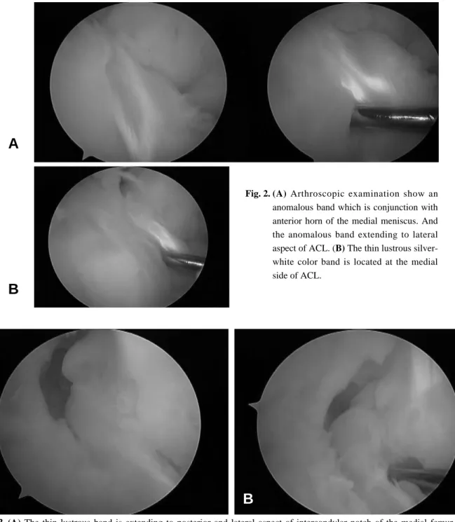
관절경 소견상 내측 반월상 연골판의 전각부 전외측에서 비정상적인 띠가 기시되어 전방십자인대 내측 옆을 지나 대 퇴골 과간 절흔 내의 후방십자인대의 외측과 전방십자인대 내측 사이를 지나 후방십자인대 부착부 뒤쪽에 부착되었다 (Fig. 2). 이 비정상적인 띠는 육안상 반월상 연골판과 비슷한 광택나는 은백색을 띠었다. 이 비정상적 띠는 기시부에서는 비교적 넓고 대퇴골쪽으로 연장될수록 얇아지는 은행잎 모양 을 보이다 전방십자인대와 후방십자인대 사이에서는 얇은 띠 소견을 보이며 내측 대퇴골의 외측면에 부착하였다(Fig. 3).
증
증상 상을 을 유 유발 발하 하는 는 내 내측 측 반 반월 월상 상 연 연골 골 전 전각 각부 부의 의 비 비정 정상 상적 적 삽 삽입 입 -
- 1 1예 예 보 보고 고 - -
이화여대 의학전문대학원 목동병원 정형외과학교실 유재두∙신상진∙김태호
Symptomatic Abnormal Insertion of the Anterior Horn of Medial Meniscus - A Case Report -
Jae-Doo Yoo, M.D., Sang-Jin Shin, M.D., Tae-Ho Kim, M.D.
Department of Orthopedic Surgery, Mok-Dong Hospital, Ewha Womans University, School of Medicine, Seoul , Koreea
We report a case of a girl with a symptomatic anomaly of medial meniscus. The complaint of the patient was pain and snapping of the knee. The anterior horn of medial meniscus has anomalous insertion which was extended to the intercondylar notch of the femur on the surface of the anterior cruciate ligament, it attached to lateral wall of medial femoral condyle. This anomalous band was not detected in MRI of knee but found during the operation. After resection of anomalous band, the symptoms completely disappeared.
KEY WORDS: Medial meniscus, Anomalous band, Resection
�Address reprint request to Jae-Doo Yoo, M.D.
Department of Orthopedic Surgery, Mok-Dong Hospital, Ewha Womans University, School of Medicine, 911-1 Mok-dong,Yangcheon gu, Seoul, Korea Tel: 82-2-2650-6142, Fax: 82-2-2634-9941 E-mail: [email protected]
이 띠는 슬관절의 굴곡과 신전시 대퇴골의 내측과와 충돌 (impingement)을 일으키는 소견을 보였는데, 약 30도에서 50도 굴곡시 충돌이 발생하였다. 대퇴내과의 원위 관절면과 내과의 외측면이 이루는 경계부위에는 반복적인 충돌로 인한 약간의 퇴행성 변화가 있었다(Fig. 4). 슬관절의 완전 굴곡시 나, 과신전시에 충돌 소견은 없었으며, 슬관절 굴곡과 신전시 충돌은 비정상적인 띠에만 국한되어, 내측 반월상 연골판의 충돌 소견과 비정상적 움직임은 없었다. 상환의 주 증상인 슬 관절 굴곡과 신전시 발생하는 통증은 비정상적인 띠와 대퇴 내과가 부딪히면서 발생하는 소견으로 고려 할 수 있었다. 이 비정상적인 띠와 전방십자인대나 후방십자인대 사이에는 유 착이 전혀 없었으며, 슬관절의 완전 굴곡시와 과신전시에 충 돌 소견 또한 없었다(Fig. 5). 이 비정상적인 띠를 기시부로 생각되는 내측 반월상 연골판 전각부에서부터 부착부인 대퇴 골 내과의 외측면까지 관절경 술식으로 제거하였다(Fig. 6).
이 비정상적인 띠의 관절경적 제거 후 수술 전 증상이 없어 졌으며, 관절 운동도 어떠한 제한없이 완전 굴곡과 과신전이 가능하였으며, 불안정성 소견이나 다른 특이 소견을 발견할 수 없었다.
고 찰
내측 반월상 연골판의 비정상적 삽입에 대해서는 Ikeuchi2) 가 1978년 variant horn of the anterior horn of the medial meniscus of knee (VAMM)의 존재에 대하여 주장 하였으며, 이 VAMM을 4가지 형태로 분류하였다. 첫번째 형태는 ante- rior cruciate ligament (ACL) type으로 내측 반월상 연골판 의 전각부가 전방십자인대에 부착되는 형태이고, 두번째 형태 는 transverse ligament type으로 내측 반월상 연골판의 전 각부가 횡인대에 부착되는 형태이며, 세번째 형태는 coronary
ligament type으로 내측 반월상 연골판의 전각부가 관상인대 에 부착되는 형태이다. 마지막으로 네번째 형태는 infrapatella fold type인데 이 형태는 내측 반월상 연골판의 전각부가 infrapatellar synovial fold에 부착되는 형태이다.
Ohkoshi 등4)은 1992년부터 1995년까지 슬관절에 관절경 을 시행한 일본인 903명의 953례에 대해서 VAMM은 103례 (10.9%)가 존재했으며, 그 중 ACL type이 39례, trans- verse ligament type이 51례, coronary type이 11례, infrapatellar fold type이 2례였다고 보고 하였다.
저자들이 보고하는 내측 반월상 연골판의 비정상적 삽입은 Ikeuchi2)가 나눈 4 가지 형태 중 하나가 아니라 정상적인 내 측 반월상 연골판의 전각부 삽입 이외에도 전각부에서 비정 상적인 은행잎 모양의 돌출된 띠가 따로 있고 이 띠가 연장되 어 전방십자인대와 후방십자인대 사이를 지나 대퇴골 내과 뒤쪽 외측면에 부착되는 형태를 띠고 있다. 이는 Nakajima 등3)이 보고한 형태와 유사하다. 그러나 Nakajama 등3)이 보 고한 형태에서는 슬관절을 과신전할 때만 충돌 소견을 보여 충돌이 유발되는 부위가 저자들이 보고하는 증례와 달랐다.
저자들이 보고하는 증례에서는 슬관절을 30도에서 50도 굴곡 시 충돌이 생겼으며, 과신전이나 완전 굴곡시는 충돌 소견이 없어 Nakajama 등3)이 보고한 증례와는 차이가 있었다. 저자 들이 보고하는 충돌 현상은 슬관절에 체중을 실어 굴곡과 신 전을 하면 발생가능 하므로 환자 계단을 오르내릴 때 호소하 는 통증을 설명하는데 적합한 소견을 보였다.
내측 반월상 연골판 전각부의 비정상적 소견의 진단을 위 해 자기 공명 영상검사등을 시행해 볼 수 있으나, 진단이 되지 않는 경우가 종종 있다3). 이는 내측 반월상 연골판 전각부의 비 정상적 삽입을 자기 공명 영상을 이용하여 진단하는 것은 어렵다는 것을 나타내고 있다. Arjun 등1)도 같은 의견을 주장 하였다. 그러나 드물게는 Nakajima 등3)이 보고한 예처럼 자
Fig. 1. These pictures are sagittal view and coronal view of MRI T2 weighted image. Anterior cruciate ligament (ACL) & posterior cruciate ligament (PCL) show nonspecific finding.
기 공명 영상의 시상면에서 반월상 연골판과 같은 신호 강도 를 갖는 띠 소견을 발견하기도 한다. 그러나 비 정상 적인 띠 가 전방 십자 인대와 후방십자 인대와 접하여 주행하는 경우, 자기 공명 영상에서 이 띠와 전방십자인대 및 후방십자인대 를 구분하기 어렵다. Soejima 등5)은 자기 공명 영상 시상면 에서 전방십자인대 전방부에“cord-like lesion”을 관찰할 수 있었다고 주장하였으며, 비정상적인 띠가 슬관절이 굴곡 할 때 팽팽해지고 신연시 느슨해져 충돌을 유발하는 소견을
슬관절 굴곡시와 신연시 시행한 자기 공명 영상 시상면에서 관찰할 수 있다고 보고하였다. 그러나 본 논문에서 보고하는 증례는 비정상적인 띠가 전방십자인대 전내측부에서 내측부 로 돌아 주행하며 전방십자인대와 접해있어서 자기 공명 영 상에서는 구분되지 않았다. 따라서 주위 정상적인 구조 즉 전 방십자인대와 후방십자인대 등에 접해있는 비정상적 띠의 경 우 그 직경이나 크기가 확연히 이상할 정도로 크지않는 한 자 기 공명 영상 검사만으로 진단하기 어려울 것으로 사료된다.
Fig. 2. (A) Arthroscopic examination show an anomalous band which is conjunction with anterior horn of the medial meniscus. And the anomalous band extending to lateral aspect of ACL. (B) The thin lustrous silver- white color band is located at the medial side of ACL.
A
B
Fig. 3. (A) The thin lustrous band is extending to posterior and lateral aspect of intercondylar notch of the medial femur condyle through medial surface of the ACL. (B) The band attached to posterior and lateral aspect of intercondylar notch of the medial femur condyle.
A B
결 론
내측 반월상 연골판 전각부의 비정상적 삽입의 경우 자기 공명 영상 등의 비침습적인 방법으로는 경우에 따라서는 진 단하기가 어려우며, 이 경우 진단적 관절경 술식이 확진에 필
요하다. 특히 자기 공명 영상검사에서 특이소견이 없으나 지 속 되는 슬관절 전내측 통증에 대해서는 진단을 위해 관절경 을 시행하는 것이 도움될 것으로 사료된다.
REFERENCES
11) Arjoun S, Takahashi S, Tang Y, Nakane N and Yonemitsu H: MR appearance of anomalous insertion of the medial meniscus. A case report.Acta Radiologica, 8;39:554-556, 1998.
12) Ikeuchi H: Appendix to arthroscopic anatomical knowl- edge of the knee joint. Part 2: The Meniscus. J Japanese Soc Orthopaedic Assoc, 52:11-24, 1978.
13) Nakajima T, Nabeshima Y, Fujii H, Ozaki A, Muratsu H, Yoshiya S: Symptomatic Anomalous Insertion of the Medial Meniscus. Arthroscopy, 21:629.e1-629.e4., 2005.
14) Ohkoshi Y, Takeuchi T, Inoue C, Hashimoto T, Shigenobu K, and Yamane S: Arthroscopic Studies of Variants of the Anterior Horn of the Medial Meniscus.
Arthroscopy, 13:725-730, 1997.
15) Soejima T, Murakami H, Tanaka N and Nagata K:
Anteromedial Meniscofemoral ligament. Arthroscopy, 19:90-95, 2003.
Fig. 5. The anomalous band was not impinged on the medial condyle of femur in hyperextension of knee.
Fig. 6. The anomalous band was resected with meniscal punch forcep.
Fig. 4. The lateral edge of medial condyle of femur shows mild degeneration due to repeated impingement of anom- alous band during knee motion.
11세 여환의 증상을 유발하는 내측 반월상 연골판의 비정상적 소견을 보고하고자 한다. 환자는 내원 4개월 전부터 지속 된 좌측 슬관절의 동통을 주소로 보존적 치료를 시행 받았으나, 치료에 반응이 없어 관절경 술식을 시행받았다. 관절경 소 견상 환자의 내측 반월상 연골판 전각에서 기시하는 비정상적인 띠를 발견 할 수 있었으며, 이 띠는 전방십자인대 옆을 지 나 대퇴골 과간 절흔으로 뻗어나가 대퇴골 내과의 외측면에 부착되는 비정상적인 소견을 보였다. 이 비정상적인 띠는 수 술 전 시행한 슬관절 자기 공명 영상에서는 확인되지 않았으며, 관절경 시행시 발견되어 절제술을 시행하였다. 절제술 시 행 후 술 전 호소하던 증상은 완전히 소실되었다.
색인 단어: 내측 반월상 연골판, 비정상적 띠, 절제 초 록