INTRODUCTION
A schwannoma is a benign nerve sheath tumor that origi- nates from the Schwann cells of the nerve sheath. It occurs usually during the third to sixth decade of life. Although it can develop in any part of the body, the most common sites include the head, neck and flexor surfaces of the extremities.
A schwannoma can develop infrequently in the gastrointesti- nal tract or retroperitoneal cavity as well. However, a prima- ry benign schwannoma of the liver is extremely rare. Only nine cases have been reported in the medical literature to date (1-8). Although a benign schwannoma of the porta hep- atis has been reported in Korea (9), there is no previous report of a case involving the liver parenchyma. We herein describe a case of a benign schwannoma of the liver parenchyma, which is the first report in a Korean patient diagnosed after surgi- cal resection, with a review of the literature.
CASE REPORT
A 36-yr-old woman was referred to our hospital for fur- ther evaluation of a 4.5 cm-sized mass in the liver detected by abdominal ultrasonography at a private clinic. The patient initially presented to the clinic with vague and constant epi- gastric discomfort for two weeks. No specific findings were noted in the medical, social and family history.
On physical examination, the vital signs on admission were a blood pressure (BP) of 110/80 mmHg, heart rate (HR) 78/
min, respiratory rate (RR) 20/min and body temperature (BT) 36.2℃. The patient appeared alert and well. There were no neck lymph nodes palpable. The patient’s sclerae were anicter- ic and the conjunctivae were not anemic. Physical examina- tion of the chest, back and extremities were unremarkable.
Physical examination of the abdomen showed normoactive bowel sounds, no tenderness or rebound tenderness of the epigastrium, and no hepatomegaly or splenomegaly. There were no cafe-au-lait spots or cutaneous neurofibromas sug- gestive of von Recklinghausen’s disease. The laboratory find- ings on admission were as follows: the hemoglobin was 13.4 g/dL, the peripheral white blood cell count was 4,590/μL and the platelet count was 173,000/μL. The total bilirubin was 0.4 mg/dL, the alkaline phosphatase (ALP) was 61 IU/L, the aspartate aminotransferase (AST) was 17 IU/dL, and the ala- nine aminotransferase (ALT) was 17 IU/dL. The serum sodi- um was 140.3 mM/L, the serum potassium was 4.2 mM/ L and the serum chloride was 102 mM/L. The blood urea nitrogen (BUN) was 12.6 mg/dL and the creatinine was 0.7 mg/dL.
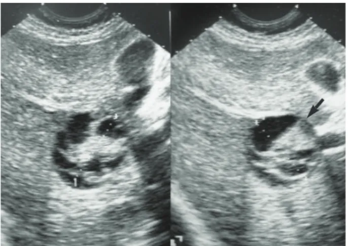
An ultrasound of the liver revealed a round multi-septat- ed irregular hypoechoic mass in the right lobe (Fig. 1). A computed tomography (CT) of the abdomen was obtained for further analysis of the mass. The CT scan demonstrated a 4.5 cm-mass in the right lobe of the liver at segment V with internal septa and hyperdense solid elements (Fig. 2).
There were no enlarged lymph nodes found near the mass and no other abnormal findings on the abdominal CT scan.
We suspected a biliary cystic neoplasm such as a biliary cys-
727
Won Hyun Lee, Tae Hyo Kim, Soong Suk You, Sun Pil Choi, Hyun Ju Min, Hyun Jin Kim, Ok Jae Lee, and Gyung Hyuck Ko*
Departments of Internal Medicine, Institute of Health Science and Pathology*, Gyeongsang National University School of Medicine, Jinju, Korea
Address for correspondence Tae Hyo Kim, M.D.
Department of Internal Medicine and Institute of Health Science, Gyeongsang National University School of Medicine, 90 Chilam-dong, Jinju 660-702, Korea Tel : +82.55-750-8726, Fax : +82.55-755-9078 E-mail : [email protected]
J Korean Med Sci 2008; 23: 727-30 ISSN 1011-8934
DOI: 10.3346/jkms.2008.23.4.727
Copyright � The Korean Academy of Medical Sciences
Benign Schwannoma of the Liver: A Case Report
A primary benign schwannoma of the liver is extremely rare. Only nine cases have been reported in the medical literature worldwide and no case has been reported in Korea previously. A 36-yr-old woman was admitted to our hospital with vague epigastric pain. The ultrasound and computed tomography scan revealed a multi- septated cystic mass in the right lobe of the liver. The mass was resected; it was found to be a 5×4×2 cm mass filled with reddish yellow fluid. The histological examination confirmed the diagnosis of a benign schwannoma, proven by positive immunoreaction with the neurogenic marker S-100 protein and a negative response to CD34, CD117 and smooth muscle actin. This is the first report of a benign schwan- noma of the liver parenchyma in a Korean patient.
Key Words : Benign Schwannoma; Liver
Received : 4 June 2007 Accepted : 24 September 2007
728 W.H. Lee, T.H. Kim, S.S. You, et al.
tadenoma or adenocarcinoma. The ultrasound-guided nee- dle biopsy obtained a specimen with spindle cells of benign appearance and clusters of hepatocytes. The overall impres- sion was a benign mesenchymal tumor of the liver.
With the suspicion of a benign mesenchymal tumor of the liver, we proceeded to a segmentectomy. The gross find- ings were a round, cystic and approximately 5×5 cm mass.
The patient’s postoperative course was uneventful. The sur- gical specimen was composed of an encapsulated mass of 5
×4×2 cm with adjacent normal liver parenchyma (Fig. 3).
Fig. 1. Ultrasound findings of the mass. A 4.5-cm sized round hypoechoic mass with multiple central echogenic bands in the right lobe of the liver (segment V).
Fig. 2. Abdominal computed tomography shows a 5 cm smooth margined low-attenuating mass in the right lobe of liver (segment V), containing multiple central septations.
Fig. 3. A photograph showing the cut section of the tumor: The specimen was 5×4×2 cm. A multi-septated cyst that was previ- ously opened, measuring 4×4 cm. The cyst was 1 cm from the resection margin. The cyst contained 10 soft tissues. The wall thickness measured 0.4 cm.
Fig. 4. (A) Spindle cell tumor with a mixture of two growth patterns, Antoni A and Antoni B, is seen. In the Antoni A pattern of growth, elon- gated cells were arranged in areas of moderate to high cellularity (black arrow). In the Antoni B pattern of growth, the tumor was less densely cellular, had a loose meshwork of cells, as well as microcysts and myxoids changes (red arrow) (H&E, ×100). (B) Spindle cell tumor with cells arranged in whorls in a storiform pattern is seen (H&E, ×200).
A B
Benign Schwannoma of the Liver 729
Microscopic examination revealed a spindle cell tumor with cells arranged in whorls in a storiform pattern surrounded by a fibrous capsule and a mixture of two growth patterns, Antoni A and Antoni B (Fig. 4). In the Antoni A pattern of growth, elongated cells were arranged in areas of moderate to high cellularity with little stromal matrix. In the Antoni B pattern of growth, the tumor was less densely cellular, had a loose meshwork of cells, as well as microcysts and myxoids changes. Atypias or mitoses of cells were not recognized. The immunohistochemical staining showed a strongly positive S-100 protein reaction (Fig. 5). However, CD34, CD117 and smooth muscle actin were all negative.
Based on the above findings, the diagnosis of a primary benign schwannoma of the liver parenchyma was made. The patient remained healthy and free from recurrence during the 18 months of follow-up.
DISCUSSION
A schwannoma is a benign mesenchymal tumor that orig- inates from Schwann cells in the peripheral nerves. The tumor is associated with neurofibromatosis in about 50% of cases.
Malignant transformation of these tumors is very rare (10).
They grow very slowly and are well encapsulated in most cases. Schwannomas are usually smaller than 5 cm at diag- nosis (11). Larger schwannomas have a tendency to undergo secondary degeneration such as pseudocystic regression, hem- orrhage and calcification.
The incidence of a schwannoma in the gastrointestinal (GI) tract is very low; it accounts for about 2-6% of all mesenchy- mal tumors that develop in the GI tract (12). There are sev- eral reports of benign schwannomas in the stomach, duode- num, rectum, and retroperitoneum. However, there is no previous report of a benign schwannoma of the liver parenchy- ma in a Korean patient.
The hepatobiliary nerves originate from the hepatic plexus in the hilum. A schwannoma of the liver can originate from a variety of hepatic sympathetic and parasympathetic nerves distributed among the intralobular connective tissues and hepatic arteries (13). The schwannoma in our case was sur- rounded by normal hepatic parenchyma. Thus, we diagnosed the tumor as a benign primary schwannoma of the liver.
In 1993, Hytiroglou et al. reported the first primary benign hepatic schwannoma diagnosed after surgical resection (3).
We reviewed the clinical characteristics, location, size and secondary degeneration of primary benign schwannomas and the findings are shown in Table 1 (1-8). They were found in eight women and two men. The age of onset was from 38 to 70 yr, and the mean age of onset was 57.2 yr. Most patients complained of pain or discomfort in the upper abdomen or epigastrium. Two patients were asymptomatic. Tumors were located in the right lobe of the liver in five cases and in the
Fig. 5. A photograph of immunochemical staining with S-100 protein: The tumor is composed of bundles of compact spindle cells that are immunoreactive to the S-100 protein (×200).
Source Sex/Age Chief complaint Location (lobe) Size (cm) Secondary degeneration
Pereira et al. 1977 (3) F/56 Swelling & pain - - Cystication
of epigastrium
Bekker 1982 (4) M/70 - Right 30×27 Necrosis
Hytiroglou et al. 1993 (5) M/67 Right flank & back pain Right 13×11×10 Hemorrhage,
necrosis cystification
Heffron et al. 1993 (6) F/38 RUQ pain Left 5 None
Yoshida et al. 1994 (7) F/56 Discomfort of Right 16 Cystification, hemorrhage
epigastrium & chest
Wada Y et al. 1997 (8) F/64 Asymptomatic Left 4 None
Wada Y et al. 1997 (8) F/69 Asymptomatic Left 15×13 Cavitation
P Flemming et al. 1998 (9) F/57 Upper abdominal pain Right Huge Cystification
Sorabh Kapoor et al. 2005 (10) F/mid-aged Epigastric lump Left 23×20×10 Central necrosis
Lee et al. 2007 (present study) F/38 Epigastric pain Right 5×4×2 Cystification
Table 1. Review of benign primary nerve sheath tumors in the liver in patients without neurofibromatosis
RUQ, right upper quadrant.
730 W.H. Lee, T.H. Kim, S.S. You, et al.
left lobe in four cases. The maximal diameter of the tumors ranged from 4 cm to 30 cm. The mean diameter was 18.9 cm. In most cases, the tumors had secondary degeneration.
Smaller tumors, approximately 4 or 5 cm in diameter, were less likely to have secondary degeneration. Radiological find- ings showed degenerative changes with cyst-like character- istics, calcification and hemorrhage as well as normal liver tissue surrounding the encapsulated mass, which was well demarcated, round or ovoid (14). In comparing our case with other cases shown Table 1, the clinical characteristics, loca- tion and secondary degeneration of the tumors are similar.
However, the tumor size was much smaller than average.
Generally, a plain CT depicts a schwannoma as a non- homogenous area; an enhanced CT shows the margins clear- ly and the inside appears as an irregular pattern (6). A defini- tive diagnosis of a hepatic schwannoma by radiological meth- ods is difficult because it is such a rare finding. Therefore, pathological examination is essential for the diagnosis.
Microscopic examination shows spindle cells like other stromal tumors. The histological diagnosis of a benign schwan- noma is usually a simple procedure with standard Hematox- ilin-Eosin staining. The distinction between a schwannoma and other spindle cell tumors or neurofibromas is based on the presence of a true capsule and Antoni A and Antoni B findings in the schwannoma (11). The type A area is dense and cellular, but the type B area is hypocellular. Immuno- histochemical staining is diffusely and strongly positive for S-100 protein in a schwannoma consistent with the finding of a nerve sheath tumor. The tumor is also positive for the glial fibrillary acidic protein and CD57 (Leu 57) (15). Even though a few gastrointestinal stromal tumors are positive for S-100, they are also positive for either CD34 or CD117. How- ever, a schwannoma is negative for both CD34 and CD117.
A leiomyoma would be negative for S-100 and positive for desmin or smooth muscle actin (16).
The treatment for a benign primary schwannoma is a sim- ple excision. The complete excision of the tumor is curative and most cases do not relapse; additional treatments are not necessary. The overall prognosis is very good (17).
In conclusion, we report the first case of a primary schwan- noma of the liver parenchyma in Korea. The diagnosis was established after surgical resection of the tumor that was identified to have spindle cells with Antoni A and Antoni B areas and was strongly positive for the S-100 protein but negative for CD34, CD117 and smooth muscle actin.
REFERENCES
1. Pereira Filho RA, Souza SA, Oliveira Filho JA. Primary neurilem-
mal tumour of the liver: case report. Arq Gastroenterol 1978; 15:
136-8.
2. Bekker GM. Neurofibroma of the liver. Sov Med 1982; 10: 120-1.
3. Hytiroglou P, Linton P, Klion F, Schwartz M, Miller C, Thung SN.
Benign schwannoma of the liver. Arch Pathol Lab Med 1993; 117:
216-8.
4. Heffron TG, Coventry S, Bedendo F, Baker A. Resection of prima- ry schwannoma of the liver not associated with neurofibromatosis.
Arch Surg 1993; 128: 1396-8.
5. Yoshida M, Nakashima Y, Tanaka A, Mori K, Yamaoka Y. Benign schwannoma of the liver: a case report. Nippon Geka Hokan 1994;
63: 208-14.
6. Wada Y, Jimi A, Nakashima O, Kojiro M, Kurohiji T, Sai K. Schwan- noma of the liver: report of two surgical cases. Pathol Int 1998; 48:
611-7.
7. Flemming P, Frerker M, Klempnauer J, Pichlmayr R. Benign schwan- noma of the liver with cystic changes misinterpreted as hydatid dis- ease. Hepatogastroenterology 1998; 45: 1764-6.
8. Kapoor S, Tevatia MS, Dattagupta S, Chattopadhyay TK. Primary hepatic nerve sheath tumor. Liver Int 2005; 25: 458-9.
9. Park MK, Lee KT, Choi YS, Shin DH, Lee JY, Lee JK, Paik SW, Ko YH, Rhee JC. A case of benign schwannoma in the porta hep- atis. Korean J Gastroenterol 2006; 47: 164-7.
10. Ducatman BS, Scheithauer BW, Piepgras DG, Reiman HM, Ilstrup DM. Malignant peripheral nerve sheath tumors. A clinicopatholog- ic study of 120 cases. Cancer 1986; 57: 2006-21.
11. Brennan MF, Singer S, Maki RG, O’sullivan B. Soft tissue sarco- ma. In: DeVita VT Jr., Hellman S, editors. Cancer: Principles &
practice of oncology. 7th edition. Philadelphia: Lippincott Williams
& Wilkins, 2000; 1595.
12. Lantz PE, Listrom MB. Gastrointestinal mesenchymal neoplasms.
In: Fenoglio-Preiser CM, Noffsinger AE, Stemmermann GN, edi- tors. Gastrointestinal pathology. 2nd edition. Philadelphia: Lippin- cott-Raven, 2002; 1185-8.
13. Williams PL, Warwick R, Dyson M, Bannister LH, editors. Gray’s anatomy. 37th edition. New York: Churchill Livingstone, 1989; 1165, 1390, 1395.
14. Cohen LM, Schwartz AM, Rockoff SD. Benign schwannomas:
pathologic basis for CT inhomogeneities. AJR Am J Roentgenol 1986; 147: 141-3.
15. Daimaru Y, Kido H, Hashimoto H, Enjoji M. Benign schwannoma of the gastrointestinal tract: a clinicopathologic and immunohisto- chemical study. Hum Pathol 1988; 19: 257-64.
16. Miettinen M, Lasota J. Gastrointestinal stromal tumors: review on morphology, molecular pathology, prognosis, and differential diag- nosis. Arch Pathol Lab Med 2006; 130: 1466-78.
17. Enzinger FM, Weiss SW. Benign tumors of peripheral nerves. In:
Enzinger FM, Weiss SW, editiors. Soft tissue tumors. 3rd edition. St.
Louis: Mosby, 1995; 829-42.