Introduction
As a chronic infectious disease, dental caries is one of the most common problems encountered in clinical den- tistry,1and shows a very high incidence of over 80% of all adults in Korea according to the 2006 national survey.2 Early and accurate diagnosis of caries is essential for clini- cians, who require exact knowledge of the depth of caries in order to determine the appropriate type of restoration and treatment planning.3,4Among the various types of methods in the diagnosis of caries, probing, visual examination, intraoral film, and digital sensors are commonly used in routine clinical practice.5Such diagnostic methods in the management of dental caries are used to determine the pre-
sence of caries and its extent, to monitor the course of car- ies progression, and to evaluate the effectiveness of treat- ment.6However, a general observation from a review was that current methods tended to be more specific than sen- sitive (that is, their use results in relatively more false nega- tive findings than false positive findings).7Actually, sev- eral studies have shown that between 25% and 42% of car- ies lesions remain undetected by clinical examination per- formed without radiographic examination.8-10
When it comes to visual examinations with the adoption of the International Caries Detection and Assessment Sys- tem (ICDAS II) clinical criteria in 2005, it has been attempt- ed to correlate the clinical image of teeth to their histolo- gical status.11However, the impact of these criteria on the diagnostic performance of visual examination is being examined on a limited number of studies, while the differ- ences in caries location, lesion extent and experimental set- up make extrapolation conduction difficult.12-15
In everyday clinical situation, if we exclude the recently introduced sophisticated methods for diagnosis of carious
Current status of dental caries diagnosis using cone beam computed tomography
Young-Seok Park, Jin-Soo Ahn*, Ho-Beom Kwon**, Seung-Pyo Lee
Department of Oral Anatomy and Dental Research Institute, School of Dentistry, Seoul National University, Seoul, Korea
*Department of Biomaterials Science and Dental Research Institute, School of Dentistry, Seoul National University, Seoul, Korea
**Department of Prosthodontics and Dental Research Institute, School of Dentistry, Seoul National University, Seoul, Korea ABSTRACT
Purpose : The purpose of this article is to review the current status of dental caries diagnosis using cone beam com- puted tomography (CBCT).
Materials and Methods : An online PubMed search was performed to identify studies on caries research using CBCT.
Results : Despite its usefulness, there were inherent limitations in the detection of caries lesions through conven- tional radiograph mainly due to the two-dimensional (2D) representation of caries lesions. Several efforts were made to investigate the three-dimensional (3D) image of lesion, only to gain little popularity. Recently, CBCT was intro- duced and has been used for diagnosis of caries in several reports. Some of them maintained the superiority of CBCT systems, however it is still under controversies.
Conclusion : The CBCT systems are promising, however they should not be considered as a primary choice of caries diagnosis in everyday practice yet. Further studies under more standardized condition should be performed in the near future. (Imaging Sci Dent 2011; 41 : 43-51)
KEY WORDS : Cone-Beam Computed Tomography; Dental Caries; Diagnosis
*This research was supported by Basic Science Research Program through the Nation- al Research Foundation of Korea (NRF) funded by the Ministry of Education, Sci- ence, and Technology (2010-0023586).
Received March 16, 2011; Revised April 19, 2011; Accepted April 29, 2011 Correspondence to : Prof. Seung-Pyo Lee
Department of Oral Anatomy, School of Dentistry, Seoul National University, 28 Yeongeon-dong, Jongno-gu, Seoul 110-749, Korea
Tel) 82-2-740-8671, Fax) 82-2-740-8674, E-mail) [email protected]
Copyright ⓒ 2011 by Korean Academy of Oral and Maxillofacial Radiology
This is an Open Access article distributed under the terms of the Creative Commons Attribution Non-Commercial License (http://creativecommons.org/licenses/by-nc/3.0) which permits unrestricted non-commercial use, distribution, and reproduction in any medium, provided the original work is properly cited.
Imaging Science in Dentistry∙pISSN 2233-7822 eISSN 2233-7830
lesion such as fiberoptic transillumination (FOTI), electri- cal conductance (EC), laser fluorescence, and so on, the ra- diographic examination is the most frequently recommend- ed method as a supplement to the clinical inspections.16,17 Regarding proximal non-cavitated caries, in spite of the variety of diagnostic modalities available, radiography is the most widely used method.11Bitewing radiography has been available for more than 80 years,12,18 while in the more recent years digital radiographic modalities attempt- ed to substitute conventional film radiography. The advan- tages of a digital system are the abilities to manipulate the image contrast and brightness and to magnify the images.
However, extensive researches regarding conventional and various digital radiographic modalities failed to detect sig- nificant differences in their diagnostic performances, only to show comparable results.19-28
Despite its usefulness, the detection of caries lesions through conventional radiograph remains rather an elusive process.29The limitations inherent in conventional radio- graphy are mainly due to the 2D representation of caries lesions, which are 3D structures in reality, and this might lead to loss of valuable information.30,31Small lesions re- main undetected when the relative amount of mineral loss is low, resulting in low subject and image contrast. More- over, the radiographic appearance of a lesion can change dramatically as a function of the chosen projection geome- try. The replacement of film by digital detectors does not address these fundamental limitations.21,28,32,33
Dentistry has largely used the same method of 2D imag- ing since the first intraoral radiograph obtained in 1896.
According to the review by Tyndall et al,34only one or two significant advances in dental imaging have been made since then in the sense of imaging geometry. These advan- ces include panoramic imaging and tomography, with the former being far more useful for dental applications, and the latter historically being limited primarily to temporo- mandibular joint and implant site imaging.34
Computed tomography (CT), which was invented by Hounsfield35in 1973, is considered as a technical break- through. It is a well-known medical technique for the nondestructive examination of internal structures and its introduction to the dentistry has been revolutionary in the sense that it can provide true 3D imaging.29 Cone beam computed tomography (CBCT) is a new application of CT that generates 3D data at lower cost and absorbed doses than conventional “fan beam” CT found in the prac- tice of medical field.36Data from the craniofacial region are often collected at higher resolution in the axial plane than those from conventional CT systems.37 In addition,
these systems do require relatively small amount of space and can easily fit into most dental clinics today. Although most of the CBCT usage has been confined to the appli- cations for dental implant placement, orthodontics, sur- gery, and temporomandibular joint disease38-44so far, sev- eral studies focusing on the diagnosis of dental caries have been reported.34
The aim of this article is to review the brief history of the usage of 3D concept in diagnosis of the dental caries and the current status of diagnosis using CBCT. This re- view builds on the findings of several recent articles relat- ed to caries diagnosis using CBCT, and seeks to outline the possible advanced applications that might be used in the future.
Materials and Methods
A PubMed search from 1965 up to February of 2011 was conducted to identify articles published in dental literature, and limited to human trials, using the search terms “caries”,
“diagnosis”, “3 dimensional”, “tomography”, and “com- puted tomography”. Manual searches of the bibliographies of all full text articles and related reviews selected from the electronic search were also performed.
Results
Early trials to realize the 3D image
An ideal diagnostic tool would enable the clinician to accurately assess the presence or absence of a lesion, to quantify its size and depth, and to determine its activity.
Whereas the physics underlying the radiographic image formation process is well suited for imaging the dental structures, the sampling level of traditional intraoral imag- ing is not sufficient to fulfill these requirements. In order to acquire 3D information, the level of sampling needs to be increased.29
Earlier attempts have been made to improve the diagno- sis of dentoalveolar conditions with 3D imaging using vari- ations of tomosynthesis.34 This has been the underlying premise of tuned aperture computed tomography (TACT), which uses a limited number of basis projections to gener- ate 2D slices at various depths.45Although TACT provid- ed some incremental benefit for periodontal and endodon- tic applications, improvements in caries detection and cha- racterization were limited to simulated recurrent caries. In case of proximal lesions, a significant increase in detec- tion rate could not be demonstrated according to several
reports.46-52
Unfortunately, TACT has provided limited application in the practice of dentistry thus far, partly due to the advent of CBCT, ironically. The development of CBCT has been innovative because complete (360�) sampling is now pos- sible without increasing the patient dose to unacceptable levels.29
A benchtop-based CT device using an intraoral detector as the image receptor, which was developed by van Daat- selaar and coworkers,53was another attempt used in caries diagnosis. It is referred as a local computed tomography (LCT), and has similar basic working principles with the commercial CBCT systems on the market, although it is not automated. They used a high resolution charge-coupled device (CCD) detector and rotating turntable with a fixed anode intraoral radiograph source. Both TACT and LCT generate a series of images that can be reconstructed into a series of cross-sectional images. Whereas TACT uses a fixed object and CCD sensor45 while moving the X-ray source, LCT uses a fixed CCD sensor and source and a rotating object.54
In their studies,53-56 they have shown the feasibility of LCT and the improved accuracy in caries detection com- pared with conventional radiography. The term, LCT is somewhat confusing with the local cone-beam computed tomography (LCBCT), which is frequently used in con- trast to full volume CBCT. Recently, Kalathingal et al29 used LCT in their research and found no difference in the detection of carious lesions, however they did find that LCT was superior for assessment of caries depth.
Researches using clinical CBCT systems
Astounding speed of development and improvement of dental imaging technology makes the proper verification difficult. When it comes to application of clinical CBCT systems in dental caries research, the study of Akdeniz et al57 was the first English literature available via Pubmed search. They found that Accuitomo 3DX (Morita Co. Ltd, Tokyo, Japan) CBCT system was superior in caries depth assessment compared with conventional film radiography or storage phosphor (SP) images. There were many stud- ies5,6,25,57-61dealing with the caries researches using clini- cal CBCT system including the study by Akdeniz et al.57 Caries researches have been traditionally classified accord- ing to the location of caries lesion. Their studies dealt with only proximal lesions,5,6,57,59occlusal lesions exclusively,60 and both proximal and occlusal lesions.25,58,61Various CBCT systems with different settings have been used, al-
though Accuitomo 3DX was the most frequently used one.
They were compared with the conventional imaging mo- dalities which included film radiography and digital intra- oral radiography using CCD and photostimulable phosphor (PSP) plates. All the studies were in vitro studies, and they used premolar and molar teeth with various stages of car- ies lesions. Intraoral radiographs were taken using paral- leling technique and rectangular collimation. For compari- son of film radiography, the film was developed automa- tically. For the rating of the caries lesions, five-scale score was used when using histology examinations.
However, aforementioned studies have not been stan- dardized in every aspect. For conventional imaging modal- ities, there were differences not only in systems but also in their X-ray taking environment such as voltage, cur- rent, exposure time, and focus to film (or receptor) distance.
Regarding these differences, the authors might follow the manufacturer’s instructions or clinical experiences. Stor- age method of teeth, the width of acrylic block mimick- ing the soft tissue, image software and comparison analy- sis were different among the studies. In addition, the sta- tus of caries lesion was diverse according to the studies.
Several studies used the image of X-ray microcomputed tomography (MicroCT) as a gold standard instead of con- ventional histology. The number of observers was also different from study to study. Detailed information about imaging modalities and environments are described in Tables 1-3.
Table 1. Product information. The manufacturer’s name follows the original articles’ description
Kodak Insight (Eastman Kodak company, Rochester, NY, USA) Kodak Ectaspeed (Eastman Kodak company, Rochester, NY, USA) Diagora-fmx (Soredex, Tuusula, Finland)
Diagora Optime (Soredex/Orion Corp., Helsinki, Finland)
#2 CCD (E2V Technologies Inc., Elmsford, NY, USA) Progeny Vision DX (Progeny Dental, Buffalo Grove, IL, USA) MAX CCD (Benlio˘glu Dental, Ankara, Turkey)
3DX Accuitomo (Morita Co. Ltd., Tokyo, Japan) NewTom 3G (Quantitative Radiology, Verona, Italy) NewTom 9000 (Quantitative Radiology, Verona, Italy) Kodak 9000 3D (Carestream Health, Inc., Rochester, NY, USA) Kodak 9500 3D (Carestream Health, Inc., Rochester, NY, USA) Promax 3D (Planmeca Oy, Helsinki, Finland)
DCT PRO (VATECH, Co., Ltd., Yongin-Si, Korea) ILUMA (Imtec Imaging, Ardmore, OK, USA)
Gendex 1000 X-ray unit (General Electric Co., Milwaukee, WI, USA) Gendex Oralix DC X-ray unit (Gendex Dental Systems, Milan, Italy) HD-70 X-ray unit (Asahi Roentgen Ind. Co., Kyoto, Japan) Trophy Trex X-ray unit (Croissy, Beaubourg, France) Trophy ETX X-ray unit (Trophy Radiologie, France) AET-Orix 70 X-ray unit (Ardet, Buccinasco, Italy)
Methods of comparing analysis
Several studies5,6,59,61 used Az value for comparison which means area under the receiver operating character-
istic (ROC) analysis. The ROC curve is a fundamental tool for diagnostic test evaluation. It allows to a complete sen- sitivity/specificity report. In a ROC curve the true positive rate (sensitivity) is plotted in function of the false positive
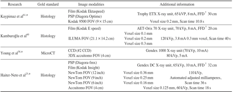
Table 2.Detailed information about imaging modalities used in studies regarding detection of lesions in approximal surfaces
Research Gold standard Image modalities Additional information
Film (Kodak Ektaspeed)
Trophy ETX X-ray unit, 65 kVP, 8 mA, FFD†30 cm Kaypimaz et al61,* Histology PSP (Diagora Optime)
Kodak 9500 FOV (9×15 cm) Voxel size 0.2 mm, Scan time 10.8 s Kodak E speed
enel et al5 Histology CCD (Progeny Vision DX) Trophy Trex X-ray unit, 65 kVP, 8 mA, FFD†20 cm PSP (Diagora Optime)
ILUMA FOV (21.1×14.2 cm) 120 kVp, 3.8 mA 0.3 mm voxel, Scan time 40 s
NewTom9000 FOV (15×15 cm) 110 kVp, 2.1 mA, Scan time 36 s, Voxel size 0.3 mm, Slice thickness 0.4 mm 3DX Accuitomo FOV (3×4 cm) 80 kVp, 5 mA, Scan time 17.5 s, Voxel size 0.125 mm, Slice thickness 1 mm Qu et al59 Histology Kodak 9000 3D FOV (5×3.7 cm) 70 kVp, 10 mA, Scan time 24 s, Voxel size 0.076 mm, Slice thickness 0.076 mm
Promax 3D FOV (8×8 cm) 76 kVp, 6 mA, Scan time 18 s, Voxel size 18 mm, Slice thickness 0.32 mm DCT Pro FOV (20×19 cm) 90 kVp, 3.5 mA, Scan time 24 s, Voxel size 0.322 mm, Slice thickness 1 mm Young et al58,* MicroCT CCD (#2 CCD) Gendex 1000 X-ray unit, 70 kVp, 10 mA
3DX accuitomo FOV (4 cm) 80 kVp, 5 mA
PSP (Diagora-fmx)
Gendex DC X-ray unit, 65 kVp, 10 mA, FFD†32 cm Film (Kodak Insight)
Haiter-Neto et al25,* Histology NewTom FOV (12 inch) Voxel size 0.36 mm 110 kVp,
NewTom FOV (9 inch) Voxel size 0.25 mm Automated adjusted milliamperes,
NewTom FOV (6 inch) Voxel size 0.16 mm Scan time 36 s
Accuitomo FOV (4 cm) Voxel size 0.125 mm, 60 kVp, Scan time 18 s Tsuchida et al6 MicroCT Film (Kodak Insight) HD-70 X-ray unit (60 kVp, 7 mA FFD†40 cm)
3DX Accuitomo FOV 4 cm 80 kVp, 4 mA, Scan time 18 s
Film (Kodak F-speed)
Gendex Oralix DC X-ray unit (60 kVp, 7 mA FFD†25 cm) Akdeniz et al57 Histology Diagora fmx (Soredex)
3DX Accuitomo FOV 4 cm 80 kVp, 1.5 mA, Scan time 17.5 s
* denotes the study dealt with both approximal and occlusal caries lesions.
†denotes the focus to film distance or focus to receptor distance.
S
Table 3.Detailed information about imaging modalities used in studies regarding detection of lesions in occlusal surfaces
Research Gold standard Image modalities Additional information
Film (Kodak Ektaspeed)
Trophy ETX X-ray unit, 65 kVP, 8 mA, FFD†30 cm Kaypimaz et al61,* Histology PSP (Diagora Optime)
Kodak 9500 FOV (9×15 cm) Voxel size 0.2 mm, Scan time 10.8 s Film (Kodak E speed) AET-Orix 70 X-ray unit, 70 kVp, 8 mA, FFD†20 cm
Voxel size 0.1 mm Kamburo˘glu et al60 Histology
ILUMA FOV (21.1×14.2 cm) Voxel size 0.2 mm 120 kVp, 3.8 mA 0.3 mm voxel, Scan time 40 s Voxel size 0.3 mm
Young et al58,* MicroCT CCD (#2 CCD) Gendex 1000 X-ray unit (70 kVp, 10 mA)
3DX accuitomo FOV (4 cm) 80 kVp, 5 mA
PSP (Diagora-fmx)
Gendex DC X-ray unit, 65 kVp, 10 mA, FFD†32 cm Film (Kodak Insight)
NewTom FOV (12 inch) Voxel size 0.36 mm 110 kVp,
Haiter-Neto et al25,* Histology
NewTom FOV (9 inch) Voxel size 0.25 mm Automated adjusted milliamperes, NewTom FOV (6 inch) Voxel size 0.16 mm Scan time 36 s Accuitomo FOV (4 cm) Voxel size 0.125 mm, 60 kVp, Scan time 18 s
* denotes the study dealt with both approximal and occlusal caries lesions.
†denotes the focus to film distance or focus to receptor distance.
rate (1-specificity) for different cut-off points of a para- meter.62Each point on the ROC curve represents a sensi- tivity/specificity pair corresponding to a particular decision threshold. The area under the ROC curve, Az, is a measure of how well a parameter can distinguish between two diag- nostic groups (diseased/normal).63 Some aforementioned studies25,58 used sensitivity and specificity for comparing image modalities, however they did not present the ROC curve and Az values.
The study of Kamburo˘glu K et al60introduced the rela- tive treatment effect (RTE) value as a statistical parameter according to the lesion depth. A ‘treatment effect’ is the average causal effect of a binary (0-1) variable on an out- come variable of scientific or policy interest. The term
‘treatment effect’ originates in a medical literature con- cerned with the causal effects of binary, yes-or-no ‘treat- ments’, such as an experimental drug or a new surgical procedure.64However, the term is now used much more generally.
The study of Akdeniz et al57 is somewhat peculiar in
that it compared the accuracy of determining the depth of proximal caries lesion, not just the accuracy of detecting the existence of lesion. Actually, this kind of studies, which deal with the defining the 3 dimensional region of the car- ies lesion, are now being actively performed by various research groups using X-ray microcomputed tomography instead of CBCT.65
Summary of comparisons
Tsuchida et al6used the noncavitated proximal incipient lesions and found that no significant differences between CBCT and film images. This result might reflect difficulty of detecting incipient lesion.
Haiter-Neto et al25compared NewTom 3G system with 3 fields of view as a full-volume CBCT with Accuitomo 3DX as a local CBCT (LCBCT). The intraoral radiography was also compared. The results showed that the NewTom 12-inches and 9-inches images had significantly lower sen- sitivities than the Accuitomo systems, whereas the New- Tom 9-inches and 6-inches images had significantly lower
Table 4.Brief summary of results from studies regarding detection of lesion in approximal surfaces
Research N Observer Image modalities Az Sensitivity Specificity Lesion depth
Film (Kodak Ektaspeed) 0.782±0.0561 Kaypimaz et al61,* 72 2 PSP (Diagora Optime) 0.689±0.0637 Kodak 9500 FOV (9×15 cm) 0.705±0.0627
Kodak E speed 0.835
enel et al5 230 3 CCD (Progeny Vision DX) 0.861 PSP (Diagora Optime) 0.823 ILLUMA FOV (21.1×14.2 cm) 0.883 NewTom9000 FOV 15×15 cm 0.541±0.033 3DX Accuitomo FOV (3×4 cm) 0.555±0.044 Qu et al59 39 7 Kodak 9000 3D FOV (5×3.7 cm) 0.577±0.038 Promax 3D FOV (8×8 cm) 0.545±0.024 DCT Pro FOV (20×19 cm) 0.549±0.028
CCD (#2 CCD) 0.18±0.15 (E) 0.96±0.08 (E)
Young et al58,* 146 8 0.33±0.06 (D) 0.96±0.02 (D)
3DX accuitomo FOV (4 cm) 0.24±0.08 (E) 0.95±0.05 (E) 0.61±0.05 (D) 0.94±0.05 (D)
PSP (Diagora-fmx) 0.17 0.91
Film (Kodak Insight) 0.18 0.92
Haiter-Neto et al25,* 100 6 NewTom FOV (12 inch) 0.13 0.88
NewTom FOV (9 inch) 0.14 0.85
NewTom FOV (6 inch) 0.18 0.84
Accuitomo FOV (4 cm) 0.21 0.89
Tsuchida et al6 50 7 Film (Kodak Insight) 0.633±0.029 3DX Accuitomo FOV 4 cm 0.625±0.018
Film (Kodak F-speed) 0.44 mm (95%CI 1.0-1.8)
Akdeniz et al57 30 2 Diagora fmx (Soredex) 0.7 mm (95%CI 1.2-2.5)
3DX Accuitomo FOV 4 cm 0.5 mm (95%CI 1.4-2.5)
* denotes the study dealt with both approximal and occlusal caries lesions.
(E) means the lesion confined to the Enamel, (D) means the lesion penetrated into the dentin.
S
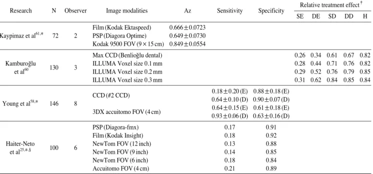
specificities than the conventional radiography in detect- ing proximal lesions. The Accuitomo images were report- ed to be comparable with the conventional radiography.
For occlusal caries detection, this LCBCT system present- ed a higher sensitivity than the other systems. In addition, it was determined to be equal to the intraoral systems, how- ever the overall true score (true positives and negatives) was not higher for detection of dentinal lesions.
Young et al58compared the efficacy of CBCT and con- ventional CCD image in detecting proximal and occlusal lesions. They used 3DX Accuitomo systems as a CBCT and found a significant difference in the average sensiti- vity score between CBCT and CCD regarding detection of proximal caries. They concluded that by using CBCT, it was able to improve the detection of proximal surface caries extending into the dentin, but not the occlusal caries.
Currently, there are two types of image detectors em- ployed in CBCT equipments.59One is the image intensi- fier with CCD, and the other is the flat panel detector in- cluding amorphous silicon flat panel and complementary metal oxide semiconductor panel. Qu et al59evaluated the diagnostic accuracy of approximal lesions among the 5 clinical CBCT systems. The systems showed no statisti- cal significant difference, and there was no significant dif- ference between the types of the detectors of CBCT sys- tems.
Kamburo ˘glu K et al60assessed the diagnostic ability of
intraoral digital CCD sensor images and CBCT images at different voxel resolutions in detection of occlusal caries.
They found that the modalities of imaging performance were different in deep enamel, superficial dentin, and deep dentin. However, there was no difference in healthy and superficial enamel caries groups. They concluded that at all voxel sizes, CBCT images could be considered a tool for use in the diagnosis of occlusal caries.
The study by enel et al5assessed the diagnostic ability of visual inspection, film, CCD sensor, PSP plate, and CBCT in the detection of proximal caries. The authors concluded that all the methods performed similarly in the detection of proximal caries.
Kayipmaz et al61compared the effectiveness of conven- tional radiograph, PSP plate, and CBCT sytems in deter- mination of occlusal and approximal caries. In determin- ing occlusal caries, CBCT was statistically superior to the other two conventional methods. However, no significant difference was verified in determining approximal caries.
Brief summary of results are described in Tables 4 and 5.
Discussion
Until now, only a small amount of research has been undertaken regarding caries diagnosis using CBCT. Altho- ugh some studies maintained the superiority or the pro- mise of using clinical CBCT system for diagnosis of den-
S
Table 5.Brief summary of results from studies regarding detection of lesion in occlusal surfaces
Research N Observer Image modalities Az Sensitivity Specificity Relative treatment effect‡
SE DE SD DD H
Film (Kodak Ektaspeed) 0.666±0.0723 Kaypimaz et al61,* 72 2 PSP (Diagora Optime) 0.649±0.0730 Kodak 9500 FOV (9×15 cm) 0.849±0.0554
Max CCD (Benlio˘glu dental) 0.26 0.34 0.61 0.67 0.82
Kamburo ˘glu
130 3 ILLUMA Voxel size 0.1 mm 0.28 0.44 0.71 0.76 0.82
et al60 ILLUMA Voxel size 0.2 mm 0.29 0.52 0.76 0.79 0.85
ILLUMA Voxel size 0.3 mm 0.31 0.62 0.84 0.85 0.84
CCD (#2 CCD) 0.18±0.20 (E) 0.88±0.18 (E)
Young et al58,* 146 8 0.64±0.10 (D) 0.90±0.07 (D)
3DX accuitomo FOV (4 cm) 0.64±0.15 (E) 0.61±0.18 (E) 0.93±0.06 (D) 0.63±0.16 (D)
PSP (Diagora-fmx) 0.17 0.91
Film (Kodak Insight) 0.18 0.92
Haiter-Neto
100 6 NewTom FOV (12 inch) 0.13 0.88
et al25,*,§ NewTom FOV (9 inch) 0.14 0.85
NewTom FOV (6 inch) 0.18 0.84
Accuitomo FOV (4 cm) 0.21 0.89
* denotes the study dealt with both approximal and occlusal caries lesions.
§The results of Haiter-Neto et al in this table is not from occlusal lesion exclusively, but from occlusal++approximal surface.
‡In the subdivisions of Relative Treatment Effect, SE: superficial enamel, DE: deep enamel, SD: superficial dentin, DD: deep dentin, H: healthy
tal caries, we could not find the consensus of the research up to now. There was a tendency among the studies to insist that the accuracy of CBCT systems was higher than the conventional methods in detecting occlusal caries and deep lesions into the dentin, however evidences are still insufficient. Sensitivity may increase, but mostly with sim- ultaneous decrease of specificity. As mentioned above, the conditions of the experiments were different from study to study and not standardized. Thus, it is appropriate to say that whether the CBCT is superior to the conventional modalities in diagnosis of dental caries is under controver- sy at this stage. Furthermore, it goes without saying that the routine use of CBCT system instead of conventional radiography should not be accepted.
When considering a comparison of different modalities, it should be reminded that an increase in efficacy or lack of thereof does not always mean superiority or inferiority.
We should consider cost, time, and effort. Above all, we cannot but consider the radiation dose despite the improve- ment of image.
The imaging of CBCT has fewer problems of geometric distortion than those of conventional methods in theory, thus the real 3D representation of caries lesion was avail- able, which was impossible before. However, there are surely several limitations in clinical situations. It should also be reminded that all the studies mentioned were in vitro studies. The images were obtained under ideal geo- metry with no closed contact, cone cut, soft tissues, and projection distortions. In addition, the presence of any metal restoration in clinical situations might affect the quality of CBCT image. Further studies are compulsory not only to elucidate the accuracy of current systems, but also to verify and keep face with the future systems includ- ing in vivo studies.
References
1. Pereira AC, Verdonschot EH, Huysmans MC. Caries detection methods: can they aid decision making for invasive sealant treatment? Caries Res 2001; 35 : 83-9.
2. Ministry of Health and Welfare. National survey of oral health in 2006. Seoul: Ministry of Health and Welfare; 2007. p. 59- 70.
3. Attrill DC, Ashley PF. Occlusal caries detection in primary teeth: a comparison of DIAGNOdent with conventional meth- ods. Br Dent J 2001; 190 : 440-3.
4. Ohki M, Okano T, Nakamura T. Factors determining the diag- nostic accuracy of digitized conventional intra-oral radiographs.
Dentomaxillofac Radiol 1994; 23 : 77-82.
5. Senel B, Kamburoglu K, Uçok O, Yüksel SP, Ozen T, Avsever H. Diagnostic accuracy of different imaging modalities in de-
tection of proximal caries. Dentomaxillofac Radiol 2010; 39 : 501-11.
6. Tsuchida R, Araki K, Okano T. Evaluation of a limited cone- beam volumetric imaging system: comparison with film radio- graphy in detecting incipient proximal caries. Oral Surg Oral Med Oral Pathol Oral Radiol Endod 2007; 104 : 412-6.
7. Bader JD, Shugars DA. A systematic review of the perfor- mance of a laser fluorescence device for detecting caries. J Am Dent Assoc 2004; 135 : 1413-26.
8. Haak R, Wicht MJ, Noack MJ. Conventional, digital and con- trast-enhanced bite-wing radiographs in the decision to restore proximal carious lesions. Caries Res 2001; 35 : 193-9.
9. Møystad A, Svanaes DB, Risnes S, Larheim TA, Gröndahl HG. Detection of approximal caries with a storage phosphor system. A comparison of enhanced digital images with dental X-ray film. Dentomaxillofac Radiol 1996; 25 : 202-6.
10. Tam LE, McComb D. Diagnosis of occlusal caries: Part II.
Recent diagnostic technologies. J Can Dent Assoc 2001; 67 : 459-63.
11. Mitropoulos P, Rahiotis C, Stamatakis H, Kakaboura A. Diag- nostic performance of the visual caries classification system ICDAS II versus radiography and micro-computed tomogra- phy for proximal caries detection: an in vitro study. J Dent 2010; 38 : 859-67.
12. Jablonski-Momeni A, Stachniss V, Ricketts DN, Heinzel- Gutenbrunner M, Pieper K. Reproducibility and accuracy of the ICDAS-II for detection of occlusal caries in vitro. Caries Res 2008; 42 : 79-87.
13. Pitts NB, Rimmer PA. An in vivo comparison of radiographic and directly assessed clinical catres status of posterior approxi- mal surfaces in primary and permanent teeth. Caries Res 1992;
26 : 146-52.
14. Peers A, Hiff FJ, Mitropoulos CM, Holloway PJ. Validity and reproducibility of clinical examination, fiber-optic transillu- mination, and bitewing radiology for the diagnosis of small approximal carious lesion: an in vitro study. Caries Res 1993;
27 : 307-11.
15. Ismail AI, Sohn W, Tellez M, Amaya A, Sen A, Hasson H, et al. The International Caries Detection and Assessment System (ICDAS): an integrated system for measuring dental caries.
Community Dent Oral Epidemiol 2007; 35 : 170-8.
16. Yang J, Dutra V. Utility of radiology, laser fluorescence, and transillumination. Dent Clin North Am 2005; 49 : 739-52.
17. Souza-Zaroni WC, Ciccone JC, Souza-Gabriel AE, Ramos RP, Corona SA, Palma-Dibb RG. Validity and reproducibility of different combinations of methods for occlusal caries detec- tion: an in vitro comparison. Caries Res 2006; 40 : 194-201.
18. Raper HR. A new kind of X-ray examination for preventive dentistry. Int J Orthod Oral Surg 1925; 11 : 76-86.
19. Tyndall DA, Ludlow JB, Platin E, Nair M. A comparison of Kodak Ektaspeed Plus film and the Siemens Sidexis digital imaging system for caries detection using receiver operating characteristic analysis. Oral Surg Oral Med Oral Pathol Oral Radiol Endod 1998; 85 : 113-8.
20. Syriopoulos K, Sanderink GC, Velders XL, van der Stelt PF.
Radiographic detection of approximal caries: a comparison of dental films and digital imaging systems. Dentomaxillofac Radiol 2000; 29 : 312-8.
21. Abreu M Jr, Mol A, Ludlow JB. Performance of RVGui sen- sor and Kodak Ektaspeed Plus film for proximal caries detec- tion. Oral Surg Oral Med Oral Pathol Oral Radiol Endod 2001;
91 : 381-5.
22. Nair MK, Nair UP. An in-vitro evaluation of Kodak Insight and Ektaspeed Plus film with a CMOS detector for natural proxi- mal caries: ROC analysis. Caries Res 2001; 35 : 354-9.
23. Khan EA, Tyndall DA, Caplan D. Extraoral imaging for proxi- mal caries detection: Bitewings vs scanogram. Oral Surg Oral Med Oral Pathol Oral Radiol Endod 2004; 98 : 730-7.
24. Wenzel A, Hintze H. The choice of gold standard for evaluat- ing tests for caries diagnosis. Dentomaxillofac Radiol 1999;
28 : 132-6.
25. Haiter-Neto F, Wenzel A, Gotfredsen E. Diagnostic accuracy of cone beam computed tomography scans compared with intra- oral image modalities for detection of caries lesions. Dento- maxillofac Radiol 2008; 37 : 18-22.
26. Møystad A, Svanaes DB, Larheim TA, Grondahl HG. Effect of image magnification of digitized bitewing radiographs on approximal caries detection: an in vitro study. Dentomaxillofac Radiol 1995; 24 : 255-9.
27. Wenzel A, Pitts N, Verdonschot EH, Kalsbeek H. Develop- ments in radiographic caries diagnosis. J Dent 1993; 21 : 131- 40.
28. White SC, Yoon DC. Comparative performance of digital and conventional images for detecting proximal surface caries.
Dentomaxillofac Radiol 1997; 26 : 32-8.
29. Kalathingal SM, Mol A, Tyndall DA, Caplan DJ. In vitro assessment of cone beam local computed tomography for proxi- mal caries detection. Oral Surg Oral Med Oral Pathol Oral Radiol Endod 2007; 104 : 699-704.
30. Hintze H, Wenzel A, Danielsen B, Nyvad B. Reliability of visual examination, fibre-optic transillumination, and bitewing radiography, and reproducibility of direct visual examination following tooth separation for the identification of cavitated carious lesions in contacting approximal surfaces. Caries Res 1998; 32 : 204-9.
31. Wenzel A. Bitewing and digital bitewing radiography for detec- tion of caries lesions. J Dent Res 2004; 83 Spec No C : C72-5.
32. Hintze H, Wenzel A, Jones C. In vitro comparison of D- and E-speed film radiography, RVG, and visualix digital radiogra- phy for the detection of enamel approximal and dentinal occlu- sal caries lesions. Caries Res 1994; 28 : 363-7.
33. Wenzel A, Borg E, Hintze H, Grondahl HG. Accuracy of car- ies diagnosis in digital images from charge-coupled device and storage phosphor systems: an in vitro study. Dentomaxillofac Radiol 1995; 24 : 250-4.
34. Tyndall DA, Rathore S. Cone-beam CT diagnostic applications:
caries, periodontal bone assessment, and endodontic applica- tions. Dent Clin North Am 2008; 52 : 825-41.
35. Hounsfield GN. Computerized transverse axial scanning (tomography). 1. Description of system. Br J Radiol 1973; 46 : 1016-22.
36. Lo EC, Zhi QH, Itthagarun A. Comparing two quantitative methods for studying remineralization of artificial caries. J Dent 2010; 38 : 352-9.
37. Ito K, Gomi Y, Sato S, Arai Y, Shinoda K. Clinical applica- tion of a new compact CT system to assess 3-D images for the
preoperative treatment planning of implants in the posterior mandible. A case report. Clin Oral Implants Res 2001; 12 : 539-42.
38. Lascala CA, Panella J, Marques MM. Analysis of the accuracy of linear measurements obtained by cone beam computed tomo- graphy (CBCT-NewTom). Dentomaxillofac Radiol 2004; 33 : 291-4.
39. Honda K, Arai Y, Kashima M, Takano Y, Sawada K, Ejima K, et al. Evaluation of the usefulness of the limited cone-beam CT (3DX) in the assessment of the thickness of the roof of the glenoid fossa of the temporomandibular joint. Dentomaxillo- fac Radiol 2004; 33 : 391-5.
40. Ziegler CM, Woertche R, Brief J, Hassfeld S. Clinical indica- tions for digital volume tomography in oral and maxillofacial surgery. Dentomaxillofac Radiol 2002; 31 : 126-30.
41. Mozzo P, Procacci C, Tacconi A, Martini PT, Andreis IA. A new volumetric CT machine for dental imaging based on the cone-beam technique: preliminary results. Eur Radiol 1998; 8 : 1558-64.
42. Danforth RA. Cone beam volume tomography: a new digital imaging option for dentistry. J Calif Dent Assoc 2003; 31 : 814-5.
43. Sukovic P. Cone beam computed tomography in craniofacial imaging. Orthod Craniofac Res 2003; 6(Suppl 1) : 31-6.
44. Scarfe WC. Imaging of maxillofacial trauma: evolutions and emerging revolutions. Oral Surg Oral Med Oral Pathol Oral Radiol Endod 2005; 100(2 Suppl) : S75-96.
45. Webber RL, Horton RA, Tyndall DA, Ludlow JB. Tuned- aperture computed tomography (TACT). Theory and applica- tion for three-dimensional dento-alveolar imaging. Dento- maxillofac Radiol 1997; 26 : 53-62.
46. Tyndall DA, Clifton TL, Webber RL, Ludlow JP, Horton RA.
TACT imaging of primary caries. Oral Surg Oral Med Oral Pathol Oral Radiol Endod 1997; 84 : 214-25.
47. Nair MK, Tyndall DA, Ludlow JB, May K, Ye F. The effects of restorative material and location on the detection of simu- lated recurrent caries. A comparison of dental film, direct digi- tal radiography and tuned aperture computed tomography.
Dentomaxillofac Radiol 1998; 27 : 80-4.
48. Abreu Júnior M, Tyndall DA, Platin E, Ludlow JB, Phillips C. Two- and three-dimensional imaging modalities for the detection of caries. A comparison between film, digital radio- graphy and tuned aperture computed tomography. Dentomax- illofac Radiol 1999; 28 : 152-7.
49. Nance RS, Tyndall DA, Levin LG, Trope M. Diagnosis of ex- ternal root resorption using TACT (tuned-aperture computed tomography). Endod Dent Traumatol 2000; 16 : 24-8.
50. Nance R, Tyndall D, Levin LG, Trope M. Identification of root canals in molars by tuned-aperture computed tomography.
Int Endod J 2000; 33 : 392-6.
51. Ramesh A, Ludlow JB, Webber RL, Tyndall DA, Paquette D.
Evaluation of tuned aperture computed tomography (TACT) in the localization of simulated periodontal defects. Dento- maxillofac Radiol 2001; 30 : 319-24.
52. Terakado M, Hashimoto K, Arai Y, Honda M, Sekiwa T, Sato H. Diagnostic imaging with newly developed ortho cubic super- high resolution computed tomography (Ortho-CT). Oral Surg Oral Med Oral Pathol Oral Radiol Endod 2000; 89 : 509-18.
53. van Daatselaar AN, Dunn SM, Spoelder HJ, Germans DM, Renambot L, Bal HE, et al. Feasibility of local CT of dental tissues. Dentomaxillofac Radiol 2003; 32 : 173-80.
54. van Daatselaar AN, Tyndall DA, van der Stelt PF. Detection of caries with local CT. Dentomaxillofac Radiol 2003; 32 : 235-41.
55. van Daatselaar AN, Tyndall DA, Verheij H, van der Stelt PF.
Minimum number of basis projections for caries detection with local CT. Dentomaxillofac Radiol 2004; 33 : 355-60.
56. van Daatselaar AN, van der Stelt PF, Weenen J. Effect of num- ber of projections on image quality of local CT. Dentomaxillo- fac Radiol 2004; 33 : 361-9.
57. Akdeniz BG, Grondahl HG, Magnusson B. Accuracy of proxi- mal caries depth measurements: comparison between limited cone beam computed tomography, storage phosphor and film radiography. Caries Res 2006; 40 : 202-7.
58. Young SM, Lee JT, Hodges RJ, Chang TL, Elashoff DA, White SC. A comparative study of high-resolution cone beam computed tomography and charge-coupled device sensors for detecting caries. Dentomaxillofac Radiol 2009; 38 : 445-51.
59. Qu X, Li G, Zhang Z, Ma X. Detection accuracy of in vitro approximal caries by cone beam computed tomography images.
Eur J Radiol (in press).
60. Kamburo˘glu K, Murat S, Yüksel SP, Cebeci AR, Paksoy CS.
Occlusal caries detection by using a cone-beam CT with dif- ferent voxel resolutions and a digital intraoral sensor. Oral Surg Oral Med Oral Pathol Oral Radiol Endod 2010; 109 : e63-9.
61. Kayipmaz S, Sezgin OS, Saricao˘glu ST, Can G. An in vitro comparison of diagnostic abilities of conventional radiography, storage phosphor, and cone beam computed tomography to determine occlusal and approximal caries. Eur J Radiol (in press).
62. Hanley JA, McNeil BJ. A method of comparing the areas under receiver operating characteristic curves derived from the same cases. Radiology 1983; 148 : 839-43.
63. McNeil BJ, Hanley JA. Statistical approaches to the analysis of receiver operating characteristic (ROC) curves. Med Decis Making 1984; 4 : 137-50.
64. Rothwell PM. Can overall results of clinical trials be applied to all patients? Lancet 1995; 345 : 1616-9.
65. Park YS, Bae KH, Chang J, Shon WJ. Theory of X-ray micro- computed tomography in dental research: application for the caries research. J Korean Acad Conserv Dent 2011; 36 : 98-107.