CASE REPORT
간문부 담관에 발생한 소세포 신경내분비종양 1예
김범철, 송태준, 이 혁, 주 미
1, 배원기, 김남훈, 김경아, 이준성
인제대학교 의과대학 일산백병원 내과학교실, 병리학교실1
A Case of Small Cell Neuroendocrine Tumor Occurring at Hilar Bile Duct
Bum Chul Kim, Tae Jun Song, Hyuk Lee, Mee Joo1, Won Ki Bae, Nam-Hoon Kim, Kyung-Ah Kim and June Sung Lee Departments of Internal Medicine and Pathology1, Ilsan Paik Hospital, Inje University College of Medicine, Goyang, Korea
Neuroendocrine tumors of the extrahepatic biliary tree are extremely rare malignancies accounting for 0.2-2.0% of all gastro- intestinal carcinoid tumors. Neuroendocrine tumors obstructing the biliary tree are extremely difficult to diagnose preoperatively and nearly impossible to differentiate from cholangiocarcinoma. Statistically, the most common anatomic location in the biliary tree is the common bile duct, followed by the perihilar region. Herein, we present a case of a small cell neuroendocrine carcinoma of the hilum in a 79-year-old man following laparotomy. To our knowledge, this is the first case of small cell type neuroendocrine carcinoma of hilar bile duct reported in Korea. (Korean J Gastroenterol 2013;62:301-305)
Key Words: Neuroendocrine tumors; Biliary tract; Klatskin’s tumor
Received January 17, 2013. Revised April 5, 2013. Accepted April 10, 2013.
CC This is an open access article distributed under the terms of the Creative Commons Attribution Non-Commercial License (http://creativecommons.org/licenses/
by-nc/3.0) which permits unrestricted non-commercial use, distribution, and reproduction in any medium, provided the original work is properly cited.
교신저자: 송태준, 411-706, 고양시 일산서구 주화로 170, 인제대학교 일산백병원 내과
Correspondence to: Tae Jun Song, Department of Internal Medicine, Inje University Ilsan Paik Hospital, 170 Juhwa-ro, Ilsanseo-gu, Goyang 411-706, Korea. Tel: +82- 31-910-7200, Fax: +82-31-910-7219, E-mail: tjsong@paik.ac.kr
Financial support: None. Conflict of interest: None.
서 론
신경내분비종양은 생물학적 성상에 따라 유암종(carcinoid tumor), 도세포종(islet cell tumor), 신경아세포종(neurobla- stoma), 폐 및 폐외 소세포암(small cell carcinoma) 등으로 구분되며 매우 다양한 임상적 특징을 가진다.1 신경내분비종 양은 상피세포 종양으로 배아신경관으로부터 호흡기관이나 위장관으로 이동하는 장크롬친화세포(enterochromaffin cell) 에서 기원하기 때문에 주로 폐와 위장관계에서 발생하게 된 다. 담관에 생기는 신경내분비종양은 전체 신경내분비종양의 0.4% 미만을 차지한다고 알려져 있으며 그 중에서도 간문부 주위 담관에 생기는 경우는 극히 드물다. 담관의 신경내분비 종양에만 특징적으로 나타나는 증상이나 영상의학적으로 감 별이 가능한 특징적인 소견이 없기 때문에 담관의 신경내분비 종양은 수술 후 조직검사를 통해서 진단되는 경우가 대부분이 다.2
저자들은 황달을 주소로 내원한 79세 남자 환자에서 간문 부 담관의 종괴에 대해 수술을 시행하였으며 수술 후 조직 소견에서 고등급 악성의 소세포 신경내분비종양으로 진단된 예가 있어 문헌고찰과 함께 보고하는 바이다.
증 례
79세 남자가 황달을 주소로 입원하였다. 환자는 내원 20일 전부터 소화불량과 복부 불편감이 있어 외부병원에서 약물치 료를 받았으나 호전이 없었고, 내원 3일 전부터 소변색이 진 하게 변하면서 공막에 황달 소견이 보이고 전신에 소양감이 발생하였다. 고혈압, 당뇨, 결핵 등의 과거력은 없었으며 가족 력에서 특이 사항은 없었다. 신체 검사 소견에서 혈압은 139/76 mmHg, 맥박 89회/분, 체온 36.1도, 호흡수 16회/분이었다.
공막에 황달이 관찰되었고, 우상복부의 압통을 호소하였다.
입원 당시 시행한 혈액검사에서 백혈구 5,450/mm3, 혈색
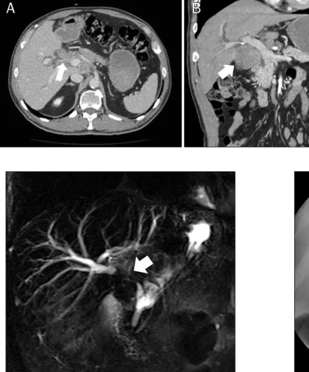
Fig. 1. (A, B) Contrast-enhanced CT image shows a diffusely infiltrating soft- tissue mass at the hilum (arrows) that extends along the gallbladder neck and cystic duct.
Fig. 2. Magnetic resonance cholangiopancreatography demon- strates low signal intensity mass involving the hepatic duct con- fluence (arrow). Both the right and left hepatic ducts are dilated.
Fig. 3. Endoscopic retrograde cholangiogram reveals malignant stricture of the common hepatic duct.
소 13.5 g/dL, 헤마토크릿 47.6%, 혈소판 196,000/mm3이었 고, 생화학검사에서 AST 287 IU/L, ALT 563 IU/L, ALP 396 IU/L, GGT 682 IU/L, 총 빌리루빈 6.5 mg/dL, 직접 빌리루 빈 4.9 mg/dL로 상승되어 있었다. 간염표지자 검사는 hep- atitis B surface antigen과 hepatitis B surface antibody, hepatitis C virus antibody 모두 음성이었고 종양표지자 검 사로는 CA 19-9이 147.3 U/mL로 상승되어 있었다. 복부 CT 에서 간문부 담관에 5.2×3.6 cm 크기의 저음영의 종괴가 관 찰되고 양쪽 간내담관이 확장되어 있었다. 총간관 및 총담관 주변으로 여러 개의 뭉쳐진 림프절 비대가 보였으며 총담관 과 주췌관의 확장은 없었다(Fig. 1). 자기공명췌담관 조영술 (magnetic resonance cholangiopancreatography) 소견에 서는 간문부에서 좌, 우 담관의 기시부가 분리되어 관찰되었 고 좌우 담관의 심한 확장이 관찰되어 Bismuth 분류상 2형 간문부 담관암의 소견을 보였다(Fig. 2). 이 종괴는 T1 강조영 상에서 저신호 강도, T2 강조영상에서 고신호 강도를 보였으
며, 조영증강영상에서 점차적인 증강을 보였다. 담관의 배액 과 조직생검을 위해 내시경 역행성 담췌관조영술(encodsco- pic retrograde cholangiopancreatography)을 시행하였다.
조영제를 주입했을 때 총간관 부위에 협착이 관찰되어 협착 부위에서 담관 내 조직 생검을 시행하고 10 Fr, 12 cm 길이의 플라스틱 배액관을 삽입하였다(Fig. 3). 조직 생검 결과 고등 급 형성이상(high grade dysplasia)을 보이는 비정형 세포들 이 관찰되었다. 양전자방출 단층촬영소견에서는 종괴와 주변 의 림프절에 표준화섭취계수(standardized uptake value) 12.3으로 현저히 높은 수치를 보였다.
고령의 환자였으나 기저질환이 없이 건강하였고 본인이 적 극적으로 수술 치료를 원하여 수술을 시행하였다. 수술은 종 양의 완전 절제를 목표로 계획하였으나 광범위한 침윤소견을 보여 완전 절제는 불가능하였고 장간막에서 조직검사 및 고식 적인 치료 목적으로 담낭절제술을 시행하였다. 수술 소견에서 종양은 주로 간문부 담관에서 기원하여 담낭관까지 침범하는 것으로 보였으며 간문맥 및 간동맥까지 침윤하는 소견을 보였
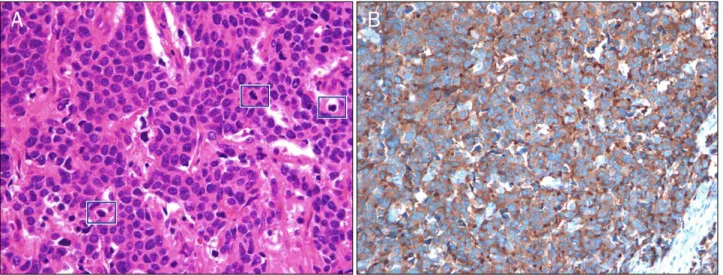
Fig. 4. Histopathological findings. (A) The tumor cells are composed predominantly of small to medium-sized round or oval cells with hyperchromatic nuclei, inconspicuous nucleoli, and scanty cytoplasm (H&E, ×400). (B) Tumor cells are stained positive for synaptophysin immunohistochemistry, which indicates the presence of a neuroendocrine tumor (×200).
다. 현미경 소견에서는 종양세포는 작고 세포질이 거의 없으 며 뚜렷한 핵소체 없이 전반적으로 과염색된 핵을 보였다 (Fig. 4A). 소세포암종의 형태로 일정한 배열양상이 없이 침 습성으로 광범위하게 증식하였고 부분적으로 괴사가 관찰되 었다. 유사분열은 현미경 400배 고배율 시야에서 3개 관찰되 고(Fig. 4A), 10군데의 400배 시야에서 관찰된 유사분열수는 30개였다. 면역조직화학 염색에서 chromogranin에서는 음 성이었지만 cytokeratin, synaptophysin, CD56에 양성 반응 을 보여 소세포암종의 신경내분비종양으로 진단할 수 있었다 (Fig. 4B). 2010년 세계보건기구에서 개정한 분류에 따르면 유사분열의 수는 10개의 고배율 시야에서 30개가 관찰되고 Ki-67 지수는 70%로 Grade 3의 분화도가 나쁜 소세포 신경 내분비종양에 해당되었다. 기능성 신경내분비 종양에서 보일 수 있는 안면 홍조, 식은땀, 설사, 저혈당, 침샘 부종, 저혈압, 저칼륨혈증 등의 증상이 없어 비기능성 신경내분비종양으로 생각되었다.2 환자는 추가적인 항암치료는 거부하여 보존적인 치료를 하면서 외래 추적 관찰 중이다.
고 찰
신경내분비종양은 정상적으로 분포하는 다양한 신경내분 비세포로부터 기원하며, 종양세포가 분비하는 호르몬에 의해 다양한 임상 소견을 보인다고 알려져 있다.3 주로 50대에 잘 생기나 연령에 관계없이 생길 수 있고 악성에 대한 잠재력을 가지고 있다. 비교적 드문 종양이지만 최근 수십 년 동안 빈도 가 점차 증가하는 양상을 보이고 있으며, 진단 및 치료에 관한 많은 논의와 발전이 있었다.2 국내 연구에 의하면 과거 30년 동안 신경내분비종양의 발생률이 인구 10만 명당 5.25명으로
4배 정도 증가했고 미국과 유럽 등의 서구에서는 20배 정도 증가하는 추세를 보이고 있다.2,4 병리소견에서는 chromogra- nin A나 synaptophysin, neuron-specific enolase 등의 면 역염색 표지자에 양성반응을 보이며 종양세포의 분화도와 크 기 및 유사분열이나 세포증식능 등을 반영하여 양성종양과 악 성종양을 구분한다.1 신경내분비종양은 발생장소, 신경내분비 세포의 분화도 및 침범의 깊이와 유사분열의 수, Ki-67 지수, 종양의 크기, 전이 여부가 예후 및 생존율과 연관성이 있는 것으로 여겨지고 있다.3
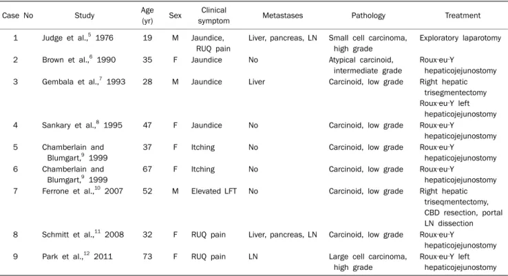
담관에서 발생하는 신경내분비종양은 매우 드물어 소화기 계에 생기는 신경내분비종양의 0.3% 미만으로 알려져 있다.1 담관계에서 가장 흔하게 발생하는 장소는 총담관이며 다음으 로 간문부와 담낭관에 발생한다고 알려져 있다.5 1976년 Judge 등6이 간문부 담관에 발생한 신경내분비종양에 대해 처음 기술한 이후 현재까지 간문부 담관에 발생한 원발성 신 경내분비종양은 세계적으로 총 9예가 보고되었다(Table 1).5-12 담관암은 성별과 관련이 없고 연령이 증가할수록 발생 위 험이 높은 반면 담관에 발생하는 신경내분비종양은 여성에서 더 흔히 나타나고 비교적 젊은 나이에 발생한다고 알려져 있 다.10 현재까지 보고된 간문부 담관의 신경내분비종양의 경우 에도 9명 중 6명이 여성으로 여성에서 더 흔하게 나타났으며 환자들의 평균 나이가 43세로 비교적 젊은 환자들에서 발생 하였다. 이번 증례에서는 고령의 남자에서 발생하여 기존의 보고들과 차이를 보였다. 담관계에 발생하는 신경내분비종양 의 예후 인자로는 2010년 세계보건기구에서 개정한 신경내분 비종양의 조직병리학적 등급과 재발 여부가 있으며 발생 위치 와는 연관이 없다. 세계보건기구(World Health Organiza- tion) 분류상 Grade 3의 분화도가 나쁜 신경내분비종양의 경
Table 2. Reported Cases of Small Cell Neuroendocrine Tumors of Bile Duct in Korea
Case No Study Age (yr) Sex Location Survival duration Treatment
1 Kim et al.,15 2000 64 M CBD Unknown Pancreaticoduodenectomy
2 Kim et al.,16 2004 57 M CBD 9 months Pancreaticoduodenectomy+adjuvant RT
3 Park et al.,17 2004 60 F Proximal CBD 5 months Transhepatic biliary drainage Roux‐en‐Y hepaticojejunostomy
4 Jeon et al.,18 2006 65 M Proximal CBD 1 year Pancreaticoduodenectomy
5 Cho et al.,19 2009 59 M Distal CBD 6 months Bile duct tumor resection Roux‐en‐Y hepaticojejunostomy M, male; F, female; CBD, common bile duct; RT, radiotherapy.
Table 1. Data on Hilar Neuroendocrine Tumors
Case No Study Age
(yr) Sex Clinical
symptom Metastases Pathology Treatment
1 Judge et al.,5 1976 19 M Jaundice, RUQ pain
Liver, pancreas, LN Small cell carcinoma, high grade
Exploratory laparotomy
2 Brown et al.,6 1990 35 F Jaundice No Atypical carcinoid,
intermediate grade
Roux‐eu‐Y
hepaticojejunostomy 3 Gembala et al.,7 1993 28 M Jaundice Liver Carcinoid, low grade Right hepatic
trisegmentectomy Roux‐eu‐Y left
hepaticojejunostomy
4 Sankary et al.,8 1995 47 F Jaundice No Carcinoid, low grade Roux‐eu‐Y
hepaticojejunostomy 5 Chamberlain and
Blumgart,9 1999
37 F Itching No Carcinoid, low grade Roux‐eu‐Y
hepaticojejunostomy 6 Chamberlain and
Blumgart,9 1999
67 F Itching No Carcinoid, low grade Roux‐eu‐Y
hepaticojejunostomy 7 Ferrone et al.,10 2007 52 M Elevated LFT No Carcinoid, low grade Right hepatic
triseqmentectomy, CBD resection, portal LN dissection 8 Schmitt et al.,11 2008 32 F RUQ pain Liver, pancreas, LN Carcinoid, low grade Roux‐eu‐Y
hepaticojejunostomy
9 Park et al.,12 2011 73 F RUQ pain LN Large cell carcinoma,
high grade
Roux‐eu‐Y left hepaticojejunostomy M, male; F, female; RUQ, right upper quadrant; LN, lymph nodes; LFT, liver function test; CBD, common bile duct.
우에는 2년 생존율이 0%인 반면 Grade 1, 2는 100%였다.13 신경내분비종양 중 소세포암종의 경우에는 96%가 주로 폐 에서 발생한다고 알려져 있고 폐외 소세포암종은 4%로 매우 드물며 담관 중에서도 특히 간문부 담관에 발생하는 소세포암 종은 아직까지 국내 보고가 없었다. 폐외 소세포암종은 위장 관이 가장 흔하나 기관지, 췌담도계, 난소, 고환, 유방, 또는 비뇨기계 등 내분비계가 존재하는 곳이면 어디든지 발생할 수
있다.14,15 그러나 담관에 원발성 소세포암종이 발생한 경우는
세계적으로 20예 정도에 불과하며 국내에서는 총 5예의 보고 가 있었다(Table 2).15-19 5예 모두 총담관에서 발생하였으며 간문부 담관에 발생한 예는 국내에서 처음이고, 세계적으로는 1976년 Judge 등6이 19세 남자에서 간문부 담관에 발생한 소세포 신경내분비종양을 보고한 이후 2번째 증례이다.
다른 신경내분비종양과는 달리 소세포 신경내분비종양은 질환의 진행이 빠르고 조기전이로 인해 진단 후 환자가 사망 에 이르는 기간이 매우 짧다고 알려져 있다.3 예후인자로는 신경주위 침윤, 종양의 병기, 발생위치가 보고되고 있으며 위 에 발생하는 소세포암종은 담관계에 발생하는 것보다 예후가 좋은 것으로 알려져 있다.3 수술이 완전 관해를 시킬 수 있는 유일한 치료 방법이며 항암화학요법이나 방사선요법에 대해 서는 효과가 명확히 입증된 바 없다.1,14 폐의 소세포암종 치료 와 유사하게 다제화학요법을 시행하고 있으나 증례가 충분하 지 않아 효과적인 항암화학요법에 대해 더 많은 연구가 필요 하다.
일반적으로 신경내분비종양은 증상이 발견되기까지 4-5년 정도 시간이 걸릴 만큼 종양의 성장속도가 느리고 생존기간이
길다고 알려져 있다.9 이전의 보고들에서도(Table 1) 9명 중 5명이 분화도가 좋고 전이가 없어 외과적 절제로 완치를 기대 할 수 있었다. 전이가 있는 경우는 4예였고, 전이가 있다 하더 라도 분화도가 좋은 비기능성 종양이 대부분이어서 총 9예 중 7예에서 외과적인 절제를 통해 완전 관해를 시킬 수 있었 다. 반면 분화도가 나쁜 소세포암종의 경우에는 일반적으로 예후가 불량하여 진단 당시 원격전이가 동반된 경우가 많고 빠른 시간 내에 종양이 진행하는 것으로 알려져 있다. 이번 증례에서도 조직학적으로 분화도가 나쁜 소세포암종의 형태 를 보이고 있었고 진단 당시 이미 주위 림프절과 장간막에 전이가 있었다.
신경내분비종양의 경우 8-35%에서 안면 홍조, 잦은 설사, 복통의 증상과 소변에서 5-hydroxyindoleacetic acid가 증가 한 소견을 보이는 카르시노이드 증후군을 보인다고 알려져 있 다.3 현재까지 보고된 간문부 담관에 발생한 신경내분비종양 의 증례들에서는 이러한 카르시노이드 증후군을 보이는 경우 는 없었고 담관암의 증상과 유사하게 상복부 동통, 오심, 구토 등의 임상 증상과 황달, 상복부 종괴 등의 소견을 보였다 (Table 1). 또한 모든 증례에서 영상 검사 소견상 담관암을 의심하여 수술이 행해진 후 병리검사에서 신경내분비종양으 로 진단되었다. 5예에서는 종양에 의한 폐쇄성 황달이 발생하 여 내시경 역행성 담췌관조영술을 통한 담관 배액을 시행하고 담즙 세포진 검사 및 담관 조직 검사를 시행했으나 수술 전 신경내분비종양으로 진단된 예는 없었다.5,7-9,12 이번 증례에서 도 담관배액을 목적으로 수술 전 시행한 내시경 역행성 담췌 관조영술에서 담관 내 조직 생검을 시행하였으나 신경내분비 종양으로 진단하지 못하였다. 신경내분비종양은 주로 점막하 종양의 형태로 발현하는 경우가 많기 때문에 주로 점막에서 조직을 얻는 담관 내 조직 생검이나 세포진 검사로는 진단이 어려울 가능성이 높다.
아직까지 국내에는 간문부 담관에 발생한 소세포 신경내분 비종양은 보고가 없다. 저자들은 황달로 내원하여 영상 검사 에서 간문부 담관의 종괴 소견을 보여 수술 후 병리검사에서 소세포 신경내분비종양으로 진단한 1예를 경험하여 문헌고찰 과 함께 보고한다.
REFERENCES
1. Klimstra DS, Modlin IR, Adsay NV, et al. Pathology reporting of neuroendocrine tumors: application of the Delphic consensus process to the development of a minimum pathology data set.
Am J Surg Pathol 2010;34:300-313.
2. Ganetsky A, Bhatt V. Gastroenteropancreatic neuroendocrine tumors: update on therapeutics. Ann Pharmacother 2012;46:
851-862.
3. Cho MY, Kim JM, Sohn JH, et al; The Gastrointestinal Pathology Study Group of Korean Society of Pathologists. Current trends of the incidence and pathological diagnosis of gastroentero- pancreatic neuroendocrine tumors (GEP-NETs) in Korea 2000- 2009: multicenter study. Cancer Res Treat 2012;44:157-165.
4. Yao JC, Hassan M, Phan A, et al. One hundred years after “carci- noid”: epidemiology of and prognostic factors for neuro- endocrine tumors in 35,825 cases in the United States. J Clin Oncol 2008;26:3063-3072.
5. Chamberlain RS, Blumgart LH. Carcinoid tumors of the extra- hepatic bile duct. A rare cause of malignant biliary obstruction.
Cancer 1999;86:1959-1965.
6. Judge DM, Dickman PS, Trapukdi S. Nonfunctioning argyrophilic tumor (APUDoma) of the hepatic duct: simplified methods of de- tecting biogenic amines in tissue. Am J Clin Pathol 1976;66:40- 45.
7. Brown WM 3rd, Henderson JM, Kennedy JC. Carcinoid tumor of the bile duct. A case report and literature review. Am Surg 1990;56:343-346.
8. Gembala RB, Arsuaga JE, Friedman AC, et al. Carcinoid of the in- trahepatic ducts. Abdom Imaging 1993;18:242-244.
9. Sankary HN, Foster P, Frye E, Williams JW. Carcinoid tumors of the extrahepatic bile duct: an unusual cause of bile duct obstruction. Liver Transpl Surg 1995;1:122-123.
10. Ferrone CR, Tang LH, D'Angelica M, et al. Extrahepatic bile duct carcinoid tumors: malignant biliary obstruction with a good prognosis. J Am Coll Surg 2007;205:357-361.
11. Schmitt TM, Bonatti H, Hagspiel KD, Iezzoni J, Northup P, Pruett TL. Carcinoid of the bile duct bifurcation. J Am Coll Surg 2008;206:399.
12. Park SY, Lee SH, Kim JY, et al. A case of large cell neuroendocrine cell carcinoma in the hilar bile duct. Korean J Med 2011;
80(Suppl 2):S137-S141.
13. Kim J, Lee WJ, Lee SH, et al. Clinical features of 20 patients with curatively resected biliary neuroendocrine tumours. Dig Liver Dis 2011;43:965-970.
14. Lee SS, Lee JL, Ryu MH, et al. Extrapulmonary small cell carcino- ma: single center experience with 61 patients. Acta Oncol 2007;46:846-851.
15. Kim SH, Park YN, Yoon DS, Lee SJ, Yu JS, Noh TW. Composite neu- roendocrine and adenocarcinoma of the common bile duct as- sociated with Clonorchis sinensis: a case report. Hepatogastro- enterology 2000;47:942-944.
16. Kim JH, Lee SH, Park J, et al. Extrapulmonary small-cell carcino- ma: a single-institution experience. Jpn J Clin Oncol 2004;34:
250-254.
17. Park HW, Seo SH, Jang BK, et al. A case of primary small cell carci- noma in the common bile duct. Korean J Gastroenterol 2004;
43:260-263.
18. Jeon WJ, Chae HB, Park SM, Youn SJ, Choi JW, Kim SH. A case of primary small cell carcinoma arising from the common bile duct. Korean J Gastroenterol 2006;48:438-442.
19. Cho SB, Park SY, Joo YE. Small cell carcinoma of extrahepatic bile duct presenting with hemobilia. Korean J Gastroenterol 2009;
54:186-190.