INTRODUCTION
Following the availability of novel technology, cere- bral digital subtraction angiography (DSA) is com- monly used clinically to diagnose cerebrovascular ab- normalities8). Cerebral DSA is a safe procedure; how-
ever, complications may occur in rare cases6)8). Considering the widespread use of cerebral DSA as a general diagnostic modality for cerebrovascular dis- ease, it is important to reduce the complications re- lated to this technique.
Among the various complications of cerebral DSA
Efficacy of dual antiplatelet therapy as premedication before diagnostic cerebral digital subtraction angiography
Yoon-Hee Choo, Young-Jin Jung, Chul-Hoon Chang, Jong-Hoon Kim
Department of Neurosurgery, Yeungnam University College of Medicine, Daegu, Republic of Korea
Objective : Several studies have reported that periprocedural dual anti- platelet therapy lowers the incidence of thromboembolic complications (TEC) associated with coiling of unruptured aneurysms. We hypothesized that preprocedural administration of dual antiplatelet agents (aspirin and cilostazol) for 7days may reduce the risk of complications associated with diagnostic cerebral digital subtraction angiography (DSA).
Methods : We retrospectively reviewed the records of patients who un- derwent diagnostic cerebral DSA between September 2015 and April 2018. Of the 419 patients included (149 men, 270 women, mean age 58.5 years), 221 (72 men, 149 women, mean age 57.8 years) who un- derwent cerebral DSA between September 2015 and June 2016 were not premedicated with antiplatelet therapy. The remaining 198 (77 men, 121 women, mean age 59.4 years) who underwent cerebral DSA be- tween July 2016 and April 2018 were premedicated with dual anti- platelet therapy (aspirin and cilostazol). We defined ischemic stroke as a cerebral DSA-induced complication identified on magnetic resonance imag- ing (MRI) among patients with neurological symptoms.
Results : Of the 221 patients who did not receive antiplatelet therapy, 210 (95.0%) showed no neurological symptoms; however, 11 (5.0%) de- veloped neurological symptoms with MRI-proven ischemic stroke, which represents a TEC. Of the 198 patients who received dual antiplatelet therapy, 196 patients (99.0%) showed no evidence of TEC. The remaining 2 (1.0%) developed diplopia and motor weakness each, and MRI con- firmed acute ischemic stroke (p=0.019).
Conclusions : The use of dual antiplatelet agents (aspirin and cilostazol) for 7 days before DSA may reduce the risk of cerebral DSA-induced TEC.
J Cerebrovasc Endovasc Neurosurg.
2019 September;21(3):131-137 Received : 13 July 2019
Revised : 18 September 2019 Accepted : 28 September 2019 Correspondence to Jong-Hoon Kim Department of Neurosurgery, Yeungnam University College of Medicine, 170 Hyeonchung-ro, Nam-gu, Daegu, Republic of Korea Tel : +82-53-620-3790, 3792 Fax : +82-53-620-3770 E-mail : [email protected]
ORCID : http://orcid.org/0000-0001-9492-6901
This is an Open Access article distributed under the terms of the Creative Commons Attribution Non- Commercial License (http://creativecommons.org/li- censes/by-nc/3.0) which permits unrestricted non- commercial use, distribution, and reproduction in any medium, provided the original work is properly cited.
Keywords digital subtraction angiography, antiplatelet therapy, premed- ication, thromboembolism
including nausea, vomiting, transient hypotension, anaphylaxis, and groin hematoma5)23), we focused on neurological complications secondary to thromboembolism.
Thromboembolism is the most significant complica- tion of cerebral DSA14), which occurs secondary to thrombus formation within catheters, within pre-exist- ing friable intravascular thrombotic plaques, or de- vice-induced microdissections11).
Antiplatelet and anticoagulant therapy effectively re- duces the risk of thromboembolism13)18). Several stud- ies have reported that periprocedural antiplatelet ther- apy effectively lowers the incidence of thromboem- bolic complications (TEC) that may occur with coiling of unruptured aneurysms15-17)24). However, to the best of our knowledge, no studies have been performed for diagnostic cerebral DSA. We hypothesized that pre- procedural administration of dual antiplatelet agents (aspirin and cilostazol) for 7 days may reduce the risk of complications associated with diagnostic cerebral DSA.
MATERIALS AND METHODS
Patients
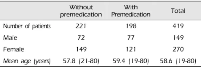
We retrospectively reviewed the records of 419 pa- tients who underwent diagnostic cerebral DSA be- tween September 2015 and April 2018 (Table 1).
Among these 419 patients, 221 patients who under- went diagnostic cerebral DSA from September 2015 to June 2016 did not received dual antiplatelet therapy as premedication. The remaining 198 patients who underwent diagnostic cerebral DSA between July 2016 and April 2018 received dual antiplatelet therapy as premedication. We changed the strategy because the incidence of TEC was higher than we thought when no premedication was given. Patient consent was waived for this study owing to the retrospective study design. Inclusion criteria for this study were non-ruptured aneurysms, arteriovenous malforma- tions, and arteriovenous fistulas, stenosis or occlusion of cerebral artery, as well as postoperative evaluation 1-2 years after coiling or clipping (Table 2). Patients with DSA performed for subarachnoid hemorrhage
(SAH), patients with SAH presenting for a week fol- low-up angiography, patients with an acute or sub- acute stroke, patients with symptoms of a transient is- chemic attack within 4 weeks of the scheduled cere- bral DSA, patients with evidence of intracerebral hemorrhage, those with a history of neurosurgical or interventional procedures 4 weeks prior to the study, and those with congestive heart disease or cardiac ar- rhythmia were excluded. We defined cerebral DSA-re- lated TEC as the occurrence of acute ischemic stroke that was confirmed on MRI within 48 hours of testing among patients presenting with neurological symptoms.
Procedures
Cerebral DSA was performed via the transfemoral arterial approach using a dedicated biplane neuro- angiography unit (RF-1000-125, Artis Zee biplane, Siemens Healthineer, Berlin, Germany) with the help of 1 type of guidewire (Terumo®, 0.035 inch, Tokyo, Japan) and 3 types of catheters-Taper (Cook, Bloomington, IN, USA), Imager (Boston Scientific, Marlborough, MA, USA), Simmons I (AngioDynamics, Queensbury, NY, USA). A non-ionic contrast medium (lomeron 300 [iomeprol 612.4 mg/mL], Bracco S.p.A., Milan, Italy) was used for all procedures with auto- matic machine injection of the contrast medium fol-
Without
premedication With
Premedication Total
Number of patients 221 198 419
Male 72 77 149
Female 149 121 270
Mean age (years) 57.8 (21-80) 59.4 (19-80) 58.6 (19-80) Table 1. Characteristics of the study population
Cases in premedication Number
Unruptured aneurysm Intervention evaluation Arteriovenous malformation Carotid carvernous fistula Moyamoya
Stenosis/occlusion/dissection Atherosclerosis
338 (80.67%) 51 (12.17%) 9 (2.15%) 5 (1.19%) 2 (0.48%) 13 (3.10%) 1 (0.24%)
Total 419
Table 2. Inclusion criteria for the study
lowed by a saline flush after each injection. The FemoSeal®, vascular closure device, was used for he- mostasis at the site of vascular access. All procedures in this series were performed by a single neuro- vascular surgeon (the author of this study).
Drug Protocols
Patients who underwent DSA between July 2016
and April 2018 received aspirin and cilostazol (both 100 mg/day) for 7 days. All antiplatelet therapy was discontinued post-procedure in premedicated patients who underwent cerebral DSA but did not develop TEC.
Statistical Analysis
Comparison between two groups was performed us-
Thromboembolic complication
Risk factor NO YES p-value
Hypertension No
Yes 109
87 1
1 0.874
Diabetes mellitus No
Yes 168
28 1
1 0.155
Hyperlipidemia No
Yes 169
27 2
0 0.572
Stroke history No
Yes 160
29 1
1 0.167
heart disease No
Yes 188
8 2
0 0.771
Smoking No
Yes 181
15 2
0 0.684
Kidney disease No
Yes 194
1 2
0 0.919
Number of vessel Examined 1 2 34 5
48 11666
1
00 11 0
0.989
Type of catheter Taper
Imager Simmon
159 9 28
10
1 0.358
Table 3-2. Correlation between risk factors and the incidence of thromboembolic complications secondary to cerebral digital sub- traction angiography in pretreated dual antiplatelet therapy as premedication
Thromboembolic complication
Risk factor NO YES p-value
Hypertension No
Yes 110
100 7
4 0.466
Diabetes mellitus No
Yes 180
30 9
2 0.720
Hyperlipidemia No
Yes 191
19 10
1 0.996
Stroke history No
Yes 171
39 10
1 0.426
heart disease No
Yes 195
15 11
0 0.359
Smoking No
Yes 185
25 9
2 0.535
Chronic kidney disease No
Yes 204
6 11
0 0.57
Number of vessel Examined 1 23 45
35 909 760
0 04 70
0.212
Table 3-1. Correlation between risk factors and the incidence of thromboembolic complications secondary to cerebral digital sub- traction angiography in non-pretreated dual antiplatelet therapy as premedication
ing Fisher’s exact test, chi-square test and in- dependent t tests (signigicant level 0.05). Statistical analysis was performed using the SPSS software, ver- sion 19 (IBM SPSS Statistics, IBM Corp., Armonk, NY).
A p-value<0.05 was considered statistically significant.
RESULTS
We analyzed the complication ratio based on risk factors including hypertension, diabetes mellitus, hy- perlipidemia, history of stroke, smoking, heart disease such as heart failure or arrhythmia, chronic kidney disease, and others that can affect the vascular status.
Our analysis showed that these risk factors did not significantly affect the incidence of DSA-induced TEC (Table 3-1, 3-2).
There was a significant difference in the incidence of TEC between patients who took dual antiplatelet ther- apy and those who did not, which is consistent with our hypothesis. Of the 221 patients who did not re- ceive antiplatelet therapy, 210 patients (95.0%) did not report neurological symptoms; however, 11 patients (5.0%) reported various neurological symptoms in- cluding motor weakness and showed MRI-proven is- chemic stroke (Fig. 1), which represents a TEC. Of the 198 patients who received dual antiplatelet therapy, 196 (99.0%) showed no evidence of TEC. However, 2 (1.0%) developed diplopia (Fig. 2) and motor weak- ness respectively, and acute ischemic stroke was con- firmed on MRI (p=0.019) (Table 4). Of the 13 patients diagnosed with DSA-induced TEC, 7 presented motor weakness, 4 reported visual disturbance, and 1 re- ported dysarthria and paresis (Table 5).
DISCUSSION
Cerebral DSA and noninvasive cerebrovascular imaging modalities have shown steady improvement over the past several decades. Presently, diagnostic cerebral DSA is commonly performed for treatment planning of endovascular or open surgical proce-
Fig. 2. Magnetic resonance image in treated premedication Image shows a man who was premedicated with dual antiplatelet therapy and was diagnosed with an acute ischemic stroke in- volving the right pretectal region. He complained of a diplopia.
Fig. 1. Magnetic resonance image in untreated premedication Diffusion-weighted magnetic resonance imaging scan obtained in a woman who developed right-sided motor weakness imme- diately after diagnostic cerebral digital subtraction angiography for an unruptured aneurysm. An acute ischemic stroke involving the left thalamus and both occipital lobes was confirmed as a thromboembolic complication. Of note, she had not been administered premedication.
dures8). However, diagnostic cerebral DSA is asso- ciated with a risk of complications including TEC. To qualify as a generalized diagnostic test, cerebral DSA should be safe with a low risk of complications6)8).
Diagnostic cerebral DSA may result in local compli- cations such as access-site hematoma formation and systemic complications including headache, nausea, vomiting, and/or transient hypotension, nephropathy, and neurological deficits23). Among the various com- plications of diagnostic cerebral DSA, we focused on TEC, which can cause neurological deficits.
Thromboembolism is the most common cause of is- chemic stroke, which can cause transient or perma- nent neurological deficit including hemiparesis, apha- sia, visual symptoms, and a diminished level of con- sciousness, among others2). Thus, thromboembolism counts as a significant complication of diagnostic cere- bral DSA. Complication rates are approximately 0.55-3.2%, although these vary across studies6)8). Most TEC occur within the first 48 hours post intervention.
We defined cerebral DSA-induced TEC as the occur- rence of acute ischemic stroke that was confirmed on MRI within 48 hours of testing in patients with neuro- logical symptoms.
Presently, the key concern is to minimize diagnostic cerebral DSA-related complications and identify strat-
egies to effectively manage those that have already occurred.
The risk factors for ischemic stroke after cerebral DSA include hypertension, diabetes mellitus, dyslipi- demia, cardiac disorders like heart failure or ar- rhythmia, a history of smoking, chronic kidney dis- ease, the number of catheters used, types of catheters used, and the volume of contrast agent used3)5)20)22)23). We analyzed the effect of each risk factor on the in- cidence of cerebral DSA-induced complications. The association between the incidence of TEC and each risk factor was non-significant.
An ischemic stroke may occur during the cerebral DSA procedure secondary to vascular spasm or neu- rovascular dissection following severe vascular steno- sis or occlusion. However, most instances of ischemic stroke following cerebral DSA may be associated with distal embolization of air or particulate matter11) sec- ondary to disruption of a calcified plaque, which ac- tually precipitates the clinical event. Air embolism can occur during catheter flushing, guidewire exchange, or contrast medium injection1)8), which are related to the operator’s skill. Thrombus formation may occur in catheters, in pre-existing friable intravascular throm- botic plaques, or device-induced microdissections17). In this study, a single neurosurgeon performed all cerebral DSA procedures, thereby minimizing the complication rate related to variations in operator’s skill. However, TEC secondary to emboli originating from an existing thrombus when the device passes in- to the vessel cannot be avoided. We concluded that this pathomechanism of thrombus formation is a ma- jor contributor to cerebral DSA-induced ischemic stroke. Therefore, we assumed that preprocedural an- tiplatelet therapy could effectively reduce cerebral DSA-induced TEC. Moreover, we hypothesized that premedication using dual antiplatelet therapy could be more effective than the use of a single antiplatelet agent.
Several studies have reported that the dual anti- platelet therapy compared to single antiplatelet ther-
Thromboembolic complication
Premedication No Yes Total p-value
NoYes (aspirin+cilostazol) Total
210 (95.0%) 196 (99.0%) 406 (96.9%)
11 (5.0%) 2 (1.0%) 13 (3.1%)
221198
419 0.019 Table 4. Correlation between the administration of premed- ication and the development of thromboembolic complications
Premedication Symptoms as
thromboembolic complication
No Yes
Motor weakness Ocular symptom Dysarthria Paresis Total
6 (46.2%) 3 (23.0%) 1 (7.7%) 1 (7.7%) 11 (84.6%)
1 (7.7%) 1 (7.7%) 0 (0.0%) 0 (0.0%) 2 (15.4%) Table 5. Patients diagnosed with thromboembolic complications
apy could significantly lower numerical or clinical findings associated with platelet aggregation that cause thrombus17)25)27)28)29). Platelet aggregation, an im- portant step in the process of thrombus formation, oc- curs through a chain of mechanisms. Antiplatelet agents are classified according to which mechanisms are inhibited. It can be divided into cyclooxygenase inhibitor, phosphodiesterase inhibitor, Adenosine dis- phosphonate receptor antagonist, glycopeptide IIb / IIIa antagonist and serotonin receptor antagonist30). Simultaneous use of antiplatelet agents with these dif- ferent mechanisms have shown the synergic effect26)28). Aspirin, an irreversible inhibitor of cyclooxygenase, inhibits the conversion of arachidonic acid to throm- boxane A2 and prevents vasospasm and platelet ag- gregation4)7)10). The effect of aspirin lasts approx- imately 7 to 10 days corresponding to the lifespan of platelet7). The premedication period was determined for 7 days in this study based on this fact.
Cilostazol inhibits phosphodiesterase 3, which is strongly expressed in platelets and vascular smooth muscle cells9). Thus, cilostazol inhibits platelet ag- gregation and vascular smooth muscle proliferation and causes vascular dilatation13)18)19)21). Furthermore, several reports have demonstrated the synergistic ef- fect of cilostazol and endogenous mediators to lower the risk of hemorrhage compared with the use of clo- pidogrel and endogenous mediators9). Therefore, we preferred cilostazol over clopidogrel as premedication to lower the risk of TEC. Patients who were pre- treated with dual antiplatelet therapy did not report hemorrhagic related complications.
This study suggests that preprocedural dual anti- platelet therapy can significantly reduce the cerebral DSA-induced TEC rate. However, there are some lim- itations on this study. First, considering the effect of learning curve of operator is the limitation of this pa- per, further reevaluation will be needed in the future.
Second, although our population is the perfect repre- sentation of our center, we could not determine whether this population was representative of the en-
tirety of population who were performed cerebral DSA. Third, this study was retrospective and con- sisted only of the available data at a single institution.
A better designed randomized controlled trial would be needed.
CONCLUSIONS
The use of dual antiplatelet agents (aspirin and cil- ostazol) over a week before the procedure may re- duce the risks of cerebral DSA-induced complications.
ACKNOWLEDGEMENTS
This work was supported by a grant from the Chunma medical research foundation, Korea, 2019.
REFERENCES
1. Bendszus M, Koltzenburg M, Bartsch AJ, Goldbrunner R, Günthner-Lengsfeld T, Weilbach FX, et al. Heparin and air filters reduce embolic events caused by in- tra-arterial cerebral angiography: a prospective, random- ized trial. Circulation. 2004;110(15):2210-5.
2. Bendszus M, Koltzenburg M, Burger R, Warmuth-Metz M, Hofmann E, Solymosi L. Silent embolism in diag- nostic cerebral angiography and neurointerventional pro- cedures: a prospective study. The Lancet. 1999;354(9190):
1594-7.
3. Britt PM, Heiserman JE, Snider RM, Shill HA, Bird C, Wallace RC. Incidence of postangiographic abnormalities revealed by diffusion-weighted MR imaging. American Journal of Neuroradiology. 2000;21(1):55-9.
4. Catella-Lawson F, Reilly MP, Kapoor SC, Cucchiara AJ, DeMarco S, Tournier B, et al. Cyclooxygenase inhibitors and the antiplatelet effects of aspirin. New England Journal of Medicine. 2001;345(25):1809-17.
5. Choudhri O, Schoen M, Mantha A, Feroze A, Ali R, Lawton MT, et al. Increased risk for complications fol- lowing diagnostic cerebral angiography in older patients:
Trends from the Nationwide Inpatient Sample (1999-2009). Journal of clinical neuroscience : official journal of the Neurosurgical Society of Australasia. 2016 Oct;32:109-14.
6. Dion JE, Gates PC, Fox AJ, Barnett HJ, Blom RJ. Clinical events following neuroangiography: a prospective study.
Stroke. 1987;18(6):997-1004.
7. Fiehler J, Ries T. Prevention and treatment of throm- boembolism during endovascular aneurysm therapy.
Clinical Neuroradiology. 2009;19(1):73-81.
8. Fifi JT, Meyers PM, Lavine SD, Cox V, Silverberg L, Mangla S, et al. Complications of modern diagnostic cerebral angiography in an academic medical center.
Journal of vascular and interventional radiology : JVIR.
2009 Apr;20(4):442-7.
9. Goto S. Cilostazol: potential mechanism of action for an- tithrombotic effects accompanied by a low rate of bleeding. Atherosclerosis Supplements. 2005 Dec 15;6(4):
3-11.
10. Gotoh F, Tohgi H, Hirai S, Terashi A, Fukuuchi Y, Otomo E, et al. Cilostazol stroke prevention study: A placebo-controlled double-blind trial for secondary pre- vention of cerebral infarction. Journal of stroke and cer- ebrovascular diseases : the official journal of National Stroke Association. 2000 Jul-Aug;9(4):147-57.
11. Hankey GJ, Warlow CP, Sellar RJ. Cerebral angiographic risk in mild cerebrovascular disease. Stroke. 1990;21(2):
209-22.
12. Jerjes-Sanchez C. Venous and arterial thrombosis: a con- tinuous spectrum of the same disease? : European Heart Jounal. 2005 Jan;26(1):3-4.
13. Kwon SU, Cho YJ, Koo JS, Bae HJ, Lee YS, Hong KS, et al. Cilostazol prevents the progression of the sympto- matic intracranial arterial stenosis: the multicenter dou- ble-blind placebo-controlled trial of cilostazol in sympto- matic intracranial arterial stenosis. Stroke. 2005 Apr;36(4):
782-6.
14. Lanterna LA, Tredici G, Dimitrov BD, Biroli F.
Treatment of unruptured cerebral aneurysms by emboli- zation with Guglielmi detachable coils: case-fatality, mor- bidity, and effectiveness in preventing bleeding-a sys- tematic review of the literature. Neurosurgery. 2004;55(4):
767-78.
15. Matsushige T, Kiura Y, Sakamoto S, Okazaki T, Shinagawa K, Ichinose N, et al. Multiple antiplatelet therapy contributes to the reversible high signal spots on diffusion-weighted imaging in elective coiling of un- ruptured cerebral aneurysm. Neuroradiology. 2013 Mar;55(4):449-57.
16. Nishikawa Y, Satow T, Takagi T, Murao K, Miyamoto S, Iihara K. Efficacy and safety of single versus dual antiplatelet therapy for coiling of unruptured aneurysms.
Journal of stroke and cerebrovascular diseases : the offi- cial journal of National Stroke Association. 2013 Jul;22(5):650-5.
17. Rahme RJ, Zammar SG, El Ahmadieh TY, El Tecle NE, Ansari SA, Bendok BR. The role of antiplatelet therapy in aneurysm coiling. Neurological research. 2014 Apr;36(4):383-8.
18. Shinohara Y, Katayama Y, Uchiyama S, Yamaguchi T, Handa S, Matsuoka K, et al. Cilostazol for prevention of secondary stroke (CSPS 2): an aspirin-controlled, dou- ble-blind, randomised non-inferiority trial. The Lancet Neurology. 2010;9(10):959-68.
19. Sudo T, Tachibana K, Toga K, Tochizawa S, Inoue Y, Kimura Y, et al. Potent effects of novel anti-platelet ag- gregatory cilostamide analogues on recombinant cyclic
nucleotide phosphodiesterase isozyme activity. Biochemical pharmacology. 2000;59(4):347-56.
20. Tanaka H, Ueda Y, Hayashi M, Date C, Baba T, Yamashita H, et al. Risk factors for cerebral hemorrhage and cerebral infarction in a Japanese rural community.
Stroke. 1982;13(1):62-73.
21. Tanaka T, Ishikawa T, Hagiwara M, Onoda K, Itoh H, Hidaka H. Effects of cilostazol, a selective cAMP phos- phodiesterase inhibitor on the contraction of vascular smooth muscle. Pharmacology. 1988;36(5):313-20.
22. Tanizaki Y, Kiyohara Y, Kato I, Iwamoto H, Nakayama K, Shinohara N, et al. Incidence and risk factors for subtypes of cerebral infarction in a general population:
the Hisayama study. Stroke. 2000;31(11):2616-22.
23. Timothy J. Kaufmann M, John Huston III M, Jay N.
Mandrekar P, Cathy D. Schleck B, Kent R. Thielen M, David F. Kallmes M. Complications of Diagnostic Cerebral Angiography: Evaluation of 19 826 Consecutive Patients1. Radiology.n 2007;243(3):812-819
24. Yamada NK, Cross DT, 3rd, Pilgram TK, Moran CJ, Derdeyn CP, Dacey RG, Jr. Effect of antiplatelet therapy on thromboembolic complications of elective coil emboli- zation of cerebral aneurysms. AJNR American journal of neuroradiology. 2007 Oct;28(9):1778-82.
25. Tepe G, Bantleon R, Brechtel K, Schmehl J, Zeller T, Claussen CD, et al. Management of peripheral arterial interventions with mono or dual antiplatelet therapy—
the MIRROR study: a randomised and double-blinded clinical trial. European radiology. 2012;22(9):1998-2006.
26. Geeganage CM, Diener H-C, Algra A, Chen C, Topol EJ, Dengler R, et al. Dual or mono antiplatelet therapy for patients with acute ischemic stroke or transient is- chemic attack: systematic review and meta-analysis of randomized controlled trials. Stroke. 2012;43(4):1058-66.
27. Wong KSL, Chen C, Fu J, Chang HM, Suwanwela NC, Huang YN, et al. Clopidogrel plus aspirin versus aspirin alone for reducing embolisation in patients with acute symptomatic cerebral or carotid artery stenosis (CLAIR study): a randomised, open-label, blinded-endpoint trial.
The Lancet Neurology. 2010;9(5):489-97.
28. Nishikawa Y, Satow T, Takagi T, Murao K, Miyamoto S, Iihara K. Efficacy and safety of single versus dual antiplatelet therapy for coiling of unruptured aneurysms.
Journal of stroke and cerebrovascular diseases : the offi- cial journal of National Stroke Association. 2013 Jul;22(5):650-5.
29. Cassar K, Ford I, Greaves M, Bachoo P, Brittenden J.
Randomized clinical trial of the antiplatelet effects of as- pirin–clopidogrel combination versus aspirin alone after lower limb angioplasty. British Journal of Surgery:
Incorporating European Journal of Surgery and Swiss Surgery. 2005;92(2):159-65.
30. Jennings LK. Mechanisms of platelet activation: need for new strategies to protect against platelet-mediated atherothrombosis. Thrombosis and haemostasis. 2009;102(08):
248-57.