ORIGINAL ARTICLE Print ISSN 1738-6586 / On-line ISSN 2005-5013 10.3988/jcn.2009.5.1.24 J Clin Neurol 2009;5:24-28
Long-Term Durability of Percutaneous Transluminal Angioplasty in Patients with Symptomatic
Middle Cerebral Artery Stenosis
Joon-Tae Kim, MDa; Seung-Han Lee, MDa; Seong-Min Choi, MDb; Man-Seok Park, MDa; Byeong-Chae Kim, MDa; Woong Yoon, MDc; Jae-Kyu Kim, MDc; Myeong-Kyu Kim, MDa; Ki-Hyun Cho, MDa
Departments of aNeurology and cRadiology, Chonnam National University Medical School, Gwangju, Korea Department of bNeurology, Chonnam National University Hwasun Hospital, Hwasun, Korea
Received December 1, 2008 Revised December 15, 2008 Accepted December 23, 2008 Correspondence Man-Seok Park, MD Department of Neurology, Chonnam National University Medical School, 8 Hak-dong, Dong-gu, Gwangju 501-757, Korea Tel +82-62-220-6174 Fax +82-62-228-3462 E-mail [email protected]
Background and PurposeaaPercutaneous transluminal angioplasty (PTA) is being increas- ingly used in the treatment of symptomatic middle cerebral artery (MCA) stenosis. We evalu- ated the long-term durability after PTA for symptomatic MCA stenosis.
MethodsaaWe analyzed consecutive patients included in our stroke database who were treat- ed with angioplasty alone. The subjects without major periprocedural complications were fol- lowed up for at least 42 months. Recurrent ischemic symptoms were defined as newly devel- oped episodes of transient ischemic attack or ischemic stroke in the territory of the treated vessel.
Stroke was defined as ischemic stroke in any vessel.
ResultsaaPTA was technically successful in 37 of the 40 included patients. Thirty-two of the 37 patients were followed up at regular intervals of 1 to 6 months in the outpatient clinic of our institution for at least 42 months. Restenosis occurred in 3 of the 32 patients (9.4%) within 2 years of PTA, and no restenosis was identified thereafter. Two of the three patients with re- stenosis had asymptomatic complications such as dissection and vasospasm during the inter- vention. The ischemic area was in the treated vessel in 1 of the 32 patients and in other vessels in 3 of the 32 patients (9.4%).
ConclusionsaaSuccessful PTA can result in a low rate of recurrent ischemic symptoms, and restenosis during a long-term follow-up appears to be more frequent in the early period.
J Clin Neurol 2009;5:24-28 Key Wordsaadurability, angioplasty, middle cerebral artery, restenosis.
Introduction
Intracranial atherosclerosis is a major cause of ischemic stroke (IS) in Asian populations. Previous studies found annual inci- dences of stroke recurrence in patients with symptomatic mid- dle cerebral artery (MCA) stenosis of 8-15%.1-5 Medical treat- ment has been widely and empircally used, but there is still a high rate of failures that result in recurrent transient ischemic attack (TIA), stroke, and death in symptomatic patients.4 Con- sequently, there is ongoing controversy on the optimal thera- peutic strategies for these patients. Nowadays endovascular therapy has emerged as an alternative potential therapeutic op- tion for patients with symptomatic MCA stenosis. With recent imrovements in microcatheter technology, percutaneous trans- minal angioplasty (PTA) is now being increasingly used in
the treatment of symptomatic MCA stenosis. There are sev- eral reports of fair results of PTA in selected patients with in- tracranial arterial disease,3,6,7 but there are few reports on the long-term durability of PTA for MCA stenosis.8 Restenosis after endovascular treatment has emerged as a significant com- plication, with a reported rate as high as 32%.3 Therefore, the present study evaluated the long-term durability of PTA for symptomatic MCA stenosis.
Methods
Subjects and data collection
We analyzed consecutive patients included in our stroke da- tabase who were treated with angioplasty alone. Between Feb- ruary 1996 and December 2004, 56 patients underwent 57 PTA
procedures for the treatment of symptomatic MCA stenosis at our institution. Exclusion criteria comprised acute interven- tion performed within 3 days of symptom onset (n=2), under- going MCA angioplasty for the treatment of acute cardio- genic embolic occlusion (n=11) or vasospasm after subara- chnoid hemorrhage (n=2), and atherosclerotic stenosis in the M2 segment of the MCA (n=1).9 All patients and their family members provided informed consents before the procedure.
All patients had severe MCA stenosis (>50% luminal narrow- ing) with ischemic symptoms despite medical treatment. Brief- ly, the stenosis percentage was quantified as the diameter of maximal narrowing (D narrow) relative to the diameter of the normal (D normal) symptomatic MCA just distal or proximal to the stenosis: {1–(D narrow/D normal)}×100%.10 The nor- mal portion of the MCA was defined as where the MCA walls appeared parallel on angiography.
Interventional technique and postprocedural management
PTA was performed by two experienced radiologists. The de- tailed method for PTA has been reported previously.9 A 6-F guiding catheter was inserted into the cervical portion of the internal carotid artery over an exchange wire. The first 30 pa- tients were treated with Stealth angioplasty balloons (Target Therapeutics, Fremont, CA), and the last 10 patients were treated with coronary balloons (HayatePro, Terumo, Tokyo, Japan). The balloon was inflated slowly with a screw-type pressure inflation device at 2-4 atm for 30-60 seconds either once or twice. Follow-up angiography was performed using a guiding catheter after the procedure, and the degree of re- sidual stenosis was recorded. We defined technical success as a residual stenosis of less than 50% on follow-up angio- grams without any serious complications. After the procedure, heparin was infused intravenously for 24 hours, and antiplate- let medication (a daily dose of 325 mg of aspirin plus 250 mg ticlopidine or 75 mg clopidogrel) was maintained contin- uously during the follow-up period.
Imaging follow-up
Imaging follow-up was performed with CT angiograms at 6 months, 12 months, and yearly or biannually thereafter. Regu- lar transcranial Doppler (TCD) follow-up was performed on 32 patients for first 6 months. After these, 19 patients under- went regular TCD follow-ups. A single-channel 2-MHz Dop- pler device was used for TCD examinations. M1 MCA was defined as an insonation depth of 45-65 mm.11,12 All record- ings were performed by an experienced sonographer who was not involved in the other aspects of the study. Patients regu- larly underwent TCD examinations on day 1 and at months 1 and 6 after PTA. We evaluated several parameters, includ-
ing the mean flow velocity (MFV) and peak systolic velocity (PSV), in order to assess flow velocity changes after PTA.
Radiological and clinical outcomes
Major complications were defined as symptomatic and severe neurological deficits related to procedures, and minor peripro- cedural complications were defined as asymptomatic or tran- sient neurological deficits. Restenosis was defined as an in- crease in the MFV of >30 cm/sec between two consecutive TCD examinations or as >50% stenosis measured on follow- up CT angiograms. If velocity changes remained below this threshold, the stenosis was considered to be stable. When a de- crease of >30 cm/s was observed, we considered MCA steno- sis to have regressed. This threshold value of 30 cm/s for son- ographic progression and regression was based on previous- ly described criteria.13 Recurrent ischemic symptoms were de- fined as newly developed episodes of TIA or ischemic stroke in the territory of the treated vessel. Stroke was defined as is- chemic stroke in any vessel.
Statistical analysis
Except where stated otherwise, data are presented as median values for continuous variables and as percentages for categor- ical variables. We compared baseline demographics and clini- cal risk factors using the chi-square test for categorical varia- bles and Student’s t-test for continuous risk factors between the TIA and ischemic stroke groups. We also used the Wilcox- on signed-rank test to compare baseline and follow-up TCD values. The Kaplan-Meyer method was used to estimate event- free survival rates. The data were analyzed using SPSS ver- sion 14.0.
Results
Clinical outcomes
PTA was technically successful in 37 of the 40 patients, with major complications (subarachnoid hemorrhage and cerebral infarction) in 2 cases and 1 case of technical failure. Other pe- riprocedural complications were asymptomatic or no neuro- logical sequelae (2 TIAs, 3 intimal dissections, and 1 vaso- spasm). The rate of minor periprocedural complications was 16.2% (6 of 37 patients). Thirty-two of the 37 patients were followed up at regular intervals of 1 to 6 months in the outpa- tient clinic of our institution for at least 42 months. There- fore, the 32 subjects included in this study underwent suc- cessful PTA without any major periprocedural complications and were followed up for at least 42 months. They compris- ed 18 men and 14 women with a mean age of 56.5 years. Pre- senting symptoms were TIA in 21 patients (65.6%) and is- chemic stroke in 11 patients (34.4%). The degree of stenosis
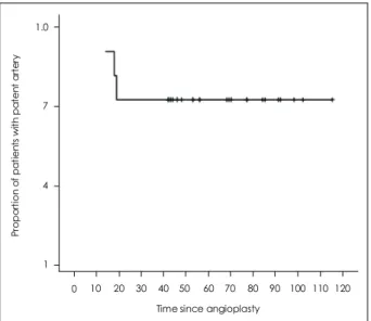
before the procedure ranged from 50% to 95% (median, 75.5%), whereas the degree of residual stenosis after the pro- cedure ranged from 9% to 48% (median, 21%). The general characteristics of the subjects (including risk factors and treat- ment) did not differ significantly between the two groups (Table 1). During the median follow-up period of 56 months (range, 42 to 115 months), restenosis occurred in 3 of the 32 patients (9.4%) within 2 years of PTA, and no restenosis was identified thereafter (Fig. 1). Two of the 3 patients with reste- nosis had asymptomatic complications such as dissection and vasospasm during the intervention (Table 2). The ischemic area was in the treated vessel in 1 of the 32 patients and in
other vessels in 3 of the 32 patients (9.4%)(Fig. 2).
Transcranial Doppler findings
TCD examinations showed a preprocedural MFV of 165.9±
47.6 cm/s (mean±SD) and a preprocedural PSV of 208.7±
61.2 cm/s (n=32), which declined to 146.2±53.9 and 186.4±
68.7 cm/s, respectively, immediately after the procedure (p<
0.05). In patients with serial TCD evaluations, the MFV and PSV gradually declined during 6 months of follow-up (n=19, p<0.05)(Table 3).
Discussion
In this study, the rates of restenosis and recurrent ischemic
Table 1. General characteristics of the subjects Characteristic TIA
(n=21)
IS (n=11)
Total (n=32)
Age* (years) 52.5 64.2 56.5
Prestenosis* (%) 73.7 75.7 74.4
Poststenosis* (%) 24.2 23.6 24.0
Follow-up* (months) 53 56 56
Risk factors† (%, n)
Hypertension 61.9 (13) 63.6 (7) 62.5 (20) Diabetes 09.5 (2)0 18.2 (2) 12.5 (4)0 Lipidemia 23.8 (5)0 36.4 (4) 28.1 (9)0 Smoking 23.8 (5)0 36.4 (4) 28.1 (9)0 History of stroke 28.6 (6)0 18.2 (2) 25.1 (8)0 Treatment† (%, n)
Single 33.3 (7)0 27.3 (3) 31.3 (10) Combination 66.7 (14) 72.7 (8) 68.7 (22)
*Student’s t-test for continuous risk factors was applied, †The chi- square test for categorical variables was applied.
TIA: transient ischemic attack, IS: ischemic stroke.
Table 2. Characteristics of subjects with restenosis
1 2 3
Site of stenosis Right, M1, mid Left, M1, mid Right, M1, mid
Presentation TIA TIA IS
Prestenosis (%) 56 60 57
Poststenosis (%) 45 12 14
Detection 19 14 18
(months)
Follow-up 79 54 54
(months) Follow-up
examination
Angiogram CT angiogram CT angiogram Procedure event Vasospasm None Asymptomatic dissection Associated
events
None None None
TIA: transient ischemic attack, IS: ischemic stroke.
Proportion of patients with patent artery
1 4 7 1.0
0
Time since angioplasty
10 20 30 40 50 60 70 80 90 100 110 120
Fig. 1. Arterial patency after angioplasty excluding patients with significant postprocedural complications. The restenosis rate was 9.37% (3 of the 32 patients) within 2 years of PTA, and no reste- nosis was identified thereafter. PTA: percutaneous transminal an- gioplasty.
Proportion of patients with patent artery
0.0 0.2 0.6 1.0
Time since angioplasty
50 75 100
0.4 0.8
Fig. 2. Ischemic events occurring after successful PTA. The ische- mic area was in the treated vessel in 1 of the 32 patients and in other vessels in 3 of the 32 patients. PTA: percutaneous transmi- nal angioplasty.
events were lower for successful PTA for MCA stenosis, with there being no patients with restenosis 2 years after PTA. Com- pared with previous data on the risk of medically treated MCA stenosis, the long-term durability and outcomes of successful PTA appear favorable.1,4 Recurrent ischemic events in the distribution of the treated vessel occurred in 3.1% of the pa- tients within a median follow-up period of 56 months. In ad- dition, major procedural complications after PTA that caused residual deficits or death were uncommon.9 Subjects with ma- jor procedural complications after PTA were excluded from the present study in order to assess the durability of success- ful PTA in patients with symptomatic MCA stenosis. Our results indicate that successful PTA can maintain the patency of the treated artery for up to 115 months. Wojak et al. attribu- ted a decrease in the rate of ischemic symptoms after angio- plasty to multiple factors such as an increase in vessel lumen, which increases blood flow, and remodeling of the endo- thelial surface to a smoother surface, which theoretically is less thrombogenic.14 In our study, the TCD data showed that the MFV at the stenotic artery did not decrease from normal values until 12 months after PTA. This result indicates that the remodeling of the vessel surface and the widening of vessel lumen can take several months after PTA, and also suggests that restenosis is more likely to develop in the early period after PTA. However, Fiorella et al. reported that the prepro- cedural velocity of 127.7 cm/s declined to 54.0 cm/s immedi- ately after the procedure.15 This difference might have been due to technical differences. Although our results differ from previous reports, TCD appears to be a favorable modality for follow-up after PTA.11,13-15 Future studies should investigate this difference. In addition, long-term durability of PTA could be favorable despite the possibility of complications or re- stenosis in the early period. The reported restenosis rate after angioplasty has ranged from 15% to 50%.7,8,14,16,17 Mazighi et al. recently reported that restenosis after treatment was asso- ciated with a vessel size of <2.5 mm or interventions that were performed in the setting of an acute stroke.8 Two of our three restenosis subjects were associated with asymptomatic procedural problems such as vasospasm and dissection. How- ever, factors associated with restenosis were not found in this study, and restenosis was not related to clinical recurrence.
Therefore, we consider restenosis to be a slowly progressing
event after PTA. Recurrent stroke occurred in four patients during the follow-up period. However, recurrent stroke in the territory of the treated artery did not occur with the excep- tion of one occurrence of TIA, which demonstrates that PTA reduces the rate of recurrent events in the territory of the treated artery.
The results of this study are subject to at least two limita- tions: 1) the use of a small sample with single-center results and 2) the use of imperfect follow-up imaging studies for evaluating restenosis, since conventional angiography (which is the reference standard) was performed in only a few patients.
Although TCD was performed on some patients during the follow-up, it has several disadvantages in the diagnosis of MCA stenosis.
From the results of this study, it is concluded that success- ful PTA in patients with symptomatic MCA stenosis is associ- ated with a low long-term rate of recurrent ischemic symp- toms and that restenosis after the procedure is more frequent in the early period. However, restenosis does not appear to be associated with ischemic symptoms. Further studies are need- ed to evaluate the long-term efficacy and safety of PTA.
REFERENCES
1. Chimowitz MI, Lynn MJ, Howlett-Smith H, Stern BJ, Hertzberg VS, Frankel MR, et al. Comparison of warfarin and aspirin for symptomat- ic intracranial arterial stenosis. N Engl J Med 2005;352:1305-1316.
2. Sacco RL, Kargman DE, Gu Q, Zamanillo MC. Race-ethnicity and de- terminants of intracranial atherosclerotic cerebral infarction. The north- ern Manhattan stroke study. Stroke 1995;26:14-20.
3. SSYLVIA Study Investigators. Stenting of symptomatic atherosclero- tic lesions in the vertebral or intracranial arteries (SSYLVIA): study results. Stroke 2004;35:1388-1392.
4. Chimowitz MI, Kokkinos J, Strong J, Brown MB, Levine SR, Silli- man S, et al. The warfarin-aspirin symptomatic intracranial disease study. Neurology 1995;45:1488-1493.
5. Thijs VN, Albers GW. Symptomatic intracranial atherosclerosis: out- come of patients who fail antithrombotic therapy. Neurology 2000;55:
490-497.
6. Mori T, Mori K, Fukuoka M, Arisawa M, Honda S. Percutaneous tran- sluminal cerebral angioplasty: serial angiographic follow-up after suc- cessful dilatation. Neuroradiology 1997;39:111-116.
7. Alazzaz A, Thornton J, Aletich VA, Debrun GM, Ausman JI, Charbel F. Intracranial percutaneous transluminal angioplasty for arteriosclero- tic stenosis. Arch Neurol 2000;57:1625-1630.
8. Mazighi M, Yadav JS, Abou-Chebl A. Durability of endovascular ther- apy for symptomatic intracranial atherosclerosis. Stroke 2008;39:1766- 1769.
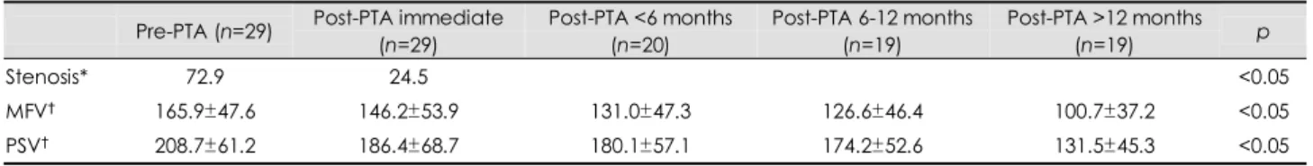
Table 3. Changes in TCD parameters after PTA Pre-PTA (n=29) Post-PTA immediate
(n=29)
Post-PTA <6 months (n=20)
Post-PTA 6-12 months (n=19)
Post-PTA >12 months
(n=19) p
Stenosis* 72.9 24.5 <0.05
MFV† 165.9±47.6 146.2±53.9 131.0±47.3 126.6±46.4 100.7±37.2 <0.05
PSV† 208.7±61.2 186.4±68.7 180.1±57.1 174.2±52.6 131.5±45.3 <0.05
*Student’s t-test was applied, †The Wilcoxon signed-rank test was applied.
TCD: transcranial doppler, PTA: percutaneous transminal angioplasty, MFV: Mean Flow Velocity, PSV: peak systolic velocity.
9. Yoon W, Seo JJ, Cho KH, Kim MK, Kim BC, Park MS, et al. Symp- tomatic middle cerebral artery stenosis treated with intracranial angi- oplasty: experience in 32 patients. Radiology 2005;237:620-626.
10. Kappelle LJ, Eliasziw M, Fox AJ, Sharpe BL, Barnett HJ. Importance of intracranial atherosclerotic disease in patients with symptomatic stenosis of the internal carotid artery. The north American symptoma- tic carotid endarterectomy trail. Stroke 1999;30:282-286.
11. Demchuk AM, Christou I, Wein TH, Felberg RA, Malkoff M, Grotta JC, et al. Specific transcranial Doppler flow findings related to the presence and site of arterial occlusion. Stroke 2000;31:140-146.
12. Demchuk AM, Christou I, Wein TH, Felberg RA, Malkoff M, Grotta JC, et al. Accuracy and criteria for localizing arterial occlusion with transcranial Doppler. J Neuroimaging 2000;10:1-12.
13. Arenillas JF, Molina CA, Montaner J, Abilleira S, González-Sánchez MA, Alvarez-Sabín J. Progression and clinical recurrence of sympto-
matic middle cerebral artery stenosis: a long-term follow-up trans- cranial Doppler ultrasound study. Stroke 2001;32:2898-2904.
14. Wojak JC, Dunlap DC, Hargrave KR, DeAlvare LA, Culbertson HS, Connors JJ 3rd. Intracranial angioplasty and stenting: long-term re- sults from a single center. AJNR Am J Neuroradiol 2006;27:1882-1892.
15. Fiorella D, Chow MM, Anderson M, Woo H, Rasmussen PA, Ma- saryk TJ. A 7-year experience with balloon-mounted coronary stents for the treatment of symptomatic vertebrobasilar intracranial athero- matous disease. Neurosurgery 2007;61:236-242; discussion 242-233.
16. Lee JH, Kwon SU, Lee JH, Suh DC, Kim JS. Percutaneous trans- luminal angi-oplasty for symptomatic middle cerebral artery stenosis:
long-term follow-up. Cerebrovasc Dis 2003;15:90-97.
17. Marks MP, Marcellus M, Norbash AM, Steinberg GK, Tong D, Al- bers GW. Outcome of angioplasty for atherosclerotic intracranial ste- nosis. Stroke 1999;30:1065-1069.