J Korean Soc Radiol 2017;76(3):198-205 https://doi.org/10.3348/jksr.2017.76.3.198
INTRODUCTION
Breast ultrasonography (US) is a widely used diagnostic mo- dality for breast lesions. US, however, has some limitations. It is heavily operator dependent (1) and represents hard and soft lesion by similar echogenecity in B-mode US (2). In general, be- nign masses have tendency to be softer than cancers but harder than normal breast tissue (2, 3). Elastography is a promising tech- nique for enhancing lesion differentiation by measuring tissue stiffness (2). Static elastography (1, 4-6) acquires relative stiffness of images in soft tissues by using US. Elastography calculates lo- cal tissue strain by comparison of the US signal gains before
and after operator’s light compression of the tissue (1, 5, 6), with hard zones demonstrating as dark and soft zones as bright (1, 5, 6).
Static elastography supplies not a quantitative estimation of the lo- cal stiffness but a map of the local tissues strain. Moreover, static elastography is easily affected by the operator’s compression pow- er, resulting in observer variability (7, 8). To be a reliable diagnos- tic method, elastography must not be easily affected by operators or interpreters.
To overcome this limitation, the quantitative elastography tech- nique of shear wave elastography (SWE) has emerged (2, 8). In SWE, estimation of the velocity of shear wave is involved in tissue elasticity imaging (1, 9-12). The mechanical vibration is remote-
Intra- and Interobserver Reproducibility of Shear Wave Elastography for Evaluation of the Breast Lesions
유방 병변에서 Shear Wave Elastography의 검사자 내, 검사자 간 재현성 평가
Min Ji Hong, MD
1, Hak Hee Kim, MD
2*
1Department of Radiology, Gil Hospital, Gachon University of Medicine and Science, Incheon, Korea
2Department of Radiology and Research Institute of Radiology, University of Ulsan College of Medicine, Asan Medical Center, Seoul, Korea
Purpose: To evaluate reproducibility of shear wave elastography (SWE) for breast lesions within and between observers and compare the reproducibility of SWE fea- tures.
Materials and Methods: For intraobserver reproducibility, 225 masses with 208 patients were included; and two consecutive SWE images were acquired by each observer. For interobserver reproducibility, SWE images of the same mass were ob- tained by another observer before surgery in 40 patients. Intraclass correlation co- efficients (ICC) were used to determine intra- and interobserver reproducibility.
Results: Intraobserver reliability for mean elasticity (Emean) and maximum elasticity (Emax) were excellent (ICC = 0.803, 0.799). ICC for SWE ratio and minimum elastici- ty (Emin) were fair to good (ICC = 0.703, 0.539). Emean showed excellent ICC re- gardless of histopathologic type and tumor size. Emax, SWE ratio and Emin repre- sented excellent or fair to good reproducibility based on histopathologic type and tumor size. In interobserver study, ICC for Emean, Emax and SWE ratio were excel- lent. Emean, Emax and SWE ratio represented excellent ICC irrespective of histo- pathologic type. ICC for Emean was excellent regardless of tumor size. SWE ratio and Emax showed fair to good interobserver reproducibility based on tumor size.
Emin represented poor interobserver reliability.
Conclusion: Emean in SWE was highly reproducible within and between observers.
Index terms Breast
Ultrasonography
Elasticity Imaging Techniques Breast Neoplasm
Received July 13, 2016 Revised October 4, 2016 Accepted November 24, 2016
*Corresponding author: Hak Hee Kim, MD Department of Radiology and Research Institute of Radiology, University of Ulsan College of Medicine, Asan Medical Center, 88 Olympic-ro 43-gil, Songpa-gu, Seoul 05505, Korea.
Tel. 82-2-3010-4390 Fax. 82-2-476-0090 E-mail: [email protected]
This is an Open Access article distributed under the terms of the Creative Commons Attribution Non-Commercial License (http://creativecommons.org/licenses/by-nc/4.0) which permits unrestricted non-commercial use, distri- bution, and reproduction in any medium, provided the original work is properly cited.
ly produced by using acoustic radiation force produced by a fo- cused US beam (1, 7, 8). The radiation force rearranges tissue, and the rearrangement causes a transient shear wave (2, 8, 13).
The shear wave carries information associated with the local vis- coelastic properties of the tissue, facilitating a quantitative ap- proach to the tissue (2, 7, 8). Shear waves move faster in stiffer area and slowly in softer area (9). The velocity information produc- es a color image of the stiffness and measures SWE features such as the minimum elasticity (Emin), mean elasticity (Emean), and maximum elasticity (Emax) and ratio of lesion-to-fat (SWE ratio) elasticity in a region of interest (1, 2, 9-13). Using quantitative elasticity data, SWE thus provides more objective information than static elastography and enhanced lesion differentiation than conventional grayscale US (14-17). Based on these characteris- tics, quantitative US elastography using SWE is expected to have higher reproducibility.
The purpose of our study was to estimate the intra- and in- terobserver reproducibility of SWE for breast masses and to de- termine which SWE features among Emin, Emax, Emean and SWE ratio was highly reproducible.
MATERIALS AND METHODS
Patients and Breast Masses
The Institutional Review Board approved this retrospective study, and informed consent was waived. Between January 2012 and February 2012, 295 patients underwent conventional US with SWE imaging in Asan Medical Center. Among them, 225 breast lesions of 208 patients (mean age, 47.20 years; range 18–82 years) were included for measurement of intraobserver repro- ducibility in our study. Inclusion criteria for intraobserver repro- ducibility included the following: 1) undergoing elastography more than twice on same lesion by one observer 2) having histo- pathologic results obtained with US-guided biopsy or surgical excision. 3) Not receiving chemotherapy or radiation therapy for any cancer. Among the 208 patients, 40 patients who underwent another US with SWE imaging by other operators during same period were included for interobserver reproducibility.
US Examination
Conventional US and elastographic imaging were obtained us- ing the Aixplorer® ultrasound system (SuperSonic Imagine, Aix-
en-Provence, France) with a 4–15 MHz linear transducer by one of five board certified radiologists who specialize in breast imag- ing with 1 to 10 years of experience interpreting images from a minimum of 1000 breast US examinations in the prior year. Each reader had experience with more than 100 cases of US breast elastography. At least two orthogonal grayscale images and elas- tography images were obtained of each solid lesion. For elastog- raphy, we used the same depth, focus position, and gain setting as for conventional images. Elastographic images were obtained by applying repetitive light compression at the skin above the target- ed breast lesion. SWE imaging was performed by setting the re- gion-of-interest quantification tool (ROI) called the Q-BoxTM, so that it included the lesion and the surrounding normal tissue.
Within the ROI, elastography values were gained by placement of a 3 mm round electronic cursor over the stiffest part of the le- sion and an area of fat. The maximum, mean stiffness and stan- dard deviation were obtained within these areas. The SWE ratio was determined by comparing the mean elasticity within the stiffest part of the lesion and fatty tissue (Fig. 1). For the assess- ment of intraobserver reproducibility, we compared SWE mea- surements performed on the same subject on the same day by the same operator. SWE measurement includes mean (Emean), mini- mum (Emin), maximum (Emax) SWE and SWE ratio. For ana- lyzing interobserver reproducibility, we recorded SWE scores individually by different two operators on different days. Per- formers were blinded to the results of others.
Statistical Analysis
The data were analysed using the statistical software (SAS ver- sion 9.1, MAGREE SAS Macro program, SAS Institute, Cary, NC, USA). Intra- and interobserver reproducibility was assessed through the calculation of the intraclass correlation coefficient (ICC). The ICC ranges between 0 and 1.00, with values closer to 1.00 representing better reproducibility. Measurement reliability was classified according to common criteria (Fleiss 1986) as ex- cellent (ICC > 0.75), fair to good (ICC = 0.40–0.75) and poor (ICC ≤ 0.40) (18).
RESULTS
In intraobserver reproducibility, among the 225 breast lesions, 174 (83.7%) were diagnosed as malignant and 51 were diagnosed
as benign on US-guided core needle biopsy or surgical excision.
The 174 malignant lesions were 133 cases of invasive ductal car- cinoma, 22 of ductal carcinoma in situ, 6 of mucinous carcino- ma, 5 of tubular carcinoma, 3 of invasive lobular carcinoma, 2 of microinvasive ductal carcinoma, 2 of invasive mammary carcino- ma, 1 of secretory carcinoma, respectively. Of the 51 benign le- sions, there were 29 cases of fibroadenoma, 5 cases of intraductal papilloma, 4 cases of adenosis, 5 cases of other proliferative breast lesion without atypia, 5 cases of nonproliferative breast change, and other benign disease. Histopathologic results of the 225 le- sions are summarized in Table 1. The mean mass size was 18.20 ± 11.55 mm in maximum diameter (range, 4–68 mm). Emean was the most reliable measurement of elasticity, with ICC = 0.803 (Table 2). About benignancy or malignancy, reliability of Emean made by same operator was excellent in malignant and benign lesion, (ICC = 0.762, 0.754 respectively). Emax of intraobserver reproducibility also showed excellent ICC values of 0.799 and was excellent (ICC = 0.769) for malignant lesions and fair to good (ICC = 0.744) for benign lesions, respectively. Overall SWE ratio represented fair to good reliability (ICC = 0.703). ICC for SWE ratio was 0.636 (fair to good) in benign lesion and 0.668 (fair to good) for malignancy. Intraobserver reproducibility of Emin was the least reliable in overall (ICC = 0.539), in benign le- sion (ICC = 0.491) and in malignancy (ICC = 0.498). Intraob- server reliability was consistently higher for all measured SWE
features of malignant masses than that of benign masses (Table 2). Ductal carcinoma in situ showed higher intraobserver repro- ducibility than invasive carcinoma in all SWE measurements (Table 3).
Intraobserver reliability based on maximum diameter of the mass is shown in Table 4. Emean showed excellent intraobserver reproducibility regardless of lesion diameter (size ≤ 10 mm; ICC
= 0.869, 10 mm < size ≤ 15 mm; ICC = 0.758, 15 mm < size ≤ 22 Table 1. Pathologic Diagnosis of 228 Breast Lesions in 208 Patients
Pathologic Diagnosis No. of Lesions Malignant lesions (n = 174)
Invasive ductal carcinoma 133
Ductal carcinoma in situ 22
Mucinous carcinoma 6
Tubular carcinoma 5
Invasive lobular carcinoma 3
Microinvasive ductal carcinoma 2
Invasive mammary carcinoma 2
Secretory carcinoma 1
Benign lesions (n = 51)
Fibroadenoma 29
Intraductal papilloma 5
Adenosis 4
Other proliferative breast lesion without atypia 5
Nonproliferative breast change 5
Columnar cell change 1
Fibroadenomatoid change 1
Pseudoangiomatous stromal hyperplasia 1 Fig. 1. Conventional US and elastographic images of invasive ductal carcinoma in a 49-year-old woman.
A. Conventional US image shows an irregular hypoechoic lesion without circumscribed margin.
B. Shear wave elastography images of quantification box (Q-BoxTM) applies on the stiffest part of mass. An additional Q-BoxTM is placed in the ad- jacent surrounding tissue.
US = ultrasonography
A B
mm; ICC = 0.760, size ≥ 22 mm; ICC = 0.804). On the other hand, Emin was the least reliable in overall size. Emean represent- ed excellent intraobserver reproducibility regardless of histo- pathologic results and tumor diameter.
Of 40 lesions, 32 were malignant and 8 were benign in in- terobserver study. Overall SWE ratio was the most reliable mea- surement of elasticity, with ICC = 0.909, and Emin was the least reliable (ICC = 0.394) (Table 5). Interobserver reproducibility was generally higher for most SWE measurements of benign masses than of malignancies, except for Emean. Emean, Emax
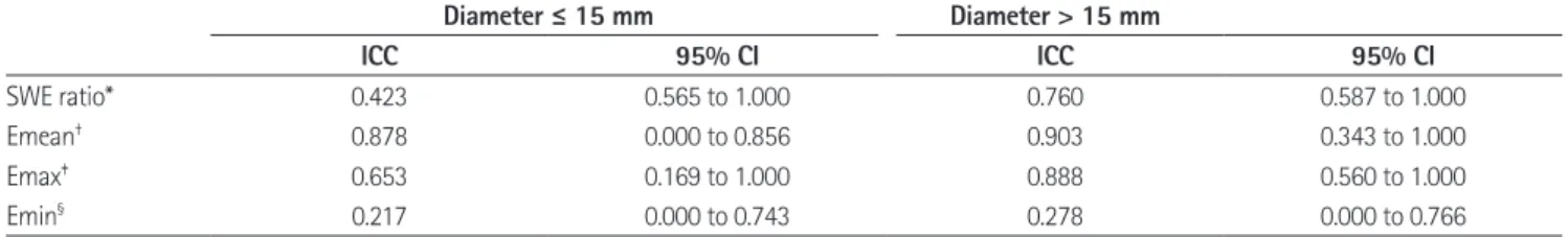
and SWE ratio showed excellent interobserver reproducibility irrespective of benignancy or malignancy. The interobserver re- producibility for all SWE measurements were better in larger le- sion than in smaller lesion (Table 6). ICC s for Emean was nearly perfect regardless of lesion diameter and benignancy or malig- nancy.
DISCUSSION
Emergence of SWE has facilitated quantitative and non-inva-
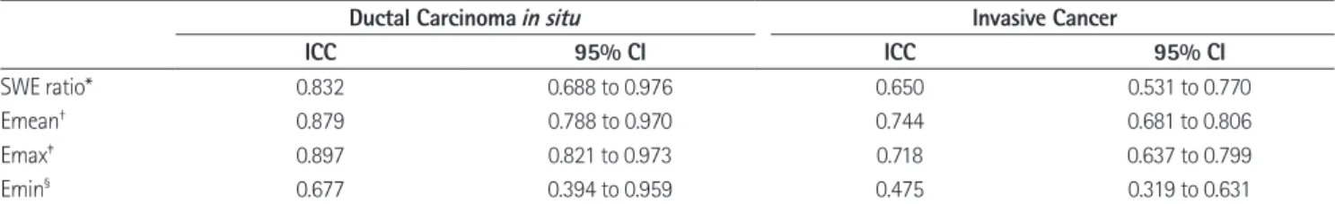
Table 3. Intraobserver Reproducibility of Quantitative Shear Wave Elastography Measurements in Patients with Invasive Cancer Versus with Ductal Carcinoma in situ
Ductal Carcinoma in situ Invasive Cancer
ICC 95% CI ICC 95% CI
SWE ratio* 0.832 0.688 to 0.976 0.650 0.531 to 0.770
Emean† 0.879 0.788 to 0.970 0.744 0.681 to 0.806
Emax‡ 0.897 0.821 to 0.973 0.718 0.637 to 0.799
Emin§ 0.677 0.394 to 0.959 0.475 0.319 to 0.631
*SWE ratio was the ratio between the mean elasticity value in the mass divided by the mean elasticity value in the fat.
†E mean was the mean value in the Q-BoxTM of the mass as calculated by the system.
‡E maximum mass was the maximum value in the Q-BoxTM of the mass as calculated by the system.
§E minimum mass was the minimum value in the Q-BoxTM of the mass as calculated by the system.
CI = confidence interval, ICC = intraclass correlation coefficients, SWE = shear wave elastography
Table 4. Intraobserver Reproducibility of Quantitative SWE Measurements according to the Mass Diameter
Diameter ≤ 10 mm 10 mm < Diameter ≤ 15 mm 15 mm < Diameter ≤ 22 mm Diameter > 22 mm
ICC 95% CI ICC 95% CI ICC 95% CI ICC 95% CI
SWE ratio* 0.718 0.585 to 0.852 0.634 0.412 to 0.856 0.746 0.622 to 0.870 0.644 0.446 to 0.842
Emean† 0.869 0.811 to 0.927 0.758 0.626 to 0.831 0.760 0.680 to 0.839 0.804 0.732 to 0.875
Emax‡ 0.891 0.848 to 0.934 0.752 0.665 to 0.839 0.740 0.656 to 0.825 0.770 0.646 to 0.893
Emin§ 0.592 0.412 to 0.772 0.603 0.443 to 0.762 0.504 0.301 to 0.707 0.435 0.157 to 0.712
Diameter was the largest measurement of the mass.
*SWE ratio was the ratio between the mean elasticity values in the mass divided by the mean elasticity value in the fat.
†E mean was the mean value in the Q-BoxTM of the mass as calculated by the system.
‡E maximum mass was the maximum value in the Q-BoxTM of the mass as calculated by the system.
§E minimum mass was the minimum value in the Q-BoxTM of the mass as calculated by the system.
CI = confidence interval, ICC = intraclass correlation coefficients, SWE = shear wave elastography Table 2. Intraobserver Reproducibility of Quantitative SWE Measurements
Overall Benign Malignancy
ICC 95% CI ICC 95% CI ICC 95% CI
SWE ratio* 0.703 0.606 to 0.801 0.636 0.452 to 0.821 0.668 0.555 to 0.780
Emean† 0.803 0.760 to 0.845 0.754 0.571 to 0.876 0.762 0.692 to 0.833
Emax‡ 0.799 0.744 to 0.855 0.744 0.648 to 0.889 0.769 0.667 to 0.822
Emin§ 0.539 0.419 to 0.660 0.491 0.115 to 0.868 0.498 0.360 to 0.637
*SWE ratio was the ratio between the mean elasticity value in the mass divided by the mean elasticity value in the fat.
†E mean was the mean value in the Q-BoxTM of the mass as calculated by the system.
‡E maximum mass was the maximum value in the Q-BoxTM of the mass as calculated by the system.
§E minimum mass was the minimum value in the Q-BoxTM of the mass as calculated by the system.
CI = confidence interval, ICC = intraclass correlation coefficients, SWE = shear wave elastography
sive measurement of tissue elasticity (19). A previous study showed that elastography had nearly the same diagnostic performance as conventional US, with 86.5% (45 of 52) sensitivity, 89.8% (53 of 59) specificity, and 88.3% (98 of 111) accuracy in the differentia- tion of benign from malignant solid breast masses (15). Because elastography is performed by free hand technique, variability is avoidable according to the performer’s compression motion and probe movement (20). Moreover, the breast is very redundant curved organ, and an application of homogenous continuous com- pression on breast lesion is difficult (21). To overcome the major limitation of elastography, subjective imaging method, quanti- tative elastography technique, SWE has been developed (14).
SWE is differentiated from conventional static elastography in automatically induced mechanical vibration via the radiation force of US beams (1, 2, 7, 8, 22). So, it less affected by the indi- vidual compression skills than static elastography (1, 2, 7, 8, 22).
To be used in daily practice as a reliable diagnostic tool, elastog- raphy must be objective. Some studies have reported intra- and interobserver reproducibility of SWE (7, 9, 22). Cosgrove et al. (9) reported that intraobserver reliability for maximum and mean
elasticity was almost perfect (ICC = 0.84 and 0.87) and was sub- stantial for the ratio of mass-to-fat elasticity (ICC = 0.77). Park et al. (7), interobserver variabilities for the elasticity score showed moderate agreement with the highest k value (k = 0.59). In phan- tom study of SWE (22), intraobserver ICCs were 0.65, 0.77, 0.92, and 0.91 for maximum elasticity; 0.70, 0.83, 0.94, and 0.94 for mean elasticity; and 0.67, 0.83, 0.92, and 0.92 for elasticity ratio for operators 1, 2, 3, and 4, respectively. Interobserver reproducibility showed good agreement with ICC values of 0.77 for maximum elasticity, 0.82 for mean elasticity, and 0.79 for elasticity ratio.
In this study, intra-and interobserver reproducibility of SWE was highly reproducible, except for Emin. Compared with pre- vious reports, outcome in our study was similar in intra- and in- terobserver reproducibility. Especially Emean showed excellent reliability in most cases except in case of invasive cancer. Emean, however, showed nearly excellent reliability even in case of inva- sive cancer (ICC = 0.744). Emean represented excellent intra-and interobserver reproducibility irrespective of lesion diameter, be- nignancy or malignancy.
Emin was the least reliable based on intra-and interobserver
Table 5. Interobserver Reproducibility of Quantitative SWE Measurements
Overall Benign Malignancy
ICC 95% CI ICC 95% CI ICC 95% CI
SWE ratio* 0.787 0.714 to 1.000 0.902 0.535 to 1.000 0.752 0.687 to 1.000
Emean† 0.909 0.558 to 1.000 0.879 0.486 to 1.000 0.906 0.481 to 1.000
Emax‡ 0.868 0.661 to 1.000 0.951 0.596 to 1.000 0.831 0.588 to 1.000
Emin§ 0.394 0.000 to 0.828 0.745 0.183 to 1.000 0.341 0.000 to 0.797
*SWE ratio was the ratio between the mean elasticity values in the mass divided by the mean elasticity value in the fat.
†E mean was the mean value in the Q-BoxTM of the mass as calculated by the system.
‡E maximum mass was the maximum value in the Q-BoxTM of the mass as calculated by the system.
§E minimum mass was the minimum value in the Q-BoxTM of the mass as calculated by the system.
CI = confidence interval, ICC = intraclass correlation coefficients, SWE = shear wave elastography
Table 6. Interobserver Reproducibility of Quantitative SWE Measurements according to the Mass Diameter
Diameter ≤ 15 mm Diameter > 15 mm
ICC 95% CI ICC 95% CI
SWE ratio* 0.423 0.565 to 1.000 0.760 0.587 to 1.000
Emean† 0.878 0.000 to 0.856 0.903 0.343 to 1.000
Emax‡ 0.653 0.169 to 1.000 0.888 0.560 to 1.000
Emin§ 0.217 0.000 to 0.743 0.278 0.000 to 0.766
Diameter was the largest measurement of the mass.
*SWE ratio was the ratio between the mean elasticity values in the mass divided by the mean elasticity value in the fat.
†E mean was the mean value in the Q-BoxTM of the mass as calculated by the system.
‡E maximum mass was the maximum value in the Q-BoxTM of the mass as calculated by the system.
§E minimum mass was the minimum value in the Q-BoxTM of the mass as calculated by the system.
CI = confidence interval, ICC = intraclass correlation coefficients, SWE = shear wave elastography
study. In intraobserver reproducibility, ICC values of Emin were 0.539 in overall, 0.491 in benign lesions and 0.498 in malignant lesions. Emin was also the least reliable based on the mass diam- eter. ICC values of Emin were 0.592 in the mass smaller than 1 cm, 0.603 in the mass between 1 cm and 1.5 cm and 0.504 in the mass between 1.5 cm and 2.2 cm and 0.435 in the mass larger than 2.2 cm. Emin was also the least reliable (ICC = 0.394) in in- terobserver reproducibility. These results of our study were con- cordant with the previous report (9) with the least reproducibility of Emin in breast masses (ICC = 0.71). The previous study ex- plained that a zero value came to the minimum on images where any pixel within the Q-BoxTM lacked SWE information, probably leading to lower reproducibility of Emin (9).
Intraobserver reproducibility of malignant lesions was consis- tently higher than that of benign lesions in all measured SWE features. Benign lesions tend to show a uniform homogeneity on the color map, as compared with malignant lesions (23). The dis- tinct color contrast in malignant lesions on color map might en- hance the intraobserver reproducibility. However, this hypothesis could not explain the higher intraobserver reproducibility in ductal carcinoma than invasive carcinoma. The number of inva- sive cancer was more than five times that of ductal carcinoma in situ in intraobserver study. Higher numbers of lesions could cause the heterogeneity in intraobserver reproducibility and the big differences in numbers between invasive cancer and ductal carcinoma in situ may influence the results of study. The study including similar numbers of each histopathologic type will be helpful in investigation of reproducibility by histopathologic type.
Contrary to results of previous studies (7, 9, 22), the ICC of in- terobserver reproducibility was superior to that of intraobserver reproducibility in our study. One previous study (22) showed the results of intraobserver variability of an operator with shorter ex- perience showed lower ICC values than those of the others. In spite of its low intrinsic changeability, SWE imaging may be in- fluenced by operators and operators’ experiences (22). The oper- ators had 1–3 years of experience in breast imaging in this study.
The operators’ relatively short experiences might affect the in- traobserver reproducibility for SWE. However, intra- and in- terobserver reproducibility of SWE for breast lesions was excel- lent irrespective of operators’ experiences in this study.
Our study has several limitations. First, variable sized lesions were included in our study (ranging from 4 to 68 mm). Regard-
ing the nodule size, a previous study (24) suggested that the care- ful decision should be made before recommending follow-up or biopsy for a small breast lesion on the basis of SWE features, to minimize false-negative results. However, several studies on SWE including subcentimeter-sized lesions also have showed good performance enough to differentiate benign lesions from malig- nant lesions (8, 23). Chang et al. (8) reported that significantly higher mean elasticity values was shown in malignant lesions than in benign lesions and even in the 4–5-mm-sized nodules, the elasticity values significantly differed between benign and malignant nodules (8). However, the SWE of small lesions mea- suring less than 5 mm might induce suboptimal results in this study. On the other hand, a previous study excluded the large breast masses (> 4 cm) that were not fully included in the maxi- mum range of SWE color overlay (25). In case of large lesions, the ROI might not fully cover the breast mass or surrounding breast parenchyma, this also may affect the results of this study.
Further studies with larger numbers of patients may provide more confirmative size cutoff that may not affect the results of SWE. Second, elastography images were obtained more than twice by same performers in one day for intraobserver reproduc- ibility, and elastography images for interobserver reproducibility were performed on different days by different operators. This may influence the results, but our study design was not for com- parison of intra-and interobserver reproducibility.
In conclusion, intra- and interobserver reproducibility of quantitative SWE for breast lesions was excellent in general. Es- pecially, Mean elasticity is the most reproducible within and across observers in SWE.
REFERENCES
1. Tanter M, Bercoff J, Athanasiou A, Deffieux T, Gennisson JL, Montaldo G, et al. Quantitative assessment of breast lesion viscoelasticity: initial clinical results using supersonic shear imaging. Ultrasound Med Biol 2008;34:1373-1386
2. Athanasiou A, Tardivon A, Tanter M, Sigal-Zafrani B, Ber- coff J, Deffieux T, et al. Breast lesions: quantitative elastog- raphy with supersonic shear imaging--preliminary results.
Radiology 2010;256:297-303
3. Sewell CW. Pathology of benign and malignant breast dis- orders. Radiol Clin North Am 1995;33:1067-1080
4. Hall TJ, Zhu Y, Spalding CS. In vivo real-time freehand pal- pation imaging. Ultrasound Med Biol 2003;29:427-435 5. Konofagou EE, D’hooge J, Ophir J. Myocardial elastogra-
phy--a feasibility study in vivo. Ultrasound Med Biol 2002;
28:475-482
6. Ophir J, Garra B, Kallel F, Konofagou E, Krouskop T, Righet- ti R, et al. Elastographic imaging. Ultrasound Med Biol 2000;26 Suppl 1:S23-S29
7. Park CS, Kim SH, Jung NY, Choi JJ, Kang BJ, Jung HS. In- terobserver variability of ultrasound elastography and the ultrasound BI-RADS lexicon of breast lesions. Breast Cancer 2015;22:153-160
8. Chang JM, Moon WK, Cho N, Yi A, Koo HR, Han W, et al.
Clinical application of shear wave elastography (SWE) in the diagnosis of benign and malignant breast diseases. Breast Cancer Res Treat 2011;129:89-97
9. Cosgrove DO, Berg WA, Doré CJ, Skyba DM, Henry JP, Gay J, et al. Shear wave elastography for breast masses is highly reproducible. Eur Radiol 2012;22:1023-1032
10. Bercoff J, Tanter M, Fink M. Supersonic shear imaging: a new technique for soft tissue elasticity mapping. IEEE Trans Ultrason Ferroelectr Freq Control 2004;51:396-409 11. Bercoff J, Tanter M, Muller M, Fink M. The role of viscosity
in the impulse diffraction field of elastic waves induced by the acoustic radiation force. IEEE Trans Ultrason Ferroelectr Freq Control 2004;51:1523-1536
12. Bercoff J, Chaffai S, Tanter M, Sandrin L, Catheline S, Fink M, et al. In vivo breast tumor detection using transient elastography. Ultrasound Med Biol 2003;29:1387-1396 13. Tozaki M, Fukuma E. Pattern classification of ShearWave™
Elastography images for differential diagnosis between be- nign and malignant solid breast masses. Acta Radiol 2011;
52:1069-1075
14. Yoon JH, Jung HK, Lee JT, Ko KH. Shear-wave elastography in the diagnosis of solid breast masses: what leads to false- negative or false-positive results? Eur Radiol 2013;23:2432- 2440
15. Itoh A, Ueno E, Tohno E, Kamma H, Takahashi H, Shiina T, et al. Breast disease: clinical application of US elastography for diagnosis. Radiology 2006;239:341-350
16. Zhu QL, Jiang YX, Liu JB, Liu H, Sun Q, Dai Q, et al. Real-time ultrasound elastography: its potential role in assessment of breast lesions. Ultrasound Med Biol 2008;34:1232-1238 17. Raza S, Odulate A, Ong EM, Chikarmane S, Harston CW. Us-
ing real-time tissue elastography for breast lesion evalua- tion: our initial experience. J Ultrasound Med 2010;29:551- 563
18. Hudson JM, Milot L, Parry C, Williams R, Burns PN. Inter- and intra-operator reliability and repeatability of shear wave elastography in the liver: a study in healthy volunteers. Ul- trasound Med Biol 2013;39:950-955
19. Ferraioli G, Tinelli C, Zicchetti M, Above E, Poma G, Di Gre- gorio M, et al. Reproducibility of real-time shear wave elas- tography in the evaluation of liver elasticity. Eur J Radiol 2012;81:3102-3106
20. Hiltawsky KM, Krüger M, Starke C, Heuser L, Ermert H, Jen- sen A. Freehand ultrasound elastography of breast lesions:
clinical results. Ultrasound Med Biol 2001;27:1461-1469 21. Yoon JH, Kim MH, Kim EK, Moon HJ, Kwak JY, Kim MJ. In-
terobserver variability of ultrasound elastography: how it affects the diagnosis of breast lesions. AJR Am J Roentgenol 2011;196:730-736
22. Mun HS, Choi SH, Kook SH, Choi Y, Jeong WK, Kim Y. Vali- dation of intra- and interobserver reproducibility of shear- wave elastography: phantom study. Ultrasonics 2013;53:
1039-1043
23. Ng WL, Rahmat K, Fadzli F, Rozalli FI, Mohd-Shah MN, Chandran PA, et al. Shearwave elastography increases diag- nostic accuracy in characterization of breast lesions. Medi- cine (Baltimore) 2016;95:e3146
24. Kim SJ, Ko KH, Jung HK, Kim H. Shear wave elastography:
is it a valuable additive method to conventional ultrasound for the diagnosis of small (≤2 cm) breast cancer? Medicine (Baltimore) 2015;94:e1540
25. Lee SH, Cho N, Chang JM, Koo HR, Kim JY, Kim WH, et al.
Two-view versus single-view shear-wave elastography:
comparison of observer performance in differentiating be- nign from malignant breast masses. Radiology 2014;270:
344-353
유방 병변에서 Shear Wave Elastography의 검사자 내, 검사자 간 재현성 평가
홍민지
1· 김학희
2*
목적: 유방 병변에서 shear wave elastography (이하 SWE)의 검사자 내, 검사자 간 재현성을 평가하고 어떤 지표가 재현 성이 높은지 알아보고자 한다.
대상과 방법: 검사자 내 재현성 평가를 위해서 208명의 225개의 유방 병변에서 연이은 두 번의 SWE 영상을 얻었다. 검 사자 간의 재현성 평가를 위해서 40명의 환자에서 같은 병변에 대해 다른 검사자에 의한 SWE 영상을 얻었다. 재현성 평 가는 급내상관계수(intraclass correlation coefficients)로 분석하였다.
결과: 검사자 내 재현성은 평균 탄력성과 최대 탄력성에서는 높은(excellent) 재현성을 보였으며, SWE 비율과 최소 탄력 성에서는 괜찮은(fair to good) 재현성을 보였다. 평균 탄력성은 병변의 병리나 크기에 상관없이 높은 재현성을 보였다. 최 대 탄력성, SWE 비율과 최소 탄력성은 병변의 병리와 크기에 따라 높은 혹은 괜찮은 재현성을 보였다. 검사자 간 재현성 연구에서는 평균, 최대 탄력성과 SWE 비율에서 높은 재현성을 보였다. 평균, 최대 탄력성과 SWE 비율은 병변의 병리에 상관없이 높은 재현성을 보였다. 평균 탄력성은 병변의 크기에도 상관없이 높은 재현성을 보였으나 최소 탄력성은 병변의 병리결과에 따라서나 병변의 크기에 따라서 낮은(poor) 재현성을 보였다.
결론: 평균 탄력성이 SWE 지표 중 검사자 내, 검사자 간 재현성 평가에서 가장 높은 재현성을 보여주었다.
1가천대학교 의과대학 길병원 영상의학과, 2울산대학교 의과대학 서울아산병원 영상의학과