© 2016 The Korean Academy of Medical Sciences.
This is an Open Access article distributed under the terms of the Creative Commons Attribution Non-Commercial License (http://creativecommons.org/licenses/by-nc/4.0) which permits unrestricted non-commercial use, distribution, and reproduction in any medium, provided the original work is properly cited.
pISSN 1011-8934 eISSN 1598-6357
Hirayama Disease with Proximal Involvement
Hirayama disease is a slowly progressing benign motor neuron disease that affects the distal upper limb. A 29-year-old man visited the hospital with a 1-year history of weakened left proximal upper limb. He was diagnosed with Hirayama disease 9 years ago, while there was no further progression of the muscle weakness afterward. Atrophy and weakness was detected in proximal upper limb muscles. Magnetic resonance imaging and somatosensory evoked potentials were normal. Needle electromyography showed abnormal findings in proximal upper limb muscles. Our patient had Hirayama disease involving the proximal portion through secondary progression. Clinical manifestation and accurate
electromyography may be useful for diagnosis. Rare cases with progression patterns as described here are helpful and have clinical meaning for clinicians.
Keywords: Electromyography; Hirayama Disease; Proximal Upper Limb; Secondary Disease Progression
Jinil Kim, Yuntae Kim, Sooa Kim, and Kiyoung Oh
Department of Physical Medicine and
Rehabilitation, Soonchunhyang University Cheonan Hospital, Cheonan, Korea
Received: 12 October 2015 Accepted: 1 March 2016 Address for Correspondence:
Kiyoung Oh, MD
Department of Physical Medicine and Rehabilitation, Soonchunhyang University Cheonan Hospital, 31 Suncheonhyang 6-gil, Dongnam-gu, Cheonan 31151, Korea E-mail: okokykkh@schmc.ac.kr
Funding: This study was supported by the Soonchunhyang University Research Fund.
http://dx.doi.org/10.3346/jkms.2016.31.10.1664 • J Korean Med Sci 2016; 31: 1664-1667
INTRODUCTION
Juvenile muscular atrophy of the distal upper extremity has been reported mainly in the Asian region (e.g., Japan and India) since 12 cases were reported by Hirayama et al. (1) in 1959. It has been called “Hirayama disease,” “benign focal amyotrophy,”
and “monomelic amytrophy” by various authors to date and is frequently observed among growing men in their teens and twenties. Hirayama disease has the clinical characteristic of in- vading only the distal portion of the muscles of a unilateral up- per extremity with accompanying symptoms of oblique atro- phy, cold paralysis, fasciculation, and tremor. Although the pathological and physiological mechanisms are currently un- clear, the major cause is anterior shifting of the dural sac, which occurs in cases of neck flexion because it causes an ischemic change in the anterior part of the spinal cord (2). Usually, there is no family history, while some researchers believe that genes such as KIAA1377 and C5ORF42 might be relevant (3). Cases with rare progress deviating from ordinary clinical aspects are occasionally reported, and they typically include bilateral symp- toms. In one Japanese study, approximately 3.1% of the patients showed asymmetric invasion in both upper extremities (2). There are even fewer cases of intrusion of the proximal upper extremity.
Our patient showed muscle weakness of proximal upper ex- tremity through secondary progression. We describe a case with atypical progression pattern, and we discuss the clinical mean- ing for clinicians.
CASE DESCRIPTION
In 15th January 2015, a 29-year-old man visited the hospital with a 1-year history of weakened muscle strength of the proxi- mal portion of the left upper extremity. The patient was diag- nosed with Hirayama disease 9 years previously in China. He had a muscle weakness in the distal portion of the left upper extremity, while there was no further progression of the muscle weakness afterward. Although he had weakness, it was not bad enough to be functional. Recently, however, he felt that the func- tionality and power of the proximal upper extremity was falling.
He reported no muscle weakness aggravation during winter, pain, dysesthesia, hypoesthesia, or dysuresia. He also had no extraordinary family history.
The physical examination results indicated the following:
blood pressure, 120/70 mmHg; pulse, 77/min; height, 170 cm;
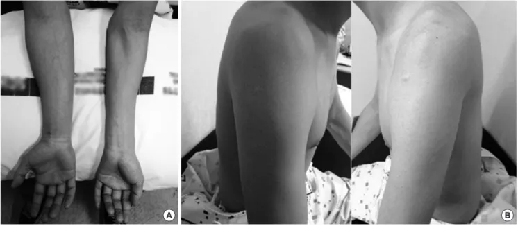
and weight, 59 kg. Apparent oblique atrophy was observed in the left forearm except for the brachioradialis muscle and in the hand muscle compared to the right side (Fig. 1A). Moreover, at- rophy was observed in the left biceps and deltoid muscles (Fig.
1B). Fasciculation in left hand muscle and irregular tremor of the fingers during finger extension were also observed. Range of motion of the joint was normal, and in an evaluation using the Medical Research Council (MRC) scale for muscle strength, left wrist flexor and extensor muscle strengths and left finger flexor, extensor, and abductor muscle strengths decreased to the 3/5 level, while those of the left elbow flexor and extensor and shoulder abductor decreased to the 4/5 level. The deep ten- CASE REPORT
Musculoskeletal Disorders
Kim J, et al. • Atypical Hirayama Disease
http://jkms.org 1665
http://dx.doi.org/10.3346/jkms.2016.31.10.1664
don reflex was normal, and no pathological reflex was observed.
Magnetic resonance imaging (MRI) of the cervical vertebrae in the neutral position showed loss of normal lordosis in the cervical vertebrae but no spinal cord atrophy. MRI of the cervi- cal vertebrae portion in maximum flexion showed neither clear- ly anterior shifting of the dural sac nor expansion of the epidur- al venous plexus behind it.
Nerve conduction study findings of bilateral upper and lower extremities were normal. A somatosensory evoked potentials (SEP) test was normal, and no extraordinary changes were ob- served in cases of maximum flexion of the cervical vertebrae.
In a needle electromyography (EMG) test performed on the left upper extremity, abnormal spontaneous activity was ob- served in the pronator teres, flexor carpi radialis, flexor carpi ul-
naris, and abductor digiti minimi muscles. In all but the bra- chioradialis muscle, polyphasic motor unit action potentials (MUAP) with increased amplitude and delayed duration were observed. Recruitment type decreased in all of the tested arm muscles (Table 1). No abnormal finding was observed in the bi- lateral paracervical muscles.
DISCUSSION
Several case reports have described Hirayama disease, a benign and characteristically slowly progressing disease. Approximate- ly 70% of patients experience disease progression within 3 years, and the disease stagnates after 5 years in approximately 95% of patients (4). Although most known cases involve invasion in a unilateral distal upper extremity, some cases show invasion in the upper extremities of both sides and some show progression within a unilateral proximal upper extremity. In particular, pro- gression within a unilateral proximal upper extremity is very rare, and according to Gourie-Devi et al. (5), only four of 44 (2.3%) patients showed apparent weakness and muscle atrophy in the proximal portion. Moreover, Huang et al. reported that only two of 40 patients had invasion of the biceps (5.0%) and only one had deltoid invasion (6). In another report, a 7-year-old Turkish girl showed weakness in the proximal upper extremity on clini- cal observation but normal EMG results (7).
To date, no report has examined additional disease progres- sion after 10 years (2). Hence, considering that our patient’s sec- ondary symptom progression occurred a long time after dis- ease onset, we believe that follow-up of the upcoming changes has great significance.
MRI findings showed no abnormal cervical vertebrae, and Fig. 1. Gross atrophy view of the patient. (A) Distal oblique portion of the left arm. (B) Proximal portion of the left arm.
A B
Table 1. Electromyography findings
Muscle IA FP R Duration Amplitude Polyphasia
ADM ↑ 2+ ↓ Long High +
FDI Normal 0 ↓ Long High +
EIP Normal 0 ↓ Long High +
EDC Normal 0 ↓ Long High +
ECR Normal 0 ↓ Long High +
FCR ↑ 1+ ↓ Long High +
FCU ↑ 2+ ↓ Long High +
PT ↑ 1+ ↓ Long High +
BR Normal 0 ↓ Normal Normal Normal
Triceps Normal 0 ↓ Long High +
Biceps Normal 0 ↓ Long High +
Deltoid Normal 0 ↓ Long High +
IA, insertional activity; FP, fibrillation potential; R, recruitment; ↑, increased; ↓, reduced;
ADM, abductor digiti minimi; FDI, first dorsal interosseous; EIP, extensor indicis pro- prius; EDC, extensor digitorum communis; ECR, extensor carpi radialis; FCR, flexor carpi radialis; FCU, flexor carpi ulnaris; PT, pronator teres; BR, brachioradialis.
Kim J, et al. • Atypical Hirayama Disease
1666 http://jkms.org http://dx.doi.org/10.3346/jkms.2016.31.10.1664 anterior shifting of the spinal cord during cervical vertebrae
flexion, which is the best-known pathophysiological cause of Hirayama disease, was not observed. This is consistent with the report stating that in cases of Hirayama disease, abnormal MRI results of the cervical vertebrae are no longer observed approxi- mately 10 years after the initial occurrence (8). However, the concept that spinal cord damage relevant to cervical vertebrae flexion is the main mechanism underlying Hirayama disease is not fully supported, and the patient may have never had such abnormal findings (9).
Thus, EMG becomes more important for patients diagnosed using medical imaging of insufficient reliability. Unless the EMG tester understands the rare progressive aspect of this disease, confusion can occur when testing the proximal upper extremity muscles. In particular, among benign motor neuron diseases, unilateral brachial amyotrophic diplegia develops from the dis- tal muscle in some cases. According to one report, symptoms of brachial amyotrophic diplegia appeared in the distal muscles, which are controlled by the middle and lower cervical vertebral spinal cord nerves, and progressed toward the proximal mus- cles, which are controlled by the upper cervical vertebral spinal cord nerves (10). This finding is similar to that of Hirayama dis- ease. As the progressive aspect and treatment plans differ be- tween the two diseases, caution is required when performing differential diagnosis.
On EMG examinations of patients with Hirayama disease, the proximal muscles of the biceps and deltoid, which are con- trolled by the C5 and C6 nerves, generally appear normal (11).
However, needle EMG findings of the biceps and deltoid can produce decreased recruitment and polyphasic MUAP with in- creased amplitude and long duration, as in this case, although it is very rare. This finding suggests recent disease progression in the proximal portion of the extremity. Similarly, a 23-year-old man in Brazil had monomelic amyotrophy that invaded the unilateral proximal upper extremity and tested malignant on both neurological and EMG screenings (12). In that case, pro- gression to the proximal portion occurred in the early disease stage, unlike the present case, in which invasion into the proxi- mal upper extremity occurred 9 years after disease onset. The SEP test, performed with the neck in a neutral and flexed posi- tion, generally shows no abnormal findings; this result was con- sistent with that observed in the present case (11).
According to Lin et al. (13), Hirayama disease can invade the proximal upper extremity, and overlooking this possibility can result in diagnostic error or confusion. Moreover, MRI findings of cervical vertebra flexion can play a critical role in early diag- nosis. However, the present case was different in that proximal invasion occurred several years after disease onset. As we could not rely on the medical imaging findings, background knowl- edge on the rare progression aspect of this disease and accurate EMG of the proximal upper extremity muscles were critical.
Hirayama disease remains a rarely reported disease with no successful treatment method other than wearing an orthosis that prevents neck flexion. Hence, an accurate early diagnosis is critical, and prevention of further aggravation is important.
However, it is difficult to diagnose nontraditional Hirayama disease subtypes because these are not frequently observed in clinics, and diagnostic caution is required to avoid wrong treat- ment. Benign motor neuron diseases, including Hirayama dis- ease, rarely become serious, and long-term observation is com- mon in most cases. Gourie-Devi et al. (5) followed 44 patients with upper monomelic amyotrophy for an average of 9.7 years (2.5-23 years), and none had newly progressed disease. How- ever, one study reported benign motor neuron disease in a pa- tient with brachial amyotrophic diplegia progressing to amyo- trophic lateral sclerosis that invaded the lower extremity and respiratory muscles over time (10). Monitoring this type of prog- ress is important because the clinical aspects can change from benign to malignant. Hence, it is necessary to track the progres- sion of Hirayama disease by carefully observing its long-term development and examining various cases.
Finally, as benign motor neuron diseases generally require long-term follow-up, we expect that cases of rare conditions such as that described here will be informative for clinicians.
DISCLOSURE
The authors have no potential conflicts of interest to disclose.
AUTHOR CONTRIBUTION
Conception and coordination of the study: Kim S, Oh K. Data acquisition and analysis: Kim J, Kim Y. Writing: Kim J, Oh K. Man- uscript approval: all authors.
ORCID
Jinil Kim http://orcid.org/0000-0002-4706-7259 Yuntae Kim http://orcid.org/0000-0003-4063-4692 Sooa Kim http://orcid.org/0000-0003-1578-0452 Kiyoung Oh http://orcid.org/0000-0002-1886-5462 REFERENCES
1. Hirayama K. Juvenile muscular atrophy of distal upper extremity (Hira
yama disease). Intern Med 2000; 39: 28390.
2. Huang YL, Chen CJ. Hirayama disease. Neuroimaging Clin N Am 2011;
21: 93950, ixx.
3. Lim YM, Koh I, Park YM, Kim JJ, Kim DS, Kim HJ, Baik KH, Choi HY, Yang GS, AlsoRallo E, et al. Exome sequencing identifies KIAA1377 and C5orf42 as susceptibility genes for monomelic amyotrophy. Neuromuscul Disord 2012; 22: 394400.
Kim J, et al. • Atypical Hirayama Disease
http://jkms.org 1667
http://dx.doi.org/10.3346/jkms.2016.31.10.1664
4. Tashiro K, Kikuchi S, Itoyama Y, Tokumaru Y, Sobue G, Mukai E, Akiguchi I, Nakashima K, Kira J, Hirayama K. Nationwide survey of juvenile mus
cular atrophy of distal upper extremity (Hirayama disease) in Japan. Amy- otroph Lateral Scler 2006; 7: 3845.
5. GourieDevi M, Nalini A. Longterm followup of 44 patients with brachi
al monomelic amyotrophy. Acta Neurol Scand 2003; 107: 21520.
6. Huang YC, Ro LS, Chang HS, Chen CM, Wu YR, Lee JD, Lyu RK. A clinical study of Hirayama disease in Taiwan. Muscle Nerve 2008; 37: 57682.
7. Yilmaz O, Alemdaroğlu I, Karaduman A, Haliloğlu G, Topaloğlu H. Benign monomelic amyotrophy in a 7yearold girl with proximal upper limb in
volvement: case report. Turk J Pediatr 2011; 53: 4716.
8. Kwon O, Kim M, Lee KW. A Korean case of juvenile muscular atrophy of distal upper extremity (Hirayama disease) with dynamic cervical cord compression. J Korean Med Sci 2004; 19: 76871.
9. Schröder R, Keller E, Flacke S, Schmidt S, Pohl C, Klockgether T, Schlegel
U. MRI findings in Hirayama’s disease: flexioninduced cervical myelopa
thy or intrinsic motor neuron disease? J Neurol 1999; 246: 106974.
10. Huh JP, Sung DH, Jo JM, Yoo JS, Kim BJ. Clinical characteristics, electrodi
agnostic, and imaging findings of atypical forms of motor neuron disease.
J Korean Acad Rehabil Med 2010; 34: 7019.
11. Hassan KM, Sahni H. Nosology of juvenile muscular atrophy of distal up
per extremity: from monomelic amyotrophy to Hirayama diseaseIndi
an perspective. Biomed Res Int 2013; 2013: 478516.
12. Neves MA, Freitas MR, Mello MP, Dumard CH, Freitas GR, Nascimento OJ. Benign monomelic amyotrophy with proximal upper limb involve
ment: case report. Arq Neuropsiquiatr 2007; 65: 5247.
13. Lin J, Zhang W, Wang N, Gao D, Chen X, Li W, Zhang L. Hirayama disease simple presenting proximal upper extremity muscular atrophy. Chin J Orthop 2011; 31: 2933.