Copyright © 2020. Anatomy & Cell Biology
Introduction
Duplication of the odontoid process remains an uncom- mon developmental pathology that has been reported rarely in the literature [1-4]. This variant has previously been linked to cases of pituitary gland duplication and Klippel- Feil syndrome, although the exact etiology and pathogenesis are incompletely understood [2, 3, 5]. Many other anatomical
variations have been reported in relation to odontoid malfor- mation, the most common being os odontoideum [1]. Other reported congenital or acquired variants of the odontoid process include hypoplasia, aplasia, anteversion, malposition, odontoid process bicornis, ossiculum terminale, and fused nonseparated odontoid process to the anterior arch of the atlas [1, 4, 5].
Most of the case reports currently available within the literature describe complete duplication of the odontoid pro- cess, most commonly in conjunction with fusion of adjacent cervical vertebrae, atlas deformities, and pituitary duplica- tion [1-4]. A single report describing a syndromic patient noted duplication of the C2 body in addition to duplication of the odontoid process [3]. We present a rare case of a pa- tient with a duplicated odontoid process in association with
Corresponding author:
Joe Iwanaga
Department of Neurosurgery, Tulane Center for Clinical Neurosciences, Tulane University School of Medicine, New Orleans, LA 70112, USA E-mail: [email protected]
Duplication of the odontoid process with
other congenital defects of the craniocervical Junction: case report and review of the
literature
Tyler Zeoli
1, Joe Iwanaga
1,5, CJ Bui
3, Aaron S. Dumont
1, R. Shane Tubbs
1,2,3,41Department of Neurosurgery, Tulane Center for Clinical Neurosciences, Tulane University School of Medicine, New Orleans, LA, 2Department of Structural & Cellular Biology, Tulane University School of Medicine, New Orleans, LA, 3Department of Neurosurgery and Ochsner Neuroscience Institute, Ochsner Health System, New Orleans, LA, USA , 4Department of Anatomical Sciences, St. George’s University, St. George’s, Grenada, West Indies, 5Department of Neurology, Tulane Center for Clinical Neurosciences, Tulane University School of Medicine, New Orleans, LA, USA
Abstract: Duplication of the odontoid process remains a rare developmental pathology that is underrepresented in the current literature. As the pivot point for the craniovertebral junction, the odontoid process is vital for the integrity of the atlanto-axial joint and the ability of the head and cervical spine to rotate correctly. The pathogenesis being incompletely understood, it has been proposed that odontoid process duplication involves faulty sclerotome migration and disruption of the axis ossification center. Patients presenting with this pathology usually have associated structural abnormalities. A detailed anatomical and embryological understanding of the odontoid process is necessary for successful management and treatment of patients presenting with odontoid process duplication. We present a rare case of a patient with a duplicated odontoid process in association with C2–C3 fusion, incomplete anterior arch of C1, variant inferior bony process of the transverse process of C1, and enlarged right jugular foramen.
Key words: Odontoid process duplication, Odontoid process, Axis, Duplicated odontoid process Received July 20, 2020; Revised September 15, 2020; Accepted September 21, 2020
C2–C3 fusion, an incomplete anterior arch of C1, variant bony process of the transverse process of C1, and an enlarged right jugular foramen.
Case Report
A two-year-old Caucasian male presented with congenital torticollis and limited head rotation. The patient was born at normal gestation and was the second son for his mother. No known medical conditions or history of surgery were noted.
On physical examination, the child moved all extremities, and had intact sensation in all major dermatomes tested.
Cranial nerves and major peripheral nerves were within nor- mal limits. His cerebellar examination was felt to be normal.
The neck was thought to be short. There was torticollis with neck deviation to the left. Rotation of the head was restricted and more so to the left. Flexion and extension of the head ap- peared to be normal. Radiographs noted a fusion anomaly at C2/C3. Computed tomography (CT) further identified a du- plicated odontoid process in association with the previously noted C2–C3 fusion, incomplete anterior arch of C1, variant bony process of the transverse process of C1, and enlarged right jugular foramen (Fig. 1).
The two separate apical ossifications centers of the odon- toid process were more or less in the same coronal plane with the left one being slightly larger than the right. The an- terior arch defect of C1 was approximately the width of the base of the combined odontoid process bases. The margins of the anterior arch were sclerosed indicating a congenital defect. The bodies of C2 and C3 were fused i.e., Klippel-Feil
anomaly. The right-sided jugular foramen was found to be three times larger than the more normally sized left jugular foramen. Lastly, the left transverse process was found to have an inferiorly pointed approximately 1 cm long, bony excres- cence. The remaining intracranial e.g., normal pituitary gland and spinal anatomy were felt to be normal.
Fig. 1. Three-dimensional reconstructed computed tomography of the craniocervical junction in the patient presented herein. Note the two ossification centers (arrow heads) of the duplicated odontoid process via a posterior view through the foramen magnum and from a posterior view (left). On this same image, the right jugular foramen (arrow) was significantly enlarged compared to the left. From an anterior view (right), note again the two ossification centers for the apical parts of the odontoid processes (arrow heads), the split anterior arch of C1 (asterisks), and the fused C2 and C3 vertebrae (black arrow). Also, note the right-sided inferior bony extension from the transverse process of C1 (white arrow).
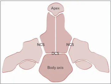
Body axis NCS DCS Apex
NCS
Fig. 2. Schematic drawing of the various ossification centers of the axis. Note the DCS between the body and odontoid process and the NCS separating the odontoid process and body of the axis from the remaining parts of C2. Smaller sits of fusion include the synchondrosis between the apex of the odontoid process and its lower parts and the midline fusion of left and right sides of the odontoid process. DCS, dentocentral synchondrosis; NCS, neurocentral synchondrosis.
Discussion
EmbryogenesisThe development of the human vertebral column has been widely explored, key research illustrating the vital role of neurulation and the subsequent formation of somites [6].
Somites are blocks of mesodermally-derived tissue that sit adjacent to the notochord bilaterally [6]. In addition, homeo- box (hox) and paired box (pax) genes are crucial in devel- opmental control and the organization of the craniocervical junction during embryogenesis [7]. Following neurulation, the mesodermal tissue adjacent the notochord creates 42–44 pairs of somites by the end of the fifth week of embryogen- esis [6, 7], each somite further differentiating into a sclero- tome, dermatome, and myotome [7]. These various subspe- cialized structures go on to form the vertebrae and ribs, skin, and muscles, respectively [6]. The proatlas is a rudimentary vertebral structure and precursor of the craniocervical junc- tion, formed from the fourth occipital sclerotome [7, 8]. This structure gives rise to the apex of the odontoid process in addition to sections of the atlas and both the apical and alar ligaments [6, 9]. However, the body of the odontoid process originates from the centrum formed from the first spinal sclerotome, which also forms most of the atlas. Therefore, while the entire odontoid process is formed via the first spi- nal and fourth occipital sclerotomes, the axis body originates
via the centrum of the second spinal sclerotome [7-9].
Embryologically, around the sixth week of gestation, the odontoid process begins to separate from the centrum of the atlas and migrates caudally to fuse with the axis [5, 9, 10].
Two cartilaginous bands, termed the neural central and den- tocentral synchondroses, serve as the initial juncture of the newly-migrated odontoid process (Fig. 2). These articulations separate the odontoid process from the axis body and neural arches, and both persist at birth [5, 9]. Specifically, the den- tocentral synchondrosis separates the base of the odontoid process from the body of the axis, whereas the neural central synchondrosis is immediately lateral to the odontoid process and axis, separating them from the neural arches bilaterally (Fig. 2) [5, 8, 9]. Ossification and eventual fusion of the odon- toid process to the C2 vertebral body has been described as occurring in three waves [8]. It begins with a primary os- sification center within the body of the axis, with eventual ossification around four months of gestation [8, 11]. This is followed by ossification from two other primary ossification centers located in the basal segment of the odontoid process, which fuse in the midline around six to seven months gesta- tion [1, 5, 8]. Lastly, ossification of the odontoid process apex begins around three to four years of age via a secondary ossi- fication center [8]. Complete fusion of the apex with the axis body occurs before 12 years of age [5].
Vertebral primordia O1
O2
O3 Proatlas
C1 C2 C3 Sclerotomes
Hypocentrum
Centrum Neural arch
Fig. 3. Schematic drawing of the embryological derivations of the craniocervical junction. Note the early sclerotome contributions to this region.
The first two occipital somites (yellow) give rise to the regions of the clivus at the skullbase and the third occipital somite (green) gives rise to parts of the occipital bone and region of jugular foramen. The proatlas (fourth occipital somite) gives rise to the apical region of the odontoid process, occipital condyle, and lateral mass of the atlas. The first cervical sclerotome gives rise to the anterior and posterior arches of the atlas, and the body of the odontoid process.
Pathogenesis
As discussed, there are numerous interconnected and simultaneous embryological events that ultimately lead to development of the odontoid process and the accompany- ing craniocervical junction. While the exact mechanism of odontoid process duplication remains unknown, current research suggests a lack of midline fusion of the two ossifica- tion centers within the basal odontoid process [1, 5]. Dys- regulation within mesenchymal development, vascular ab- normalities, and even abnormal migration or segmentation of sclerotomes have been suggested as the pathogenesis of this condition [1]. Furthermore, since nearly all case reports involve some combination of associated syndromes, verte- bral defects, pituitary duplications, and midline abnormali- ties, some suggest that the etiology lies in faulty interactions among the notochord, prechordal plate, and surface ecto- derm [4, 5]. Other possible causes include aberrant fusion or interactions across the cartilaginous synchondrosis between the odontoid process and axis, although these need to be explored further. Proatlantal derailments have been used to explain entities such as an epitransverse process. Although our case presented with an inversely positioned aberrant process on the transverse process, such a variant might be related embryologically. The apex of the odontoid has been attributed to the proatlas so the duplicated odontoid process ossification centers might relate to dysembyrology of the at- las as well. The basis of anterior arch of C1 defects, analogous to the duplicated odontoid process, can be tied to abnormal ossification center fusion or development [12]. Lastly, the re- gion of the jugular foramen is also attributed to arising from the proatlas (Fig. 3).
Taken together, third occipital somite (e.g., enlarged jugu- lar foramen), proatlantal (e.g., duplicated odontoid process- es), and first cervical sclerotome (e.g., anterior arch defect of C1) involvement would all most likely be involved in the present case.
Management
While cervical trauma leading to fracture or instability of the odontoid process can warrant immediate surgical in- tervention [13], management strategies for odontoid process duplication are less clear. All of the duplicated odontoid process cases presented symptomatically, either with limited head rotation, numbness and paresthesias, or with extensive craniofacial abnormalities. While 50% of cases had normal neurological exams with no deficits appreciated during the
physical exam, the other half showed clear syndromic abnor- malities associated with Klippel Feil syndrome and midline craniofacial defects [1-4]. The anatomy and narrowing of the differential can be evaluated in detailed with CT and mag- netic resonance imaging [1, 14]. Detailed management strat- egies regarding symptomatic and surgical approaches have not been fully explored. Related odontoid process variations such as os odontoideum strategize treatment based on odon- toid process stability and fracture integrity, which could help guide the management of related odontoid process abnor- malities [13]. Further research could analyze best practices among patients with odontoid process duplication.
In conclusion, odontoid process duplication is an ex- tremely rare anomaly that is underrepresented in the cur- rent literature. To our knowledge, only five cases have been reported to date, including the case presented here [1-4]. This case was also found to have Klippel Feil anomaly of C2/C3, incomplete anterior arch of C1, unusual inferior process of the transverse process, and an enlarged right jugular fora- men. To our knowledge, this is the first report in the litera- ture including this specific constellation of abnormalities.
Patients presenting with this pathology usually have asso- ciated structural abnormalities within the adjacent cervical vertebrae, Klippel Feil syndrome, pituitary gland duplication, or other craniofacial abnormalities. Thus, it is difficult to ascertain the exact pathogenesis leading to odontoid process duplication, although numerous embryological processes have been implicated. A detailed anatomical and embryolog- ical understanding of the odontoid process is necessary for successful management and treatment of patients present- ing with odontoid process duplication. Our case of odontoid process duplication with associated abnormalities can help bolster the current understanding of this rare pathology, and hopefully spark continued research into the etiology.
ORCID
Tyler Zeoli: https://orcid.org/0000-0002-3929-5964 Joe Iwanaga: https://orcid.org/0000-0002-8502-7952 CJ Bui: https://orcid.org/0000-0001-5760-1382
Aaron S. Dumont: https://orcid.org/0000-0002-8077-8992 R. Shane Tubbs: https://orcid.org/0000-0003-1317-1047
Author Contributions
Conceptualization: JI, ASD, RST. Data acquisition: TZ,
RST. Data analysis or interpretation: TZ, JI. Drafting of the manuscript: TZ, JI. Critical revision of the manuscript: CJB, ASD, RST. Approval of the final version of the manuscript:
all authors.
Conflicts of Interest
No potential conflict of interest relevant to this article was reported.
References
1. Garant M, Oudjhane K, Sinsky A, O'Gorman AM. Duplicated odontoid process: plain radiographic and CT appearance of a rare congenital anomaly of the cervical spine. AJNR Am J Neu- roradiol 1997;18:1719-20.
2. Dilettoso S, Uccello M, Dilettoso A, Gelardi S, Dilettoso B. Du- plicated odontoid process and atlas clefts associated to Klippel- Feil syndrome. Spine J 2012;12:449-50.
3. Manjila S, Miller EA, Vadera S, Goel RK, Khan FR, Crowe C, Geertman RT. Duplication of the pituitary gland associated with multiple blastogenesis defects: duplication of the pituitary gland (DPG)-plus syndrome. Case report and review of litera- ture. Surg Neurol Int 2012;3:23.
4. Usta Y, Sakha F, White WL, Little AS, Knecht L. Duplicated pi- tuitary gland and odontoid process. A case report. Neuroradiol J 2012;25:360-3.
5. Akobo S, Rizk E, Loukas M, Chapman JR, Oskouian RJ, Tubbs RS. The odontoid process: a comprehensive review of its anato-
my, embryology, and variations. Childs Nerv Syst 2015;31:2025- 34.
6. Kaplan KM, Spivak JM, Bendo JA. Embryology of the spine and associated congenital abnormalities. Spine J 2005;5:564-76.
7. Offiah CE, Day E. The craniocervical junction: embryology, anatomy, biomechanics and imaging in blunt trauma. Insights Imaging 2017;8:29-47.
8. Bambakidis NC, Dickman CA, Spetzler RF, Sonntag VKH.
Surgery of the craniovertebral junction. 2nd ed. New York:
Thieme; 2012.
9. Muhleman M, Charran O, Matusz P, Shoja MM, Tubbs RS, Loukas M. The proatlas: a comprehensive review with clinical implications. Childs Nerv Syst 2012;28:349-56.
10. Sureisen M, Achannan R, Chong KC, Wong CC. What the mind does not know, the eyes do not see: a rare congenital fu- sion of the odontoid process to the atlantal hemiarch. BMJ Case Rep 2015;2015:bcr2015212748.
11. Britton E, Barry M. Fractures of the spine in children. In: Ar- esti N, Barry M, Paterson M, Ramachandran M, editors. Paedi- atric orthopaedic trauma in clinical practice. London: Springer;
2015. p.57-70.
12. Bonneville F, Jacamon M, Runge M, Jacquet G, Bonneville JF.
Split atlas in a patient with odontoid fracture. Neuroradiology 2004;46:450-2.
13. O'Brien WT Sr, Shen P, Lee P. The dens: normal development, developmental variants and anomalies, and traumatic injuries.
J Clin Imaging Sci 2015;5:38.
14. Jain N, Verma R, Garga UC, Baruah BP, Jain SK, Bhaskar SN.
CT and MR imaging of odontoid abnormalities: a pictorial re- view. Indian J Radiol Imaging 2016;26:108-19.