Valgus deformity with lateral compartment knee osteoarthritis is much less common than varus deformity with medial compart
ment knee osteoarthritis. Thus, there are fewer studies reporting outcomes of distal femoral varus osteotomy (DFO)1,2) than of high tibial osteotomy (HTO) for medial osteoarthritis. Unfortu
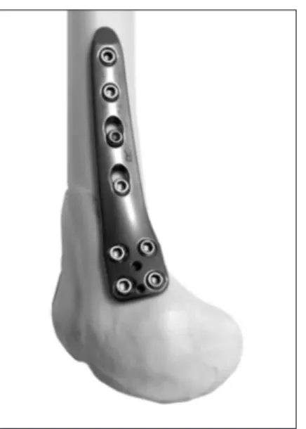
nately, before the TomoFix Medial Distal Femoral Plate (TomoFix MDF; DePuy Synthes GmbH, Solothurn, Switzerland) (Fig. 1) was launched in Japan, plates specifically designed for medial closed wedge distal femoral varus osteotomy (CWDFO) were not available. Therefore, from 2010 to 2014, we performed CWDFO
Closed Wedge Distal Femoral Osteotomy with a Polyaxial Locking Plate Designed for the Proximal Tibia: Minimum 5Year Outcomes
Ryuichi Nakamura, MD
1, Kenji Fujita, MD
2, Rei Omi, MD
1, Kazunari Kuroda, MD
1, Masaki Takahashi, MD
1, Kazumi Ikebuchi, MD
1, Hitoshi Nishimura, MD
3, and Yasuo Katsuki, MD
11Department of Orthopaedic Surgery, Yawata Medical Center, Komatsu; 2Department of Orthopaedic Surgery, Kanazawa University, Kanazawa; 3Department of Rehabilitation, Yawata Medical Center, Komatsu, Japan
Since distal femoral varus osteotomy (DFO) specific plates had not been available in Japan before 2015, we performed DFO using a plate for tibia. The purpose of this study was to elucidate the efficacy and problems associated with the nonspecific plate in DFO. We used NCBPT plates (Zimmer Inc.) in the upsidedown position and the minimum 5year outcomes were evaluated. The mean preoperative weight bearing line ratio and Japanese Orthopaedic Association score improved from 97.6%±35.8% and 68.0±11.5, respectively, to 44.0%±16.1% and 82.0±7.6, respectively, 1 year postoperatively and to 42.8%±15.7% and 86.0±8.2, respectively, 5 years postoperatively. The flexion range decreased from 149.0°±6.5° to 138.0°±5.7°
1 year postoperatively and to 135.0°±20.9° 5 years postoperatively. Although DFO using the NCBPT plate provided midterm benefits, it resulted in a loss of knee flexion, possibly due to excessive coverage of the medial femoral epicondyle.
Keywords: Knee, Osteoarthritis, Osteotomy, Plate, Complication pISSN 2234-0726 · eISSN 2234-2451
Knee Surgery & Related Research
Received February 13, 2017; Revised (1st) April 8, 2017;
(2nd) May 13, 2017; Accepted May 13, 2017 Correspondence to: Ryuichi Nakamura, MD, PhD
Department of Orthopaedic Surgery, Yawata Medical Center, 127 Yawata, Komatsu, Ishikawa 9238551, Japan
Tel: +81761471212, Fax: +81761471941 Email: ryu[email protected]
232
This is an Open Access article distributed under the terms of the Creative Commons Attribution NonCommercial License (http://creativecommons.org/licenses/bync/4.0/) which permits unrestricted noncommercial use, distribution, and reproduction in any medium, provided the original work is properly cited.
Copyright © 2017 KOREAN KNEE SOCIETY www.jksrr.org
Fig. 1. Fitting of the TomoFix Medial Distal Femoral Plate (MDF; DePuy Synthes GmbH) on the medial distal femur. The distal end of the plate is located above the medial femoral epicondyle.
with the NonContact Bridging Plate for Proximal Tibia (NCB
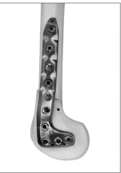
PT; Zimmer Inc., Winterthur, Switzerland), placing the plate upside down. Although not specifically designed for CWDFO, when used in this position, the NCBPT provided good contact with the medial distal femur (Fig. 2), and its polyaxial locking system3) was easy to use and allowed for correct screw placement.
The purpose of this study was to evaluate the midterm results of a minimum 5year followup of CWDFO using the NCBPT and to elucidate the problems associated with the use of a plate not specifically designed for CWDFO. TomoFix MDF has been avail
able in Japan since 2015, but we believe that some of the pitfalls of CWDFO may be more evident and more easily clarified by our description of the use of a plate not specifically designed for CWDFO.
Case Report
Five patients who were treated with CWDFO using the NCB
PT between 2010 and 2012 and had a minimum of 5 years of followup were investigated. We aimed for a corrected weight bearing line (WBL) percentage4) of 40%–50%, except in case 2.
Patients began active and passive range of motion on the first postoperative day and the standard protocols for partial and full weight bearing were started 4 and 8 weeks after surgery, respec
tively. All plates were removed one year after surgery.
A fulllength anteroposterior radiograph of the lower extremity with full weight bearing was obtained preoperatively and 1 and 5 years postoperatively. The Japanese Orthopaedic Association (JOA) score5) and range of knee flexion were assessed preopera
tively and 1 and 5 years after surgery. Both the radiographic and clinical outcomes at 1 year after surgery were evaluated before plate removal. The data for each case are presented in Table 1.
The preoperative mechanical proximal tibial angle (mMPTA)6)
Table 1. Details of Each Case Case Sex Age
(yr) BMI
(kg/m2) F/U time
(yr) mMPTA
(°)
mLDFA (°)a) Weight bearing line (%)b) Japanese Orthopaedic Association score (points)b) Preop Postopc) Preop 1 year 5 years Preop 1 year 5 years
1 F 74 20.5 6.2 88 84 97 60 17 17 65 75 80
2 F 58 25.7 5.8 90 80 94 156 60 60 50 80 80
3 F 77 19.9 5.4 89 83 93 100 43 43 70 80 85
4 F 58 18.7 5.0 90 81 90 81 50 50 75 80 85
5 M 62 23.0 5.0 88 81 90 91 50 44 80 95 100
Mean 65.7 21.6 5.5 89.0 81.8 92.8 97.6 44.0 42.8 68.0 82.0 86.0
SD 8.9 2.8 0.5 1.0 1.6 2.9 35.8 16.1 15.7 11.5 7.6 8.2
pvalued) <0.001 <0.005 NS <0.005 NS
BMI: body mass index, F/U: followup, mMPTA: mechanical medial proximal tibial angle, mLDFA: mechanical lateral distal femoral angle, Preop:
preoperative, Postop: postoperative, SD: standard deviation, NS: not significant.
a)A paired ttest was used to compare preoperative and postoperative values for mLDFA.
b)A paired ttest and Wilcoxon signedrank test were used to compare preoperative and 1 and 5year postoperative values for weight bearing percentage, Japanese Orthopaedic Association score, and range of knee flexion.
c)Postoperative measurement of the mLDFA was taken 4 weeks after surgery.
d)Differences with pvalues <0.05 were considered statistically significant.
Fig. 2. Fitting of the NonContact Bridging Plate for Proximal Tibia (NCBPT; Zimmer Inc.) in the upsidedown position on the medial distal femur. The plate fits well on the modeled bone after medial closed
wedge distal femoral osteotomy. It fully covers the medial femoral epi
condyle.
and mechanical lateral distal femoral angle (mLDFA)6) were 89.0°±1.0° and 81.8°±1.6°, respectively (Table 1). This means there was no proximal tibial deformity, and all knees had a valgus deformity at the distal femur. The mLDFA was significantly cor
rected to 92.8°±2.9° (p<0.001) (Table 1) and it was unchanged for 5 years after CWDFO in all patients (data not shown). This means no bonerelated correction loss or overcorrection was noted during the first 5 years postoperatively. The mean WBL percentage and JOA score were corrected from 97.6%±35.8% and 68.0±11.5 points to 44.0%±16.1% and 82.0±7.6 points (p<0.005 and p<0.05), respectively one year after surgery and maintained for the 5year followup period (Table 1). However, the mean range of flexion decreased significantly from 149.0°±6.5° preop
eratively to 138.0°±5.7° at 1 year after surgery (p<0.001). Despite plate removal, the range of flexion improved to the preoperative level in only one case (case 5) at the 5year followup. No patient required conversion to total knee arthroplasty (TKA) during the followup period.
In case 1, there was a 6° overcorrection compared with preop
erative planning. The overcorrection was not noted during sur
gery despite the intraoperative alignment check using the cable method7). At the 5year followup, the patient complained of in
creased discomfort in the left knee. Arthroscopy was performed and a horizontal tear of the medial meniscus was partially re
moved. Case 2 demonstrated the most severe valgus deformity, with a WBL percentage of 156% (Fig. 3A). The mMPTA, joint line convergence angle6) and mLDFA were 90°, –10°, and 80°, respectively. Therefore, the deformity was solely caused by the
distal femur and the remaining problem was severely stretched medial soft tissue laxity. If a TKA were chosen for such cases, a constrained type of TKA would be chosen due to the medial lax
ity8). Therefore, in spite of the medial and patellofemoral osteo
arthritic changes observed on arthroscopy, DFO was selected to avoid a constrained type TKA at a young age. To prevent osteo
arthritis progression, we aimed for a corrected WBL percentage of 60%, and the planned alignment was achieved (Fig. 3B). The mLDFA was corrected from 80° to 94°, and it was preserved dur
ing the 5year followup (Fig. 3C). Case 3 suffered an intraopera
tive lateral cortex fracture. Because bone union was delayed, full weight bearing was postponed until 3 months after surgery. The osteotomized site was completely fused 9 months after surgery, without correction loss or overcorrection. Despite the absence of intraoperative complication, case 4 had a 10° loss of flexion at the 5year followup as well. The flexion range of cases 1–4 was de
creased one year after surgery, resulting in a loss of flexion at the 5year follow up. In case 5, based on the flexion losses described above, we attempted deep knee flexion in the patient intraop
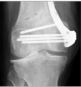
eratively after plate fixation. The medial patellofemoral ligament (MPFL) was too tight during knee flexion because the plate head encroached on the medial femoral epicondyle (Fig. 4). After re
leasing the MPFL, full knee flexion was achieved and no patellar subluxation was seen intraoperatively.
Discussion
As the improvement in the JOA score was maintained for 5
Fig. 4. Case 5. Anteroposterior view showing complete coverage of the medial femoral epicondyle by the plate.
A B C
Fig. 3. Case 2. Standing wholeleg radiographs taken preoperatively (A), 1 year postoperatively (B) and 5 years postoperatively (C).
years without TKA conversion in all cases, CWDFO with the NCBPT may provide midterm benefits for patients with lateral OA. Despite good longterm results of TKA for severely valgus knees, achieving adequate softtissue balance remains challeng
ing8). There may be limitations to correction of extraarticular bony valgus deformity solely via intraarticular correction during TKA; a constrained prosthesis, which can be used if appropriate balance is not obtained8), can result in problems such as implant failure. Therefore, for younger patients like case 2 (Fig. 3), even when there is moderate medial and patellofemoral osteoarthritis, timesaving CWDFO could be a possible option to avoid TKA with a constrained prosthesis.
The healing of the lateral cortex fracture in case 3 required 9 months. Therefore, preserving an intact lateral cortex is crucial to the success of medial CWDFO. For open wedge HTO, a lateral cortex fracture is classified as Takeuchi type I or II9). According to the “fibulaassupportingstrut theory” of Nakamura et al.10), a type I lateral cortex fracture in which the fracture portion is sup
ported by the fibula is considered stable, whereas a type II frac
ture, in which the fracture portion is not supported by the fibula, is considered unstable10). Because there is no supporting structure similar to the fibula in the distal femur, lateral cortex fractures that occur during CWDFO should always be considered un
stable.
More than 10º of decrease in flexion was found in only one case (case 2) and the other cases showed relatively wellpreserved flex
ion angles. However, the mean flexion range decreased one year after surgery, resulting in permanent loss of flexion in four of our five cases (cases 1–4). Therefore, for more objective evaluation of these results, despite the lack of midterm followup, we inves
tigated six patients treated by CWDFO using a TomoFix MDF with a minimum of 1year followup (range, 1.2 to 2.2 years).
In that series, the JOA score improved from 70.0±13.8 points to 96.7±2.6 points (p<0.005, paired ttest) one year after surgery (data not shown in Table). In contrast to CWDFO with the NCB
PT, the mean range of flexion also significantly improved from 130.8°±3.8° to 144.2°±10.7° (p<0.01, paired ttest) one year after surgery (data not shown in Table). These results might indicate that flexion loss may be a plateinduced complication. Figures 1 and 2 show the difference in plate position between the TomoFix MDF and the NCBPT. The distal end of the former is located above the medial femoral epicondyle so as not to cover the joint capsule (Fig. 1), while the latter fully covers the medial femoral epicondyle (Fig. 2). This difference may imply that putting a plate on the medial femoral epicondyle causes flexion loss after CWDFO due to the immobilization of the medial capsule and/or
MPFL. The fact that only the patient with MPFL release during CWDFO (case 5) recovered full range of motion may support this hypothesis. Therefore, a capsulefriendly CWDFO plate may be required to acquire a good range of flexion. Even when DFO
specific plates are used, special care should be taken not to place the plate on the medial femoral epicondyle. An intraoperative check of the flexion range may be useful for prevention of the flexion loss.
In conclusion, the use of the NCBPT in the upsidedown posi
tion for CWDFO for the treatment of valgus knee deformities provided midterm benefits for patients with lateral unicompart
mental osteoarthritis. Although the number of patients was small for statistical analysis, the outcomes indicate that using the NCB
PT plate may cause loss of knee flexion.
Conflict of Interest
No potential conflict of interest relevant to this article was re
ported.
References
1. Stahelin T, Hardegger F, Ward JC. Supracondylar osteotomy of the femur with use of compression. Osteosynthesis with a malleable implant. J Bone Joint Surg Am. 2000;82:71222.
2. Kosashvili Y, Safir O, Gross A, Morag G, Lakstein D, Back
stein D. Distal femoral varus osteotomy for lateral osteo
arthritis of the knee: a minimum tenyear followup. Int Orthop. 2010;34:24954.
3. Jockel JA, Erhardt J, Vincenti M, Reissig J, Hoffmann R, Hu
sain B, Tager G, Partenheimer A, Lill H, Gebhard F, Roderer G. Minimally invasive and open surgical treatment of proxi
mal tibia fractures using a polyaxial locking plate system: a prospective multicentre study. Int Orthop. 2013;37:7018.
4. Takeuchi R, Ishikawa H, Aratake M, Bito H, Saito I, Kum
agai K, Akamatsu Y, Saito T. Medial opening wedge high tibial osteotomy with early full weight bearing. Arthroscopy.
2009;25:4653.
5. Okuda M, Omokawa S, Okahashi K, Akahane M, Tanaka Y.
Validity and reliability of the Japanese Orthopaedic Association score for osteoarthritic knees. J Orthop Sci. 2012;17:7506.
6. Paley D, Herzenberg JE, Tetsworth K, McKie J, Bhave A.
Deformity planning for frontal and sagittal plane corrective osteotomies. Orthop Clin North Am. 1994;25:42565.
7. Yoon SD, Zhang G, Kim HJ, Lee BJ, Kyung HS. Comparison of cable method and miniaci method using picture archiving
and communication system in preoperative planning for open wedge high tibial osteotomy. Knee Surg Relat Res.
2016;28:2838.
8. Ranawat AS, Ranawat CS, Elkus M, Rasquinha VJ, Rossi R, Babhulkar S. Total knee arthroplasty for severe valgus defor
mity. J Bone Joint Surg Am. 2005;87 Suppl 1:27184.
9. Takeuchi R, Ishikawa H, Kumagai K, Yamaguchi Y, Chiba N, Akamatsu Y, Saito T. Fractures around the lateral cortical
hinge after a medial openingwedge high tibial osteotomy:
a new classification of lateral hinge fracture. Arthroscopy.
2012;28:8594.
10. Nakamura R, Komatsu N, Murao T, Okamoto Y, Nakamura S, Fujita K, Nishimura H, Katsuki Y. The validity of the clas
sification for lateral hinge fractures in open wedge high tibial osteotomy. Bone Joint J. 2015;97:122631.