• 검색 결과가 없습니다.

Temporomandibular Disorder and Disuse Atrophy of the Masticatory Muscles after Surgical Resection of a Schwannoma: A Case Report JOMP

N/A
N/A
Protected

Academic year: 2021

Share "Temporomandibular Disorder and Disuse Atrophy of the Masticatory Muscles after Surgical Resection of a Schwannoma: A Case Report JOMP"

Copied!
5
0
0

로드 중.... (전체 텍스트 보기)

전체 글

(1)

pISSN 2288-9272 eISSN 2383-8493 J Oral Med Pain 2018;43(4):147-151 https://doi.org/10.14476/jomp.2018.43.4.147

Temporomandibular Disorder and Disuse Atrophy of the Masticatory Muscles after Surgical Resection of a Schwannoma: A Case Report

Yeon-Hee Lee 1 , Hye-Ji Park 1 , Mi-Jin Hwang 2 , Q-Schick Auh 1

1 Department of Orofacial Pain and Oral Medicine, Kyung Hee University Dental Hospital, Seoul, Korea

2 Department of Orofacial Pain and Oral Medicine, Seongnam Ye Dental Hospital, Seongnam, Korea

Received November 15, 2018 Revised December 12, 2018 Accepted December 12, 2018

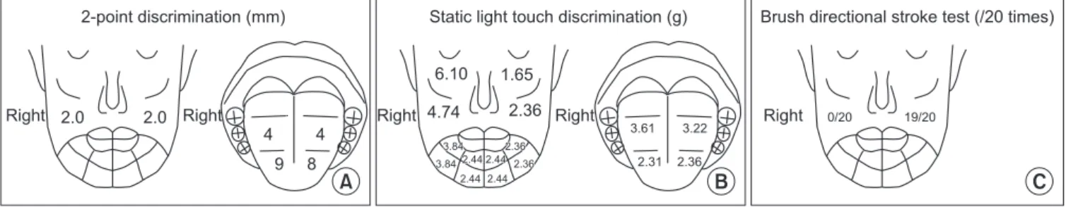
Disuse atrophy involves gradual muscle weakening due to inadequate usage and can cause temporomandibular disorder (TMD). A 45-year old man with TMD symptoms on the left side, who had disuse atrophy of the masticatory muscles on the right side following surgical remov- al of a trigeminal schwannoma on the right side, first visited the Department of Orofacial Pain and Oral Medicine at Kyung Hee University Dental Hospital with left jaw pain and difficulty in opening mouth and chewing. He had been experiencing difficulties in cognitive function, decrease in visual acuity, impaired speech, and writing deficits after brain surgery. Further- more, he complained of abnormal occlusion on the right side, which interfered with his ability to chew comfortably and open his mouth effectively. Herein, we describe a contralateral TMD case due to ipsilateral disuse atrophy after brain surgery for a trigeminal schwannoma and our successful treatment with medication, physical therapy, and stabilization splint.

Key Words: Disuse atrophy; Stabilization splint; Temporomandibular disorder; Trigeminal schwannoma

Correspondence to:

Yeon-Hee Lee

Department of Orofacial Pain and Oral Medicine, Kyung Hee University Dental Hospital, 26 Kyungheedae-ro, Dongdaemun-gu, Seoul 02447, Korea Tel: +82-2-958-9454

Fax: +82-2-962-8124 E-mail: [email protected]

JOMP Journal of Oral Medicine and Pain

Copyright Ⓒ 2018 Korean Academy of Orofacial Pain and Oral Medicine. All rights reserved.

CC

This is an open-access article distributed under the terms of the Creative Commons Attribution Non-Commercial License (http://creativecommons.org/licenses/by-nc/4.0/), which permits unrestricted non-commercial use, distribution, and reproduction in any medium, provided the original work is properly cited.

INTRODUCTION

Temporomandibular disorders (TMDs) occur when the dy- namic equilibrium and balance between the components of the temporomandibular joint (TMJ) and masticatory system are disrupted, resulting in morphological and functional de- formities. The etiology of TMD is complex and multifactori- al. Micro- and/or macro-trauma, behavioral factors, occlu- sal factors, and psychological factors are common etiologi- cal factors of TMD. 1)

There has been a few reported cases of TMD secondary to brain tumor surgery. During brain tumor removal, dam- age to the trigeminal nerve, which innervates the TMJ, may result in secondary complications, such as sensory abnor- malities, paralysis, and TMDs. 2-6) TMD may also occur due to disuse muscle atrophy, owing to malfunctioning of mo- tor nerves associated with the masticatory muscles. 7-9) To

compensate for muscle atrophy, overuse of the normal and functioning muscles may occur. Atrophy of the mandibular masticatory muscles may result in fatigue of the overused muscles, leading to decrease in the range of motion of the mandible, along with pain during function. 10-12)

TMD therapies include various conservative modali- ties, such as medication, physical therapy, and stabilization splints. 13) Stabilization splints may assist in re-establish- ing optimal orthopedic stability with reconstruction of the myoclonic reflexes and by maintaining the jaw in the most stable rest position. Thus, stabilization splints may enhance muscle functions and reduce abnormal activity. 14,15)

In this case report, we described a case of left TMD after

right trigeminal schwannoma surgery. The patient experi-

enced sensory and motor neuron dysfunction of the right

temporomandibular area. During a 1 year and 9 months fol-

low-up period at the Department of Orofacial pain and Oral

(2)

Medicine, the patient was successfully treated with medica- tion, physical therapy, and stabilization splint therapy.

CASE REPORT

A 45-years-old man presented to the Department of Oral Medicine at Kyung Hee University Dental Hospital, com- plaining of soreness in his left jaw during chewing, and ex- perienced difficulty in opening his mouth. His mouth open- ing was limited, and the opening pathway showed marked deflection. The patient reported that his TMD symptoms de- veloped one month before he presented at the hospital. The pain intensity was 7/10 on the visual analogue scale (VAS) (0: no pain, 10: maximum pain imaginable). The pain qual- ity was expressed as throbbing, rather than a sharp pain.

The patient’s past medical history revealed that a tri- geminal schwannoma on the right side was diagnosed and managed with total surgical resection at the neurosurgery department of another hospital 8 years ago. Following sur- gery, the patient reported cognitive impairment, difficulty in sleeping, decreased vision, impaired speech, and reduced writing capability. In addition, he complained of symp- toms in the orofacial region. In particular, his maxillary and mandibular teeth on the right side did not contact during occlusion. Two years ago, the patient completed orthodon- tic treatment at a local dental clinic to correct the abnor- mal occlusion. In the last 5 years, he has had hypertension, which has been managed with daily oral medication.

As part of the diagnostic process, various clinical and imaging tests were performed at our hospital. The initial

palpation test indicated tenderness of the left TMJ, includ- ing the lateral capsule, posterior capsule, TM ligament, and retrodiscal tissue. Vertical mandibular movements were lim- ited. The measurements for comfortable mouth opening and maximum mouth opening (MMO) were 15 mm and 25 mm, respectively. Bilateral movements were also limited to 4 mm on the right side and 3 mm on the left and accompanied by pain during movement to the right side. Furthermore, right posterior openbite of about 2 mm was observed.

Multiple diagnostic images of the TMJ were evaluated.

The panoramic view did not demonstrate any changes in the mandibular condyles. However, the cone beam comput- ed tomography analysis demonstrated degenerative chang- es, including flattening and erosion of the left mandibular condyle. The bone scan showed an increased uptake of the radioactive tracer in the left TMJ joint. On the magnetic res- onance imaging (MRI), structural changes were observed on both sides, and the condyle discs on both sides were located comparatively anterior to the respective condyle on open- ing and closing of the mouth (Fig. 1). The MRI also showed that the volumes of masticatory muscles were smaller on the right than left; in particular, atrophic changes of the lat- eral pterygoid muscle and masseter muscle were noted. To summarize, the patient was diagnosed with disc displace- ment without reduction and osteoarthritis of the left TMJ, and disuse atrophy of the right masticatory muscles.

In order to determine the threshold at which senso- ry changes and sensory inhibition occurred, the brush- ing motion direction, two-point discrimination, and a static touch test were performed. The results of these tests

Fig. 1. Magnetic resonance imaging images of temporomandibular joints:

(A) right and (B) left, sagittal T2 weighted images in closed mouth position.

A B

(3)

revealed hypoesthesia on the right maxillary (V2) and mandibular (V3) divisions of the trigeminal nerve (Fig. 2).

Electromyography (EMG) was performed under three con- ditions: resting, clenching, and chewing. The left side was used as the reference. In all conditions, the right mastica- tory muscles, e.g. temporalis and masseter, showed a lower EMG level than the left masticatory muscles. In resting con- dition, the EMG values of the right masseter and tempora- lis muscles were 75.5% and 76.6%, respectively. In the cot- ton roll clenching and chewing tests, the values of the right masseter and temporalis were lower than those of the left side (Table 1). The Epworth sleepiness scale, Pittsburgh sleep quality index, STOP-BANG questionnaire, and Symptom Checklist 90 Revised were used to evaluate sleep distur- bances and psychological status. The scores were all within the normal range.

During the first visit, the patient was shown how to main- tain a stable jaw position, and analgesics and anti-inflam- matory drugs were prescribed as part of the initial treatment plan. During the two-month follow-up period, 50% reduc- tion in pain was observed with the use of medications and physical therapy. However, discomfort due to the posterior openbite on the right side persisted. The patient complained that the force of the right masticatory muscle was weaker than that of the left, and that touch sensation on the right facial region was less sensitive compared to the preopera- tive level of 60%-70%.

Two months after the initial visit, the patient was ad- vised to wear a stabilization splint every night. Pain in the left jaw improved following the use of the splint for one and a half months. The VAS score decreased from 7 to 2.

Subsequently, masticatory muscle pain disappeared, and the

Table 1. EMG score of each masticatory muscle during initial examination

Muscle Rest

position

Posterior clenching Gum chewing

Right Left Right Left

Masseter

Right 0.74 (76) 7.8 (9) 7.1 (10) 9.3 (50) 3.7 (17)

Left (reference) 0.98 (100) 88.7 (100) 74.0 (100) 18.8 (100) 22.4 (100)

Temporalis

Right 1.37 (76) 5.8 (8) 5.1 (7) 5.4 (24) 5.9 (25)

Left (reference) 1.81 (100) 70.1 (100) 74.9 (100) 22.3 (100) 23.7 (100)

SCM

Right 0.91 (108) 8.4 (68) 8.6 (72) 8.6 (151) 6.0 (97)

Left (reference) 0.84 (100) 12.4 (100) 12.0 (100) 5.7 (100) 6.2 (100)

EMG, electromyography; SCM, sternocleidomastoid muscle.

Values are presented as number (%).

Rest position; when the patient is sitting on a chair and resting, EMG scores of masticatory muscles including masseter, temporalis, and SCM;

posterior clenching: EMG measurements of the muscles on the same side (right or left) when clenching a cotton roll posteriorly; gum chewing:

EMG measurements with chewing gum on the same side as the muscles being tested (right or left).

When the EMG scores of muscles on the left were taken as a reference, the right EMG scores were significantly lower in the neck muscles, including masseter, temporalis, and SCM.

Fig. 2. Results of the quantitative sensory tests: (A) two-point discrimination test, (B) static light touch discrimination test, and (C) directional discrimination test (brush directional stroke test). Tests B and C showed that sensitivity of the right side in the V1, V2, and V3 regions (maxilla and mandible) was lower than that of the left. Tests A and C did not check the chin area.

2-point discrimination (mm)

Right Static light touch discrimination (g)

6.10 1.65

4.74 2.36

3.61 3.22 2.36

0/20 19/20

2.0

4

9 8

A B C

2.0

4

Brush directional stroke test (/20 times)

2.31

3.84 2.36

3.84 2.44 2.44 2.44

2.44 2.36

Right Right

Right

Right

(4)

patient was able to chew on the right side. Partial improve- ment in his mouth opening was also observed. The patient was followed-up at intervals of 1 to 2 months for 8 months.

Following the initial conservative treatment (drug therapy, stabilization splint, physical therapy), the patient’s mouth opening and pain intensity improved significantly. His MMO increased to 37 mm, and the VAS score decreased to 1.

After one year, MMO increased to 40 mm. The left TMJ was painless, although mild stiffness was detected upon stimu- lation. Osteoarthritis in the left TMJ was still evident in the follow-up bone scan taken at 1 year and 9 months after the initial visit. Activity values decreased (right: 59 → 49; left:

80 → 74). In the follow-up MRI, significant improvement was observed in the irregularity of the cortical line and ero- sion of condyle. The patient has been using a stabilization splint for a period of 2 years, and his TMD symptoms have significantly improved.

DISCUSSION

Jaw closing involves the temporal, masseter, and medial pterygoid muscles. Jaw opening utilizes the lateral ptery- goid muscle. These muscles are considered as masticatory muscles, and they are innervated by the mandibular branch of the trigeminal nerve. 16) An intact trigeminal nerve is critical because this nerve is responsible for muscle func- tions, such as chewing and speaking, as well as the sensa- tion of the orofacial area. In addition, the morphologic or functional characteristics of masticatory muscles are con- tinuously altered by physiological changes, such as perioral habit and/or pathological processes, such as disuse related to nerve damage. 17) Thus, trigeminal nerve damage may be associated with disuse atrophy of the masticatory muscle, which ultimately impairs the individual’s quality of life.

Disuse atrophy refers to a type of muscle atrophy that oc- curs when the muscles are no longer used as usual. A lack of energy intake, unbalanced nutritional status, and inactiv- ity of muscles all lead to a reduction in muscle mass, which is associated with disuse atrophy and loss of function.

Loss of muscle mass occurs in common diseases including cancers 18) and neurodegenerative disorders. 19) Masticatory muscle is not only crucial for mastication but also impor- tant in speech, and aesthetic reasons. An understanding of

the mechanisms of disuse atrophy is crucial for designing countermeasures and/or rehabilitation protocols. 20)

Schwannoma is a benign tumor of the nerve sheath from the Schwann cells. Schwannomas commonly occurred in the vagus and the cervical sympathetic chain, whereas tri- geminal schwannomas are rare. 21) We presented a case of contralateral TMD after surgical removal of a trigeminal schwannoma. This brain tumor is usually detected as a large mass because of painless undisturbed growth, leading to diagnostic delay. 22) Treatment depends on the size, loca- tion, and stage of the mass, but surgical excision is usually performed. 23) Malignant transformation is rare in schwan- noma, and the prognosis after complete excision is remark- able. 24) However, facial dysesthesia, paresthesia, and hy- peralgesia are frequently observed. Chewing disturbance after surgical treatment has also been reported. According to Ramina et al. 25) , trigeminal hypoesthesia and facial pain are common, affecting up to 53% of patients after surgery for their trigeminal schwannoma. This may be due to nerve damage resulting from the surgical approach.

The stabilization splint is a removable hard acrylic splint that provides a temporary ideal occlusion. 26) Providing an ideal occlusion by the use of a stabilization splint reduces neuromuscular imbalance. The masticatory muscles can be relaxed over several visits until a consistent jaw relation- ship is reached. Previous clinical studies have demonstrated the efficacy of stabilization splint in the treatment of myo- fascial pain. 27,28) The patient in our study had significantly improved symptoms after TMD treatment with a stabiliza- tion splint. It was more effective than other previous treat- ment. The mechanisms of action of stabilization splint ther- apy include biofeedback, visual feedback, relaxation of the masticatory muscle, and resolution of the neuromuscular imbalance. Only a few cases of TMD after postoperative tri- geminal nerve injury have been reported in the literature.

Therefore, in order to clarify our findings, future multi-cen- tered studies with larger sample sizes and longer follow-ups are needed.

CONFLICT OF INTEREST

No potential conflict of interest relevant to this article

was reported.

(5)

ACKNOWLEDGEMENTS

The authors extend their special thanks to Jung-Pyo Hong at the department of Orofacial Pain and Oral Medicine, Kyung Hee University Dental Hospital for providing valu- able insights.

REFERENCES

1. Oral K, Bal Küçük B, Ebeoğlu B, Dinçer S. Etiology of temporo- mandibular disorder pain. Agri 2009;21:89-94.

2. Al-Mefty O, Ayoubi S, Gaber E. Trigeminal schwannomas: re- moval of dumbbell-shaped tumors through the expanded Meckel cave and outcomes of cranial nerve function. J Neurosurg 2002;96:453-463.

3. Costa AL, Yasuda CL, França M Jr, et al. Temporomandibular dysfunction post-craniotomy: evaluation between pre- and post- operative status. J Craniomaxillofac Surg 2014;42:1475-1479.

4. Kang GC, Soo KC, Lim DT. Extracranial non-vestibular head and neck schwannomas: a ten-year experience. Ann Acad Med Sin- gapore 2007;36:233-238.

5. Wanibuchi M, Fukushima T, Zomordi AR, Nonaka Y, Friedman AH. Trigeminal schwannomas: skull base approaches and opera- tive results in 105 patients. Neurosurgery 2012;70(1 Suppl Opera- tive):132-143; discussion 143-144.

6. Yasuda CL, Costa AL, Franca M Jr, et al. Postcraniotomy tem- poralis muscle atrophy: a clinical, magnetic resonance imaging volumetry and electromyographic investigation. J Orofac Pain 2010;24:391-397.

7. Schellhas KP. MR imaging of muscles of mastication. AJR Am J Roentgenol 1989;153:847-855.

8. Shi Z, Lv J, Xiaoyu L, Zheng LW, Yang XW. Condylar degrada- tion from decreased occlusal loading following masticatory muscle atrophy. Biomed Res Int 2018;2018:6947612.

9. Davis SB, Mathews VP, Williams DW 3rd. Masticator muscle en- hancement in subacute denervation atrophy. AJNR Am J Neuro- radiol 1995;16:1292-1294.

10. Chiba M, Echigo S. Unilateral atrophy of the masticatory muscles and mandibular ramus due to pure trigeminal motor neuropa- thy: a case report. Oral Surg Oral Med Oral Pathol Oral Radiol 2012;113:e30-34.

11. Mejersjö C, Kiliaridis S. Temporomandibular dysfunction in adult patients with myotonic dystrophy (DM1). J Oral Rehabil 2017;44:749-755.

12. Song Y, Forsgren S, Yu J, Lorentzon R, Stål PS. Effects on contra- lateral muscles after unilateral electrical muscle stimulation and exercise. PLoS One 2012;7:e52230.

13. Murphy MK, MacBarb RF, Wong ME, Athanasiou KA. Temporo- mandibular disorders: a review of etiology, clinical management, and tissue engineering strategies. Int J Oral Maxillofac Implants 2013;28:e393-414.

14. Alajbeg IZ, Boric Brakus R, Brakus I. Comparison of amitriptyline with stabilization splint and placebo in chronic TMD patients: a pilot study. Acta Stomatol Croat 2018;52:114-122.

15. Al-Ani MZ, Davies SJ, Gray RJ, Sloan P, Glenny AM. Stabilisa- tion splint therapy for temporomandibular pain dysfunction syn- drome. Cochrane Database Syst Rev 2004;(1):CD002778.

16. Imauchi Y, Sakamoto T, Abe K. A case of atrophy of the mastica- tory muscles due to a masticatory habit. Eur Arch Otorhinolaryn- gol 2002;259:551-553.

17. Song HS, Park CG. Masseter muscle atrophy after ostectomy of the mandibular angle in rabbits. Plast Reconstr Surg 1997;99:51- 60.

18. Stephens NA, Gallagher IJ, Rooyackers O, et al. Using transcrip- tomics to identify and validate novel biomarkers of human skel- etal muscle cancer cachexia. Genome Med 2010;2:1.

19. Verdijk LB, Dirks ML, Snijders T, et al. Reduced satellite cell num- bers with spinal cord injury and aging in humans. Med Sci Sports Exerc 2012;44:2322-2330.

20. Reggiani C. Not all disuse protocols are equal: new insight into the signalling pathways to muscle atrophy. J Physiol 2015;593:5227-5228.

21. Saydam L, Kizilay A, Kalcioglu T, Gurer I. Ancient cervical vagal neurilemmoma: a case report. Am J Otolaryngol 2000;21:61-64.

22. Servadei F, Romano A, Ferri A, Magri AS, Sesenna E. Giant tri- geminal schwannoma with parapharyngeal extension: report of a case. J Craniomaxillofac Surg 2012;40:e15-18.

23. Agrawal A, Singh V, Rohilla S, Sharma B. Trigeminal schwan- noma. Natl J Maxillofac Surg 2017;8:149-152.

24. Rasbridge SA, Browse NL, Tighe JR, Fletcher CD. Malignant nerve sheath tumour arising in a benign ancient schwannoma. Histopa- thology 1989;14:525-528.

25. Ramina R, Mattei TA, Sória MG, et al. Surgical management of trigeminal schwannomas. Neurosurg Focus 2008;25:E6; discus- sion E6.

26. Gray RJ, Davies SJ. Occlusal splints and temporomandibular dis- orders: why, when, how? Dent Update 2001;28:194-199.

27. Tsuga K, Akagawa Y, Sakaguchi R, Tsuru H. A short-term evalu- ation of the effectiveness of stabilization-type occlusal splint therapy for specific symptoms of temporomandibular joint dys- function syndrome. J Prosthet Dent 1989;61:610-613.

28. Davies SJ, Gray RJ. The pattern of splint usage in the manage- ment of two common temporomandibular disorders. Part III:

long-term follow-up in an assessment of splint therapy in the

management of disc displacement with reduction and pain dys-

function syndrome. Br Dent J 1997;183:279-283.

수치

Fig. 1. Magnetic  resonance  imaging  images of temporomandibular joints:
Table 1. EMG score of each masticatory muscle during initial examination

참조

관련 문서

Modern Physics for Scientists and Engineers International Edition,

If both these adjustments are considered, the resulting approach is called a bootstrap-BC a -method (bias- corrected-accelerated). A description of this approach

③ A student who attended Korean course at KNU Korean Language Program and holds TOPIK Level 3 or a student who completed Korean course Level 4 at the KNU Korean Language

· 50% exemption from tuition fee Ⅱ for the student with a TOPIK score of level 3 or higher or completion of level 4 or higher class of the Korean language program at the

Since every classical or virtual knot is equivalent to the unknot via a sequence of the extended Reidmeister moves together with the forbidden moves, illustrated in Section 2,

웹 표준을 지원하는 플랫폼에서 큰 수정없이 실행 가능함 패키징을 통해 다양한 기기를 위한 앱을 작성할 수 있음 네이티브 앱과

_____ culture appears to be attractive (도시의) to the

- quadriceps tendon 이 슬개골 하연에서 tibial tuberocity에 부착.