163 Introduction
Double aortic arch (DAA) is a congenital anomaly, which is formed from the dividing of the ascending aorta into two limbs that pass around the trachea and esophagus, then enter a single descending aorta.1)2) In most cases, this anomaly can be diagnosed during infancy or childhood due to symptoms caused by esophageal or tracheal obstruction. For this reason, case reports of adults, especially asymptomatic cases, are rare.3) We report the case of a 36-year-old male with balanced type DAA, diagnosed coincidentally by echocardiography on a health check up.
Case
A 36-year-old man was admitted to our hospital for health check-up. The patient had no medical and family history of cardiac disease. Besides, the patient reported no medical ab- normality in vital signs, physical examination, and laboratory findings. Chest radiography showed bilateral aortic notches at the level of aortic arch, suggesting aortic arch anomaly (Fig. 1).
Two aortic arches on suprasternal view were seen on transtho-
racic echocardiography (Fig. 2). The diameter of the right aor- tic arch was 1.86 × 1.93 cm, while the left aortic arch was
pISSN 1975-4612/ eISSN 2005-9655 Copyright © 2011 Korean Society of Echocardiography www.kse-jcu.org http://dx.doi.org/10.4250/jcu.2011.19.3.163
CASE REPORT J Cardiovasc Ultrasound 2011;19(3):163-166
A Case of Balanced Type Double Aortic Arch Diagnosed Incidentally
by Transthoracic Echocardiography in an Asymptomatic Adult Patient
Han Seok Seo, MD, Yong Hyun Park, MD, Ju Hyoung Lee, MD, So Chong Hur, MD, Yu Jin Ko, MD, So Yeon Park, MD, Jun Hwan Kim, MD, Young Jung Kim, MD,
So Yon Kim, MD and Nak Hyun Kwon, MD
Department of Internal Medicine, National Police Hospital, Seoul, Korea
A 36-year-old male patient with no remarkable medical history was admitted to our hospital for a health check up. On chest radiography, bilateral aortic notches at the level of aortic arch were shown suggesting aortic arch anomaly without any clinical symptoms. Two aortic arches were almost same-in-size on suprasternal view of transthoracic echocardiography. In addition, multidetector computed tomography showed balanced type double aortic arch forming a complete vascular ring which encircled the trachea and esophagus. The trachea was slightly compressed by the vascular ring whereas the esophagus was intact. Nevertheless, the pulmonary function test was normal. The patient was discharged from hospital with instructions for periodic follow-up.
KEY WORDS: Double aortic arch · Echocardiography · Vascular ring.
• Received: May 6, 2011 • Revised: July 8, 2011 • Accepted: August 17, 2011
• Address for Correspondence: Nak Hyun Kwon, Department of Internal Medicine, National Police Hospital, 123 Songi-ro, Songpa-gu, Seoul 138-708, Korea Tel: +82-2-3400-1225, Fax: +82-2-3400-1164, E-mail: [email protected]
• This is an Open Access article distributed under the terms of the Creative Commons Attribution Non-Commercial License (http://creativecommons.org/licenses/by-nc/3.0) which permits unrestricted non-commercial use, distribution, and reproduction in any medium, provided the original work is properly cited.
Fig. 1. Chest radiography showed bilateral aortic notches at the level of aortic arch, suggesting aortic arch anomaly. Right aortic arch (large arrow) and left aortic arch (small arrow) can be seen.
Journal of Cardiovascular Ultrasound 19 | September 2011
164
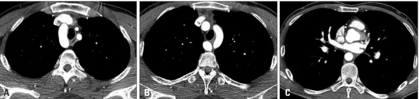
1.46 × 1.51 cm in size. Multidetector computed tomography (CT) showed nearly same sized DAA forming a complete vas- cular ring encircling the trachea and esophagus. The right aortic arch was slightly larger and higher than the left aortic arch (Fig. 3). The esophagus had no compression within the vascular ring, while the trachea was slightly compressed by the vascular ring (Fig. 4).
The patient had no abnormal breathing sound and any oth- er symptoms associated with compression of trachea or esoph- agus such as dyspnea, cough, dysphagia, and regurgitation. To evaluate the effect of the tracheal compression, pulmonary function test was done and showed normal findings. The pa- tient was discharged from hospital with instructions for peri- odic follow-up.
Discussion
DAA is reported to be found in less than 1% of the congen- ital heart disease.4) In DAA, both the aortic arches form a complete vascular ring encircling the trachea and esophagus due to failure of regression of the right aortic arch.5)6) Each aor- tic arch passes the ipsilateral mainstem bronchus superiorly and enters the descending aorta, which is more commonly lo- cated on the left side than on the right side of the spine. Both aortic arches are often different in size and position. The right aortic arch is usually larger and located higher than the left
aortic arch.7) The left aortic arch may be degenerated and so become atretic or remain as a fibrous band.8) The left and right common carotid and subclavian arteries come from each of the aortic arches.9) In our case, both aortic arches fuse into de- scending aorta located in front of the spine and the right aor- tic arch was slightly larger and located higher than the left aortic arch.
Classically, DAA is classified into three types depending on the relative size of the two arches and partial atresia of one arch; right dominant aortic arch, left dominant aortic arch, and balanced type aortic arch. Seventy-five percent of the DAA pa- tients have right dominant aortic arch, and 20% have left dominant aortic arch. The dominance of either arch cannot be determined in the remaining 5%, called balanced type DAA.
Accompanying intracardiac defect may be found in about 20% of the DAA patients.10-13) Our study patient had a bal- anced type of DAA. Although the right aortic arch was slight- ly larger than the left aortic arch, we could assume both aortic arches were nearly the same size and had no atresia of arch.
DAA patients may complain of respiratory symptoms or gastrointestinal symptoms caused by vascular ring formation.
If the vascular ring compresses the trachea, the respiratory symptoms such as noisy breathing, dyspnea, cough, recurrent respiratory infections, apnea and cyanosis, which appear espe- cially in neonates, could occur. Gastrointestinal symptoms
Fig. 2. Transthoracic echocardiography showed double aortic arch. Descending aorta which is located in the middle can be seen posterior to left atrium on parasternal short axis view (arrow) (A). Right aortic arch (left arrow) and left aortic arch (right arrow) (B) can be found together and respectively, right aortic arch (C), left aortic arch (D) and their branches on suprasternal view. RAA: right aortic arch, RCCA: right common carotid artery, RSCA: right subclavian artery, LAA: left aortic arch, LCCA: left common carotid artery, LSCA: left subclavian artery.
A B C
Fig. 3. Chest computed tomography showed double aortic arch. Right aortic arch (A) and left aortic arch (B) can be seen and both aortic arches fuse into descending aorta located in front of the spine (C).
A B C D
Silent Double Aortic Arch in Adult | Han Seok Seo, et al.
165 such as feeding difficulty, emesis, and failure to thrive occur
by esophageal compression.6) Symptoms can appear later in life when aorta enlarges due to atherosclerotic change.14) Our patients had no symptoms as mentioned above and normal findings in pulmonary function test in spite of slight compres- sion of the trachea.
Chest radiographic features of the DAA patients include a deviation or compression of the trachea, or identification of a right aortic arch contour.15) We could see not only left aortic notch but also right aortic notch formed by right aortic arch in chest radiography of our patient.
Transthoracic echocardiography of the DAA patients can re- veal aortic bifurcation on the subcostal view and the two de- scending aortic flow patterns on the suprasternal view.15)16) The echocardiography of our patient showed descending aor- ta, located in the middle, posterior to left atrium on paraster- nal short axis view and DAA on suprasternal view.
Some study reports that delineation of aortic arch abnor- malities is difficult by conventional 2-dimensional echocar- diography hence CT or magnetic resonance imaging (MRI) has been the diagnostic tool of choice.17) The transthoracic echocardiography in diagnosis of DAA patients has limited ability to image clearly the atretic aortic arch structures and ligamentum arteriosum. However, echocardiography remains a powerful, useful, non-invasive, and easily available modality
which enables complete and accurate anatomic delineation and the exclusion of other major intracardiac defects.15)18)19)
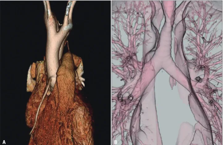
The exact anatomy and location of an aortic arch malforma- tion and adjacent structures can be accurately defined by CT and MRI, which allow 3 dimensional display of the malfor- mations.9) The CT imaging of our patient revealed balanced type DAA resulting in only slight compression of the trachea and no compression of the esophagus.
A surgical approach is the only choice for the symptomatic DAA patients. Surgical intervention with division of the minor arch is recommended for those adult patients who are symp- tomatic and can be approached via either left posterolateral thoracotomy or median sternotomy.4)20)21) Because our patient had no symptoms, we did not perform any surgical interven- tion, however, we explained possible complications of DAA to the patient and recommended periodic follow-up.
In conclusion, balanced-type DAA is a rare condition. We report the case of a 36-year-old male with balanced-type DAA, diagnosed coincidentally by echocardiography on a health check up.
References
1. Yilmaz M, Tok M, Cengiz M. Asymptomatic balanced-type double aortic arch in an elderly patient: a case report. Heart Surg Forum 2007;10:
E297-8.
Fig. 4. Reconstructed images of chest computed tomography showed balanced type double aortic arch (A) with only slight compression of the trachea (B).
A B
Journal of Cardiovascular Ultrasound 19 | September 2011
166
2. Ito H, Konishi A, Kon-Nai T, Ishibashi T, Takahashi S. Double aor- tic arch with atresia, tapering and aneurysm of the left arch. Br J Radiol 2006;79:e71-4.
3. Ikenouchi H, Tabei F, Itoh N, Nozaki A. Images in cardiovascular medicine. Silent double aortic arch found in an elderly man. Circulation 2006;114:e360-1.
4. Sariaydin M, Findik S, Atici AG, Ozkaya S, Uluisik A. Asymptomatic double aortic arch. Int Med Case Rep J 2010;3:63-6.
5. Stewart JR, Kincaid OW, Edwards JE. An atlas of vascular rings and related malformations of the aortic arch system. Springfield: Thomas; 1963.
p.579-80.
6. Jeeyani HN, Prajapati VJ, Patel NH, Shah SB. Imaging features of double aortic arch shown by multidetector computed tomography angiogra- phy. Ann Pediatr Cardiol 2010;3:169-70.
7. Lowe GM, Donaldson JS, Backer CL. Vascular rings: 10-year review of imaging. Radiographics 1991;11:637-46.
8. Schlesinger AE, Krishnamurthy R, Sena LM, Guillerman RP, Chung T, DiBardino DJ, Fraser CD Jr. Incomplete double aortic arch with atresia of the distal left arch: distinctive imaging appearance. AJR Am J Roentgenol 2005;184:1634-9.
9. Kellenberger CJ. Aortic arch malformations. Pediatr Radiol 2010;
40:876-84.
10. Jaffe RB. Radiographic manifestations of congenital anomalies of the aortic arch. Radiol Clin North Am 1991;29:319-34.
11. Moes CA, Freedom RM. Rare types of aortic arch anomalies. Pediatr Cardiol 1993;14:93-101.
12. Baraldi R, Sala S, Bighi S, Mannella P. Vascular ring due to double aor- tic arch: a rare cause of dysphagia. Eur J Radiol Extra 2004;52:21-4.
13. Emmel M, Schmidt B, Schickendantz S. Double aortic arch in a pa- tient with Fallot’s tetralogy. Cardiol Young 2005;15:52-3.
14. Kim HY, Jung HY, Yun TJ, Lee SS, Lee EY, Ko KH, Kim YM, Myung SJ, Yang SK, Hong WS, Kim JH, Min YI. Three cases of dysphagia due to vascular ring in adults. Korean J Gastrointest Endosc 2000;21:735-40.
15. Lee ML. Diagnosis of the double aortic arch and its differentiation from the conotruncal malformations. Yonsei Med J 2007;48:818-26.
16. Koz C, Yokusoglu M, Uzun M, Tasar M. Double aortic arch suspected upon transthoracic echocardiography and diagnosed upon computed tomogra- phy. Tex Heart Inst J 2008;35:80-1.
17. Lotz J, Macchiarini P. Images in clinical medicine. Double aortic arch di- agnosed by magnetic resonance imaging. N Engl J Med 2004;351:e20.
18. van Son JA, Julsrud PR, Hagler DJ, Sim EK, Puga FJ, Schaff HV, Danielson GK. Imaging strategies for vascular rings. Ann Thorac Surg 1994;57:604-10.
19. Lillehei CW, Colan S. Echocardiography in the preoperative evaluation of vascular rings. J Pediatr Surg 1992;27:1118-20; discussion 1120-1.
20. Kypson AP, Anderson CA, Rodriguez E, Koutlas TC. Double aortic arch in an adult undergoing coronary bypass surgery: a therapeutic dilem- ma? Eur J Cardiothorac Surg 2008;34:920-1.
21. Alsenaidi K, Gurofsky R, Karamlou T, Williams WG, McCrindle BW. Management and outcomes of double aortic arch in 81 patients. Pedi- atrics 2006;118:e1336-41.