The Effect of Pharmacopuncture of Mori Cortex on Galactosamine-induced Liver Injury in Rats
Wi Jun*, Kim Jae-hong*, Yoon Yeo-choong*, Wei Tung-shuen* and Yoon Dae-hwan**
*Dept. of Acupuncture & Moxibustion, College of Oriental Medicine, Dongshin University
**Dept. of Meridians & Acupoints, College of Oriental Medicine, Dongshin University
Objectives : This study was designed to investigate the effect of pharmacopuncture of Mori Cortex on galactosamine-induced liver injury in rats.
Methods : Male Sprague-Dawley rats were divided into 5 groups; Normal, liver injury not induced and not treated group. Control, the liver injury-induced and not treated group. Saline group, the liver injury-induced and saline injection at BL
18. HA-1 and HA-2 group, the liver injury-induced and pharmacopuncture of Mori Cortex applied to BL
18, each 1.3μg/g, 2.6μg/g. Then we observed the changes of γ-GTP, GOT, GPT, LDH, total cholesterol, triglyceride, total bilirubin.
Results : Pharmacopuncture of Mori Cortex treatment significantly inhibited the activities of γ-GTP, GOT, total cholesterol, triglyceride and total bilirubin in HA-1 group. γ-GTP, GTP, LDH, γ-GTP levels were significantly inhibited in HA-2 group.
Conclusions : These results demonstrate that the reduce of hepatic enzyme activation and lipid accumulation by pharmacopuncture of Mori Cortex may be by an antioxidant properties of Mori Cortex.
Key words : liver injury, galactosamine, pharmacopuncture, Mori Cortex
1)
肝兪(BL 18 ) 桑白皮약침이 Galactosamine에 의해 유발된 흰쥐의 肝손상에 미치는 영향
위준*․김재홍*․윤여충*․위통순*․윤대환**
*동신대학교 한의과대학 침구학교실
**동신대학교 한의과대학 경혈학교실
․접수 : 2009. 9. 10. ․수정 : 2009. 9. 20. ․채택 : 2009. 9. 20.
․교신저자 : 김재홍. 광주광역시 남구 월산3동 377-12 동신대학교 부속광주한방병원 침구과 Tel. 062-350-7209 E-mail : [email protected]
원 저
Abstract
Ⅰ. 서 론
간질환은 전 세계의 약 3억 5천만 명이 시달리고 있 는 질환으로 한국, 중국 등 아시아 일부 국가는 간질환 사망률이 매우 높다. 특히 우리나라는 B형 간염 바이 러스 감염률이 높고 이에 따라 만성적 간질환의 높은 유병률을 나타내므로
1,2)이를 치유 혹은 예방하기 위한 약제 및 방법에 대한 연구가 필요한 실정이다.
한의학적으로 肝은 藏血과 疏泄을 주관하고 인체 의 기혈과 장부의 활동을 조절하여 대사, 해독, 담즙 의 분비와 배설작용 및 정지활동 등을 정상적으로 유 지하는 역할을 수행하고 있다. 이러한 肝이 내외 요인 으로 인하여 그 기능을 상실하면 여러 가지 간장 질 환이 발생하게 되며 특히 음주, 고지방식, 감염, 중독 등이 간질환의 가장 흔한 원인이 된다
3).
藥鍼療法은 경락학설의 원리에 의거하여 약물을 선택해서, 유관한 穴位나 壓痛點에 주입하여 약물효 과 및 경혈자극효과를 통하여 생체의 기능을 조정하 고 병리상태를 개선시켜 질병치료의 목적을 달성하는 新鍼療法 중의 하나이다
4,5).
肝兪穴은 족태양방광경의 18번째 經穴로서 補營血, 調氣滯, 除肝膽濕熱, 能寧神明目하는 穴性을 가지고 있으며, 主治症은 黃疸, 肋間神經痛, 神經衰弱, 急慢性 肝炎, 眼病 등이다
6).
桑白皮는 뽕나무과(桑科 : Moraceae)에 속한 낙엽 교목인 뽕나무 및 동속 근연식물의 근피로, 性은 寒, 無毒하고 味는 甘하며, 肺․大腸으로 歸經한다
7-9).
간기능에 대한 상백피의 효능 연구로는 홍
10)은 지 질과산화 및 간독성에 대한 효과를 보고하였고, 김
11)은 간기능 보호효과를 보고하였으며, 김 등
12)은 사염 화탄소에 의해 유발된 간독성에 대한 간 보호효과 등 을 보고하였으나, 약침제제에 대한 간기능 효능 연구 는 아직 미진하다.
이에 저자는 桑白皮 약침요법이 간손상에 미치는 효과를 확인하기 위하여 백서에 D-galactosamine을 이용하여 간손상을 유발 시킨 후 상백피 약침을 肝兪 에 시술하여 혈청 γ-GTP, GOT, GPT, LDH, Total cholesterol, Triglyceride, Total Bilirubin의 수치 변화 및 간조직의 관찰 등을 통하여 유의한 결과를 얻었기 에 보고하는 바이다.
Ⅱ. 재료 및 방법
1. 실험재료
1) 동물
실험동물은 삼육동물센터로부터 구입한 260~300g, 8주령의 수컷 Sprague-Dawley계 흰쥐로, 일주일간 실험실 환경(온도 22±3℃, 습도 50±10%)에 적응시킨 후 사용하였다. 각 cage당 2~3마리씩 넣었으며, 물과 사료(고형사료, 삼양유지, 한국)를 자유로이 섭취하도 록 하였다.
2) 약침액 제조
桑白皮 300g을 증류수 1,000ml과 함께 24시간 동안 증류수를 보충해가며 끓인 다음, 여과지로 여과한 후 고속원심분리기(Centricon T-42K, Italy)로 3,500rpm 에서 20분간 원심분리하여 상등액을 취하였다. 상등 액은 rotary vaccum evaporator(Buchi, Netheland)로 수분을 증발시켜 100ml로 감압 농축하였으며, 농축된 검액을 동결건조기(삼원, 한국)로 -70℃에서 동결건조 시켜 최종적으로 11.8g의 시료를 얻었다. 얻어진 시료 는 약침시술을 위하여 각각의 농도별로 saline에 희석 하여 Cellulose Nitrate Membrane Filters(0.45μm, Whatman, England)로 여과한 후 시술에 사용하였다.
3) 약침 주입기
Insulin syringe(29G×12.7mm, 유일, 한국)을 이용 하여 주입하였다.
2. 실험방법
1) 간손상 유발 및 군 분류
간손상 유발은 Jonker 등
13)의 방법을 이용하여 D-galactosamine(GalN, Sigma, USA)을 흰쥐의 체중 kg당 500mg 농도로 1회 복강 주사하였다.
각 군은 정상군(Normal, n=8), 간 손상을 유발한
후 무처치한 군은 대조군(Control, n=8), 간손상을 유
발한 후 생리식염수로 시술한 생리식염수군(Saline, n=8),
간손상을 유발한 후 1.3μg/g 농도의 桑白皮 약침을
시술한 약침1군(HA-1, n=8), 간손상을 유발한 후 2.6
μ g/g 농도의 桑白皮 약침을 시술한 약침2군(HA-2,
n=8) 등 총 5개군으로 분리하였다.
2) 혈위 및 약침처치
약침이 시술된 혈위는 인체의 肝兪에 상응하는 부 위로 백서의 9․10흉추 극돌기간 양측에서 취하였다.
약침 처치는 간 손상 유발 후 대조군을 제외한 실험 군들을 대상으로 다음날부터 각 농도별로 시행되었으 며, 2일 간격으로 1회씩 20일간 총 10회에 걸쳐 각 혈 위 양측에 insulin syringe(29G×12.7mm, 유일, 한국) 을 이용하여 20μl의 약침이 시행되었다.
3) 채혈 및 혈청분리
채혈은 각 군들의 흰쥐들을 약침시술 후 21일째에 단두하여 혈액 3ml을 얻었으며, 이를 고속원심분리기 (Centricon T-42K, Italy)에서 3,500rpm으로 20분간 원심분리를 시행하여 혈청을 얻었다.
4) 혈청분석
약침시술 후 21일째에 각 군들의 백서로부터 얻은 혈액 중, 약 100μl를 EDTA-bottle에 넣은 후 곧바로 혈구측정기(K-800, Sysmax, Japan)에 주입하여 white blood cell(WBC), red blood cell(RBC), hemoglobin (HGB), hematocrit(Hct) 등을 각각 측정하였다. 나머 지 혈액은 혈청 분리에 사용하였는데, 고속원심분리 기(Centricon T-42K, Italy)에서 5,000rpm으로 10분간 시행하여 혈청을 얻었다.
분리된 혈청으로 γ-GTP, GOT, GPT, LDH, total cholesterol, triglyceride, total bilirubin 등을 측정하였 으며, 측정하기 전까지는 -70℃에 보관하였다. γ- GTP는 diagnostic kits(AM158-K, Asan, Korea)을 사용하여 spectrophotometer(Unikon-922, Kontron, Italy) 로 635nm 파장에서 측정하였으며, GOT는 GOT-SL Kit(ELITech, France)를 이용하여 340nm 파장에서 측정하였으며, GPT는 GPT-SL Kit(ELI Tech, France) 를 이용하여 340nm 파장에서 측정하였으며, LDH는 LDH-SL Kit(ELI Tech, France)를 이용하여 340nm 파장에서 측정하였으며, total cholesterol는 T. cholesterol- SL Kit(ELI Tech, France)를 이용하여 546nm 파장에 서 측정하였으며, triglyceride는 triglyceride-SL Kit (ELITech, France)를 이용하여 505nm 파장에서 측정 하였으며, total Bilirubin은 T. Bilirubin-SL Kit(ELI Tech, France)를 이용하여 505nm 파장에서 측정하였 다. γ- GTP를 제외한 혈청분석은 photometer(5010, Robert Riele GmbH & Co. Germany)를 이용하였다.
5) 조직학적 관찰
광학현미경 관찰을 위하여 실험동물의 간조직을 적출하여, 적출한 조직을 vonin solution(Sigma, Ger- many)을 사용하여 24시간 동안 고정시킨 다음, 70%, 80%, 90%, 100%, ethanol/xylol(1 : 2)액에 각각 2시간 통과시켜 탈수하고 55℃ incubator에서 xylol/hard paraffin (2 : 1), xylol/hard paraffin(1 : 2), 및 hard paraffin 용 액으로 3시간씩 처리한 후 paraffin 포매하였다. 이를 microtome을 사용하여 5μm 두께로 절편을 제작한 후 hematoxylineosin으로 염색하고, 200배율 광학현미경 (olympus BX51TF, Japan)을 이용하여 관찰하였다.
3. 통계처리
실험 성적은 평균값과 표준오차(mean±S.E.)로 표 시하였다. 각 실험군 간의 통계학적 분석은 Window 용 SPSS(version 10.05, SPSS)를 이용하여, 비모수적 방법 중 Mann-Whitney u test를 시행하였다. 전체 실험의 통계적인 유의성은 신뢰구간 p <0.05에서 의미 를 부여하였다.
Ⅲ. 결 과
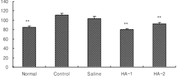
1. γ-GTP에 미치는 효과
肝兪에 대한 桑白皮약침이 간손상 흰쥐의 γ-GTP
0 20 40 60 80 100 120 140
Normal Control Saline HA-1 HA-2
r-GTP(mU/ml)
** ** **
Fig. 1. Effects of Cortex Mori radicis pharmaco- puncture according to dosage on serum γ-GTP in the liver injury rats induced by D-galactosamine
Normal, intactness. Control, the liver injury-induced and not treated group. Saline, the liver injury-induced and injected saline at acupoints of BL18. HA-1 and HA-2, Cortex Mori radicis pharmacopuncture of 1.3μg/g and 2.6μg/g at acu- points of BL18 in the rats. Results are shown as mean±S.E.
**, p<0.01 as compared with the corresponding data of control group.
에 미치는 영향을 비교 관찰한 결과, normal군은 84.9±2.84(mU/ml), control군은 111.1±3.67(mU/ml), saline 군은 103.4±4.69(mU/ml), HA-1군은 80.3±1.92 (mU/ml), HA-2군은 92.13.00(mU/ml)를 나타내었다.
각 군별의 변화 비교에서 control군에 비하여 normal 군, HA-1군과 HA-2군들에서 유의한 감소를 보였다 ( p <0.01)(Fig. 1).
2. GOT에 미치는 효과
肝兪에 대한 桑白皮약침이 간손상 흰쥐의 GOT에 미치는 영향을 비교 관찰한 결과, normal군은 146.5±
11.90(U/l), control군은 203.0±11.33(U/l), saline군은 232.3±33.83(U/l), HA-1군은 175.7±5.95(U/l), HA-2군은 166.0±15.44(U/l)을 나타내었다. 각 군별의 변화 비교 에서 control군에 비하여 normal군( p <0.01), HA-1군 ( p <0.05)들에서 유의한 감소를 보였다(Fig. 2).
0 40 80 120 160 200 240 280 320
Normal Control Saline HA-1 HA-2
GOT(U/1)
** *
Fig. 2. Effects of Cortex Mori radicis phar- macopuncture according to dosage on serum GOT in the liver injury rats induced by D-galactos- amine
Normal, intactness. Control, the liver injury- induced and not treated group. Saline, the liver injury-induced and injected saline at acupoints of BL18. HA-1 and HA-2, Cortex Mori radicis phar- macopuncture of 1.3μg/g and 2.6 μg/g at acupoints of BL18 in the rats. Results are shown as mean±S.E. *, p<0.05, **, p<0.01 as compared with the corresponding data of control group.
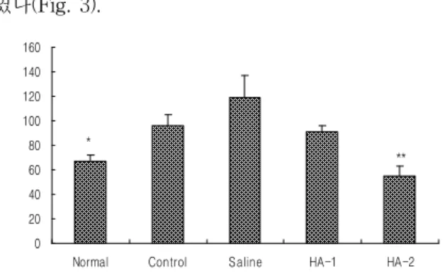
3. GPT에 미치는 효과
肝兪에 대한 桑白皮약침이 간손상 흰쥐의 GPT에 미치는 영향을 비교 관찰한 결과, normal군은 67.0±
5.40(U/l), control군은 95.6±9.63(U/l), saline군은 119.2±
18.29(U/l), HA-1군은 91.2±5.26 (U/l), HA-2군은 55.5±
7.73(U/l)을 나타내었다.
각 군별의 변화 비교에서 control군에 비하여 normal 군( p <0.05), HA-2군( p <0.01)들에서 유의한 감소를 보
였다(Fig. 3).
0 20 40 60 80 100 120 140 160
Normal Control Saline HA-1 HA-2
GPT(U/1)
**
*
Fig. 3. Effects of Cortex Mori radicis pharmaco- puncture according to dosage on serum GPT in the liver injury rats induced by D-galactosamine
Normal, intactness. Control, the liver injury-induced and not treated group. Saline, the liver injury-induced and injected saline at acupoints of BL18. HA-1 and HA-2, Cortex Mori radicis pharmacopuncture of 1.3μg/g and 2.6μ g/g at acupoints of BL18 in the rats. Results are shown as mean±S.E.
*, p<0.05, **, p<0.01 as compared with the correspond- ing data of control group.
4. LDH에 미치는 효과
肝兪에 대한 桑白皮약침이 간손상 흰쥐의 LDH에 미치는 영향을 비교 관찰한 결과, normal군은 1552.8±
282.24(U/l), control군은 2670.4±162.81(U/l), saline군은 3149.3±611.53(U/l), HA-1군은 2762.8±174.20(U/l), HA-2군은 1958.2±129.26(U/l)을 나타내었다.
각 군별의 변화 비교에서 control군에 비하여 normal 군, HA-2군들에서 유의한 감소를 보였다( p <0.01) (Fig. 4).
0 500 1000 1500 2000 2500 3000 3500 4000
Normal Control Saline HA-1 HA-2
LDH(U/1)
** **
Fig. 4. Effects of Cortex Mori radicis pharmaco- puncture according to dosage on serum GPT in the liver injury rats induced by D-galactosamine
Normal, intactness. Control, the liver injury-induced and not treated group. Saline, the liver injury-induced and injected saline at acupoints of BL18. HA-1 and HA-2, Cortex Mori radicis pharmacopuncture of 1.3μg/g and 2.6μg/g at acupoints of BL18in the rats. Results are shown as mean±S.E. **, p<0.01 as compared with the corresponding data of control group.
5. Total cholesterol에 미치는 효과
肝兪에 대한 桑白皮약침이 간손상 흰쥐의 total cholesterol에 미치는 영향을 비교 관찰한 결과, normal 군은 53.0±10.72(mg/dl), control군은 93.2±7.97 (mg/dl), saline군은 78.5±9.26(mg/dl), HA-1군은 70.0±4.72(mg/dl), HA-2군은 88.3±6.90(mg/dl)를 나타내었다.
각 군별의 변화 비교에서 control군에 비하여 normal 군과 HA-1군들에서 유의한 감소를 보였다( p <0.05) (Fig. 5).
0 20 40 60 80 100 120
Normal Control Saline HA-1 HA-2
Total cholesterol (mg/dl)
*
*
Fig. 5. Effects of Cortex Mori radicis pharmaco- puncture according to dosage on serum total cholesterol in the liver injury rats induced by D- galactosamine
Normal, intactness. Control, the liver injury-induced and not treated group. Saline, the liver injury-induced and injected saline at acupoints of BL18. HA-1 and HA-2, Cortex Mori radicis pharmacopuncture of 1.3μg/g and 2.6μ g/g at acupoints of BL18in the rats. Results are shown as mean±S.E. *, p<0.05 as compared with the corresponding data of control group.
6. Triglyceride에 미치는 효과
肝兪에 대한 桑白皮약침이 간손상 흰쥐의 tri-
0 10 20 30 40 50 60 70
Normal Control Saline HA-1 HA-2
Triglyceride(mg/dl)
**
**
*
Fig. 6. Effects of Cortex Mori radicis pharmaco- puncture according to dosage on serum triglyceride in the liver injury rats induced by D-galactosamine
Normal, intactness. Control, the liver injury-induced and not treated group. Saline, the liver injury-induced and injected saline at acupoints of BL18. HA-1 and HA-2, Cortex Mori radicis pharmacopuncture of 1.3μg/g and 2.6 μg/g at acupoints of BL18 in the rats. Results are shown as mean±S.E. *, p<0.05, **, p<0.01 as compared with the corresponding data of control group.glyceride에 미치는 영향을 비교 관찰한 결과, normal 군은 43.5±8.39(mg/dl), control군은 64.2±2.24(mg/dl), saline군은 35.0±2.45(mg/dl), HA-1군은 44.8±4.48(mg/dl), HA-2군은 57.0±8.08(mg/dl)를 나타내었다.
각 군별의 변화 비교에서 control군에 비하여 normal 군( p <0.05), saline군( p <0.01)과 HA-1군( p <0.01)들에서 유의한 감소를 보였다(Fig. 6).
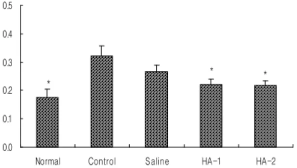
7. Total Bilirubin에 미치는 효과
肝兪에 대한 桑白皮약침이 간손상 흰쥐의 total bilirubin에 미치는 영향을 비교 관찰한 결과, normal 군은 0.2±0.03(mg/dl), control군은 0.3±0.04(mg/dl), saline 군은 0.3±0.02(mg/dl), HA-1군은 0.2±0.02(mg/dl), HA- 2군은 0.2±0.02(mg/dl)를 나타내었다.
각 군별의 변화 비교에서 control군에 비하여 normal 군, HA-1과 HA-2들에서 유의한 감소를 보였다 ( p <0.05)(Fig. 7).
0.0 0.1 0.2 0.3 0.4 0.5
Normal Control Saline HA-1 HA-2
Total Bilirubin(mg/dl)
*
* *
Fig. 7. Effects of Cortex Mori radicis pharmaco- puncture according to dosage on serum total bilirubin in the liver injury rats induced by D- galactosamine
Normal, intactness. Control, the liver injury-induced and not treated group. Saline, the liver injury-induced and injected saline at acupoints of BL18. HA-1 and HA-2, Cortex Mori radicis pharmacopuncture of 1.3μg/g and 2.6 μg/g at acupoints of BL18 in the rats. Results are shown as mean±S.E. *, p<0.05, as compared with the cor- responding data of control group.
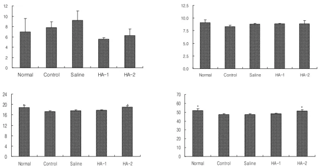
8. 혈액 내 WBC, RBC, HGB, Hct 변화
WBC를 비교 관찰한 결과, normal군은 7.0±2.58 (103/㎕), control군은 7.8±1.08(103/㎕), saline군은 9.2±
1.85(103/㎕), HA-1군은 5.6±0.30(103/㎕), HA-2군은 6.2±1.28(103/㎕)를 나타내었다(Fig. 8 : upper left).
RBC를 비교 관찰한 결과, normal군은 9.1±0.59
(106/㎕), control군은 8.3±0.26 (106/㎕), saline군은
0 2 4 6 8 10 12
Normal Control Saline HA-1 HA-2
WBC (x10000/ul)
0.0 2.5 5.0 7.5 10.0 12.5
Normal Control Saline HA-1 HA-2
RBC (x10000000/ul)
0 4 8 12 16 20 24
Normal Control Saline HA-1 HA-2
HGB (g/dl)
*
*
0 10 20 30 40 50 60 70
Normal Control Saline HA-1 HA-2
Hct (%)
* *
Fig. 8. Effects of Cortex Mori radicis pharmacopuncture according to dosage on blood WBC(upper left), RBC(upper right), HGB(lower, left), Hct(lower right) in serum in liver injury rats induced by D-galactosamine
Normal, intactness. Control, the liver injury-induced and not treated group. Saline, the liver injury-induced and injected saline at acupoints of BL18. HA-1 and HA-2, Cortex Mori radicis pharmacopuncture of 1.3μg/g and 2.6μg/g at acupoints of BL18in the rats. Results are shown as mean±S.E. *, p<0.05, as compared with the corresponding data of control group.
A B C D E
Fig. 9. Light micrographs of the hepatic tissue from Cortex Mori radicis pharmacopuncture according to dosage in the liver injury rats induced by D-galactosamine.
A, Normal group; B, Control group; C, Saline group; D, HA-1 group; E, HA-2 group. Normal, intactness. Control, the liver injury-induced and not treated group. Saline, the liver injury-induced and injected saline at acupoints of BL18. HA-1 and HA-2, Cortex Mori radicis pharmacopuncture of 1.3μg/g and 2.6μg/g at acupoints of BL18 in the rats. Hematoxylin-Eosin stain. × 200.
8.8±0.19(106/㎕), HA-1군은 8.9±0.10(106/㎕), HA-2 군은 8.9±0.58(106/㎕)를 나타내었다(Fig. 8 : upper right).
HGB를 비교 관찰한 결과, normal군은 18.9±0.79(g/dl), control군은 17.2±0.33(g/dl), saline군은 17.6±0.38(g/dl), HA-1군은 17.8±0.15(g/dl), HA-2군은 19.0±0.54(g/dl) 를 나타내었다(Fig. 8 : lower left).
Hct를 비교 관찰한 결과, normal군은 52.0±1.85(%), control군은 47.6±1.00(%), saline군은 47.2±0.93(%), HA-1군은 48.3±0.45(%), HA-2군은 51.6±1.04(%)를 나타내었다(Fig. 8 : lower right).
각 군별의 변화 비교에서 control군에 비하여 HGB, Hct는 normal군과 HA-2군에서 유의한 증가를 보였 다( p <0.01).
9. 조직학적 관찰