대한외과학회지:제 67 권 제 3 호
□ Case Report □
Vol. 67, No. 3, September, 2004
253
INTRODUCTION
The low rate of blunt trauma to the gallbladder (GB) is due to its recessed anatomic location, which confers protection from surrounding viscera and ribs.
GB injuries are rarely isolated and are usually asso- ciated with other intra-abdominal injuries, including those to the liver, duodenum, and spleen.(1-3) Blunt abdominal trauma is often overlooked because there may be no visible signs on the abdominal wall.(4) Computed tomographic (CT) scan plays an impor- tant role in the evaluation of blunt abdominal trau- ma. We report a rare case of an isolated GB per- foration after violent blunt abdominal trauma and emphasize the importance of close observation and a CT scan to confirm the diagnosis of this relatively rare but serious injury.
CASE REPORT
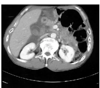
A 56-year-old man was admitted with a chief com- plaint of right upper quadrant pain radiating to the right shoulder immediately after violent blunt abdo- minal trauma. The patient was alcoholic, intoxicated at the time of admission. He had undergone previous surgery of subtotal gastrectomy with gastro-jejunos- tomy for early gastric cancer 4 years before. Clinical examination revealed stable vital signs and some right upper quadrant abdominal tenderness with peritoneal irritation sign. His initial white blood cell count was 17,200/mm3. Liver function tests showed AST 863 IU/l, ALT 167 IU/l and total bilirubin 0.9 mg/dl. Contrast-enhanced CT scan demonstrated multiple increased densities in the GB, suggesting hematoma within the lumen of the GB and moderate
복부 둔상 후 발생한 담낭의 단독 손상
김영환․조용필1․한명식1․정승문2․강길현3 장혁재1․김용호1․최윤백1
An isolated injury of the gallbladder rarely occurs after blunt abdominal trauma and is usually associated with damage to other intra-abdominal organs, which clearly necessitating surgical intervention. Blunt abdominal trauma is often overlooked because there may be no visible signs on the abdominal wall. It is important to closely follow up patients and look for early signs of organ damage as an isolated injury of the gallbladder often follows a vague and insidious clinical course. A combination of special investigations, including a contrast enhanced computed tomographic scan, may be required to confirm the diagnosis of this relatively rare, but serious injury, and if a lesion is suspected, a laparoscopy can be successfully used to confirm the diagnosis and treat this condition without the usual requirement of open exploration. Unfortunately, in our case, a laparoscopy could not be performed due to the patient having undergone previous surgery for early gastric cancer.
Herein, the case of a 56-year-old male presenting with an isolated gallbladder injury immediately after violent blunt abdominal trauma, diagnosed on the basis of a computed tomographic scan, which was treated successfully, is reported. (J Korean Surg Soc 2004;67:253-255)
Key Words: Gallbladder, Abdominal injuries, Trauma 중심 단어: 담낭, 복부 손상, 외상
ꠏꠏꠏꠏꠏꠏꠏꠏꠏꠏꠏꠏꠏꠏꠏꠏꠏꠏꠏꠏꠏꠏꠏꠏꠏꠏꠏꠏꠏꠏꠏꠏꠏꠏꠏꠏꠏꠏꠏꠏꠏꠏꠏꠏꠏꠏꠏꠏꠏꠏꠏꠏꠏ
울산대학교 서울아산병원 외과 및 강릉아산병원 1외
과, 2진단방사선과, 3진단병리과
Isolated Gallbladder Injury after Blunt Abdominal Trauma
Department of Surgery, University of Ulsan College of Medicine, Seoul Asan Hospital, 1Department of Surgery, 2Diagnostic Radiology and 3Diagnostic Pathology, University of Ulsan College of Medicine, Gangneung Asan Hospital, Republic of Korea Young Hwan Kim, M.D., Yong Pil Cho, M.D.1, Myoung Sik Han, M.D.1, Seung Mun Jung, M.D.2, Gil Hyun Kang, M.D.3, Hyuk Jai Jang, M.D.1, Yong Ho Kim, M.D.1 and Youn Baik Choi, M.D.1
Corresponding to: Yong Pil Cho, Department of Surgery, Gangneung Asan Hospital, 415 Bangdong-ri, Sacheon-myeon, Gangneung 210-711, Korea (Tel) 82-33-610-3229, (Fax) 82-33-641-8120, (E-mail) [email protected]
Received April 1, 2004, Accepted July 8, 2004
254 대한외과학회지:제 67 권 제 3 호 2004
ꠏꠏꠏꠏꠏꠏꠏꠏꠏꠏꠏꠏꠏꠏꠏꠏꠏꠏꠏꠏꠏꠏꠏꠏꠏꠏꠏꠏꠏꠏꠏꠏꠏꠏꠏꠏꠏꠏꠏꠏꠏꠏꠏꠏꠏꠏꠏꠏꠏꠏꠏꠏꠏꠏꠏꠏꠏꠏꠏꠏꠏꠏꠏꠏꠏꠏꠏꠏꠏꠏꠏꠏꠏꠏꠏꠏꠏꠏꠏꠏꠏꠏꠏꠏꠏꠏꠏꠏꠏꠏꠏꠏꠏꠏꠏꠏꠏꠏꠏꠏꠏꠏꠏꠏꠏꠏꠏꠏꠏꠏꠏꠏꠏꠏꠏ
amount of fluid collection in the pericholecystic space (Fig. 1).
Laparotomy was performed immediately and this showed gross bilious contamination of the perichole- cystic space. GB rupture was discovered during the exploration. There was no evidence of any other intra-abdominal organ injuries. Cholecystectomy was done. Blood clots were found within the lumen of the GB. The pathology report confirmed traumatic GB perforation with hemorrhage and acute gan- grenous cholecystitis. The postoperative course was uneventful and the patient was discharged without complications.
DISCUSSION
Isolated injury of the GB rarely occurs after blunt abdominal trauma. A direct force applied to the right upper quadrant injuring the GB usually causes concomitant damage to other intra-abdominal organs clearly necessitating surgical intervention.(1-3) Iso- lated injury of the GB often follows a vague and insidious clinical course. Sterile bile within the peri- toneal cavity may accumulate for days or weeks before being clinically apparent. Consequently, GB injuries commonly go undiagnosed until exploratory laparotomy.(5) Early diagnosis is essential, because
trauma to the GB is typically treated surgically, and delay in treatment can result in considerable mor- tality and morbidity.(6,7) Contrast enhanced CT scan is a sensitive tool for the diagnosis of abdominal in- jury and is therefore an important adjunct for diag- nosing GB injury.(8-12) Considering that GB injury due to blunt abdominal trauma is usually associated with other intra-abdominal organ injuries and the clinical presentation is variable, resulting in a delay in diagnosis and treatment, contrast enhanced CT scan after blunt trauma may be important in establi- shing the diagnosis of this relatively rare but serious injury.
Penn et al. classified four major groups of GB in- jury: contusion, avulsion, laceration and traumatic cholecystitis.(13) Delayed perforation following blunt abdominal trauma can also occur, following a hematoma of the GB wall developing into an area of necrosis, or a blood clot occluding the cystic duct and precipitating infection, gangrene and late per- foration.(13,14) In our case, the exact etiology re- mains obscure but we believe that multiple factors may be responsible for this injury. Blunt abdominal trauma leading to a damage of the GB wall followed by acute hemorrhage within the lumen of the GB leading to acute gangrenous cholecystitis with perforation may be one of the possible explanations for this rare case. The pathology report confirmed traumatic GB perforation with hemorrhage and acute gangrenous cholecystitis. Factors that predispose the GB to perforation in blunt trauma include a thin- walled normal GB and GB distension. Alcohol intoxi- cation was probably a contributing factor for this patient's GB rupture because alcohol induces GB distension. The more the GB is distended, the more it is likely to rupture.
Diagnostic laparoscopy is now unanimously accep- ted as the preferred approach to many disease pro- cesses. This technique averts many unnecessary lap- arotomies and often preempts other costly and time- consuming noninvasive studies. In cases with blunt abdominal trauma, the role of laparoscopy has re- ceived much attention in recent times with the advent of video laparoscopy. Unfortunately, there is still a paucity of prospective randomized trials to clearly define its role in both penetrating and blunt abdominal injuries. However, in selected cases such
Fig. 1. Contrast-enhanced computed tomographic scan demons- trated multiple increased densities in the gallbladder, suggesting hematoma within the lumen of the gallblad- der and moderate amount of fluid collection in the peri- cholecystic space.
Young Hwan Kim, et al:Isolated Gallbladder Injury after Blunt Abdominal Trauma 255
ꠏꠏꠏꠏꠏꠏꠏꠏꠏꠏꠏꠏꠏꠏꠏꠏꠏꠏꠏꠏꠏꠏꠏꠏꠏꠏꠏꠏꠏꠏꠏꠏꠏꠏꠏꠏꠏꠏꠏꠏꠏꠏꠏꠏꠏꠏꠏꠏꠏꠏꠏꠏꠏꠏꠏꠏꠏꠏꠏꠏꠏꠏꠏꠏꠏꠏꠏꠏꠏꠏꠏꠏꠏꠏꠏꠏꠏꠏꠏꠏꠏꠏꠏꠏꠏꠏꠏꠏꠏꠏꠏꠏꠏꠏꠏꠏꠏꠏꠏꠏꠏꠏꠏꠏꠏꠏꠏꠏꠏꠏꠏꠏꠏꠏꠏ as rupture of the GB where laparoscopy could fulfill
both a diagnostic and therapeutic role, there would be little doubt of this being a cost effective ap- proach.(4) In our case, laparoscopy could not be per- formed because he had undergone previous surgery for early gastric cancer.
In summary, GB lesions occur rarely in blunt ab- dominal trauma, but they are often associated with other intra-abdominal organ injuries. We report an extremely rare case of isolated GB injury after violent blunt abdominal trauma.
REFERENCES
1) Soderstrom CA, Maekawa K, DuPriest RW Jr, Cowley RA.
Gallbladder injuries resulting from blunt abdominal trauma.
Ann Surg 1981;193:60-6.
2) Sharma O. Blunt gallbladder injuries: presentation of twenty- two cases with a review of the literature. J Trauma 1995;
39:576-80.
3) Burgess P, Fultan RL. Gallbladder and extrahepatic biliary duct injury following abdominal trauma. Injury 1992;23:
413-4.
4) Kohler R, Millin R, Bonner B, Louw A. Laparoscopic treat- ment of an isolated gallbladder rupture following blunt
abdominal trauma in a schoolboy rugby player. Br J Sports Med 2002;36:378-9.
5) Posner MC, Moore EE. Extrahepatic biliary tract injury: oper- ative management plan. J Trauma 1985;25:833-7.
6) Thibodeau M, Illescas FF. Sonographic diagnosis of isolated gallbladder trauma. Can Assoc Radiol J 1988;39:280-1.
7) Fletcher WS. Nonpenetrating trauma to the gallbladder and extrahepatic bile ducts. Surg Clin North Am 1972;52:711-7.
8) Gottesman L, Marks RA, Khoury PT, Moallem AG, Wichern WA. Diagnosis of isolated perforation of the gallbladder fol- lowing blunt trauma using sonography and CT scan. J Trau- ma 1984;24:280-1.
9) Federle MP, Crass RA, Jeffrey RB, Trunkey DD. Computed tomography in blunt abdominal trauma. Arch Surg 1982;117:
645-50.
10) Meredith JW, Trunkey DD. CT scanning in acute abdominal injuries. Surg Clin North Am 1988;68:255-68.
11) Goldstein AS, Sclafani SJ, Kupferstein NH, Bass I, Lewis T, Panetta T, et al. The diagnostic superiority of computerized tomography. J Trauma 1985;25:938-46.
12) Wing VW, Federle MP, Morris JA Jr, Jeffrey RB, Bluth R. The clinical impact of CT for blunt abdominal trauma. Am J Roentgenol 1985;145:1191-4.
13) Penn I. Injuries to the gallbladder. Br J Surg 1966;49:636-41.
14) Wiener I, Watson LC, Wolma FJ. Perforation of the gallblad- der due to blunt abdominal trauma. Arch Surg 1982;117:
805-7.