INTRODUCTION
Tracheobronchial disruption is an uncommon injury asso- ciated with blunt chest trauma (1). According to several arti- cles dealing with radiologic findings of tracheobronchial rup- ture after blunt chest trauma (2-4), secondary findings of an air leak (pneumomediastinum, subcutaneous emphysema, and persistent pneumothorax despite suction drainage) can be critical hints for the diagnosis of a tracheobronchial rup- ture. However, these findings are nonspecific in polytrau- matized patients, sometimes making an early diagnosis dif- ficult (5, 6).
We report CT features and pathologic findings of two pedi- atric cases in which a bronchial injury was unnoticed initially but was diagnosed later by appearance of delayed broncho- stenosis with distal atelectasis after blunt chest trauma in recent motor vehicle accidents.
CASE REPORTS Case 1
A 7-yr-old boy was admitted to our hospital after a motor vehicle-pedestrian accident. Initial chest radiograph and chest CT scan obtained using a helical CT scanner (HiSpeed Advan-
tage; General Electric Medical Systems, Milwaukee, WI, U.S.A.) on the first hospital day showed extensive pneumo- mediastinum associated with massive subcutaneous emphy- sema in the chest wall (Fig. 1A, B). Brain CT scan showed subdural and intracerebral hemorrhage with a skull fracture.
Conservative management was done in the intensive care unit.
Ten days later, follow-up anteroposterior chest radiograph showed total collapse of the left lung (Fig. 1C), which there- after persisted for five days. Contrast-enhanced chest CT scan, which was performed in order to find out the cause of total collapse, revealed airway obliteration at the mid-portion of the left main bronchus with distal collapse (Fig. 1D). The previously noted pneumomediastinum and subcutaneous emphysema were no longer seen.
Total collapse of the left lung persisted at daily routine chest radiographs for six days more, and a bronchoscopic examina- tion was performed, which also showed near-complete obst- ruction of the left main bronchus. The next day (the 22nd hospital day) we performed bronchial segmental resection and end-to-end anastomosis. At surgery, approximately 5- mm-long, stenotic segment was found at the mid-portion of the left main bronchus.
Pathologic specimen obtained from bronchial segmental resection showed obliteration of the bronchial lumen by fibrous scar and granulation tissue (Fig. 1E), which seemed to be secondary to recent transmural injury of the bronchus. The
Hye-Kyung Yoon, Tae Sung Kim, Joungho Han*, Kang Mo Ahn�, Young Mog Shim�
Departments of Radiology and Center for Imaging Science, Pathology*, Pediatrics�, and Thoracic and Cardiovascular Surgery�, Samsung Medical Center, Sungkyunkwan University School of Medicine, Seoul, Korea
Address for correspondence Tae Sung Kim, M.D.
Department of Radiology, Samsung Medical Center, Sungkyunkwan University School of Medicine, 50 Ilwon-dong, Gangnam-gu, Seoul 135-710, Korea Tel : +82.2-3410-2518, Fax : +82.2-3410-2559 E-mail : [email protected]
555 J Korean Med Sci 2006; 21: 555-8
ISSN 1011-8934
Copyright � The Korean Academy of Medical Sciences
Delayed Bronchostenosis After Blunt Chest Trauma in Children:
CT and Pathologic Findings
Tracheobronchial disruption is an uncommon injury associated with blunt chest trau- ma. We report CT features and pathologic findings of two pediatric cases in which a bronchial injury was unnoticed initially but was diagnosed later by appearance of delayed bronchostenosis with distal atelectasis after blunt chest trauma in recent motor vehicle accidents. Pathologically, obliteration of the bronchial lumen was caused by dense fibrous overgrowth and granulation tissue.
Key Words : Bronchial Injuries; Wounds and Injuries; Computed Tomography; Trauma; Multiple Trauma
Received : 28 April 2005 Accepted : 17 June 2005
556 H.-K. Yoon, T.S. Kim, J. Han, et al.
left lung was fully re-expanded after surgery, and the recovery was uneventful. Follow-up brain CT obtained on the 38th hospital day showed decreased amount of subdural hemor- rhage and a cystic change of intracerebral hemorrhage.
Case 2
A 2-yr-old boy was admitted to our hospital after a motor vehicle-pedestrian accident. Initial chest CT scan obtained
on the first hospital day showed bilateral hemopneumotho- races, pneumomediastinum, and subcutaneous emphysema in the chest wall with multiple rib fractures. A large consol- idation containing multiple cavities was also noted in the right upper lobe, suggestive of traumatic lung cysts. Conser- vative management including bilateral chest tube placement was done in the intensive care unit.
Ten days later, a follow-up anteroposterior chest radiograph showed atelectasis of the left upper lobe, which thereafter
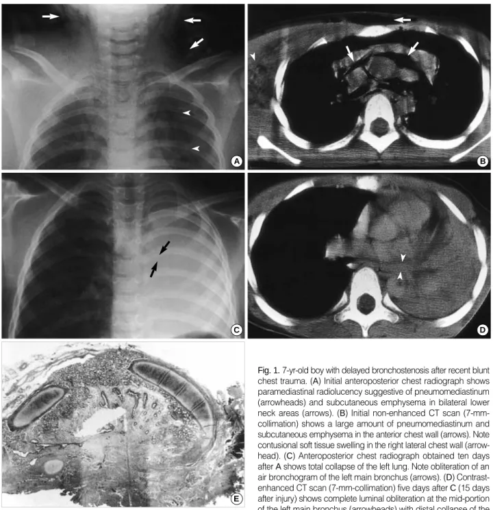
Fig. 1. 7-yr-old boy with delayed bronchostenosis after recent blunt chest trauma. (A) Initial anteroposterior chest radiograph shows paramediastinal radiolucency suggestive of pneumomediastinum (arrowheads) and subcutaneous emphysema in bilateral lower neck areas (arrows). (B) Initial non-enhanced CT scan (7-mm- collimation) shows a large amount of pneumomediastinum and subcutaneous emphysema in the anterior chest wall (arrows). Note contusional soft tissue swelling in the right lateral chest wall (arrow- head). (C) Anteroposterior chest radiograph obtained ten days after Ashows total collapse of the left lung. Note obliteration of an air bronchogram of the left main bronchus (arrows). (D) Contrast- enhanced CT scan (7-mm-collimation) five days after C(15 days after injury) shows complete luminal obliteration at the mid-portion of the left main bronchus (arrowheads) with distal collapse of the left lung. (E) Photomicrograph of pathologic specimen obtained from bronchial segmental resection and anastomosis 22 days after blunt chest trauma shows near-complete obliteration of the bronchial lumen by fibrous scar and granulation tissue, which seem to be secondary to recent transmural injury (H and E, ×10).
A B
C
E
D
Delayed Bronchostenosis After Blunt Chest Trauma 557
persisted for five days at daily routine chest radiographs. Con- trast-enhanced chest CT scan, which was performed in order to find out the cause of lobar atelectasis, revealed obliteration of the proximal portion of the left upper lobe bronchus (Fig.
2). The previous bilateral hemopneumothoraces, pneumome- diastinum, and subcutaneous emphysema were no longer seen, and the extent of the cavitary consolidation in the right upper lobe has decreased. Since left upper lobe atelectasis persisted for eight days more, a bronchoscopic examination was performed, which showed near-complete obstruction of the left upper lobar bronchus.
Four days after the bronchoscopic examination (the 27th hospital day), bronchial segmental resection and end-to-end anastomosis of the left upper lobar bronchus was performed.
At surgery, approximately 5-mm-long, stenotic segment was found at the proximal portion of the left upper lobe bronchus, showing complete luminal obstruction by fibrotic change.
Pathologic specimen obtained from bronchial segmental resection showed obliteration of the bronchial lumen by dense fibrous overgrowth and granulation tissue, which was identi- cal to the pathologic finding of Case 1. The previously atelec- tatic left upper lobe was fully re-expanded after surgery, and the recovery was uneventful.
DISCUSSION
In most cases, a tracheobronchial rupture is suspected radi- ologically in front of pneumomediastinum, cervical and tho- racic subcutaneous emphysema, and persistent pneumotho- rax and bronchopleural air leak despite chest tube placement (2). A bronchoscopic examination can confirm the presence
and location of the airway rupture, which is reconfirmed and repaired by surgery. Although chest CT scan sometimes visu- alizes directly the presence of tracheobronchial rupture, it can generally show only secondary findings of an air leak (2).
Secondary findings of an air leak are consequences of free communication between the site of the tracheobronchial dis- ruption and the pleural cavity, which results in a large per- sistent pneumothorax despite tube thoracotomy (6). If the bronchial transection is incomplete and there is little com- munication between the proximal transected bronchus and the mediastinum and/or the pleural space, a once-developed pneumomediastinum or pneumothorax will subside well either spontaneously or after chest tube placement. In this circum- stance, the tracheobronchial injury will not be noticed radio- logically until delayed atelectasis resulting from fibrotic bron- chostenosis appears as in our cases (6). In our cases, because the initial pneumomediastinum and subcutaneous emphy- sema and/or pneumothoraces subsided well either sponta- neously or after chest tube placement within 15 days with the vital signs of the patients being stable, the bronchial injury remained unnoticed over a period of two weeks.
On review of literature regarding bronchial rupture after blunt chest trauma, one of two patients reported by Epelman et al. (7) was similar to our cases, in whom a bronchial injury was identified as delayed atelectasis of the left lower lobe with bronchial obliteration at CT ten days after blunt chest trau- ma. Ozcelik et al. (8) reported a case with combined right main bronchial disruption and chylothorax manifesting as a consolidated right lung and a small pleural effusion that were diagnosed 75 days after blunt chest trauma.
In our cases, the cause of delayed atelectasis detected at chest radiographs and CT was histopathologically proved to be focal bronchial wall thickening due to exuberant tissue reaction to recent bronchial wall injury. The subclinical injury to the bronchial wall (bronchial mucosal tear, undisplaced and/or incomplete bronchial rupture) was unnoticed initially but became evident later after development of an active healing process in the traumatized region. The healing process result- ed in bronchial wall thickening with resultant airway narrow- ing, which manifested at chest CT as luminal obliteration of the involved bronchus with distal atelectasis of the lung. We think it is noteworthy that the appearance of atelectasis was seen ten days after the injury in both cases of our series. As for the initial CT diagnosis of a bronchial rupture, multiplanar reformation- or three-dimensional images of multi-detector row CT scan could demonstrate an airway injury better than axial CT images.
In conclusion, we report two pediatric cases of delayed bron- chostenosis with distal atelectasis after recent blunt chest trau- ma with pathologic correlation. We suggest that, when delay- ed pulmonary atelectasis is encountered in children who sus- tained a blunt chest trauma, a possibility of subclinical bron- chial wall injury should be considered in addition to simple mucoid plugging.
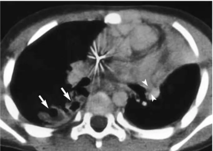
Fig. 2.2-yr-old boy with delayed bronchostenosis after recent blunt chest trauma. Contrast-enhanced chest CT scan (3-mm-collima- tion) obtained 15 days after blunt chest trauma shows left upper lobe atelectasis. Note complete luminal obliteration of the proximal portion of the left upper lobe bronchus (arrowheads). Also note cavitary consolidations in the right upper lobe, suggestive of resolv- ing traumatic lung cysts (arrows).
558 H.-K. Yoon, T.S. Kim, J. Han, et al.
REFERENCES
1. Baumgartner F, Sheppard B, de Virgilio C, Esrig B, Harrier D, Nel- son RJ, Robertson JM. Tracheal and main bronchial disruptions after blunt chest trauma: presentation and management. Ann Tho- rac Surg 1990; 50: 569-74.
2. Kunisch-Hoppe M, Hoppe M, Rauber K, Popella C, Rau WS. Tra- cheal rupture caused by blunt chest trauma: radiological and clini- cal features. Eur Radiol 2000; 10: 480-3.
3. Unger JM, Schuchmann GG, Grossman JE, Pellett JR. Tears of the trachea and main bronchi caused by blunt trauma: radiologic find- ings. AJR Am J Roentgenol 1989; 153: 1175-80.
4. de la Rocha AG, Kayler D. Traumatic rupture of the tracheobron- chial tree. Can J Surg 1985; 28: 68-71.
5. Tocino IM, Miller MH. Mediastinal trauma and other acute medi- astinal conditions. J Thorac Imaging 1987; 2: 79-100.
6. Hood RM, Sloan HE. Injuries of the trachea and major bronchi. J Thorac Cardiovasc Surg 1959; 38: 458-80.
7. Epelman M, Ofer A, Klein Y, Best LH, Guralnik L, Bentur L, Traubi- ci J. CT diagnosis of traumatic bronchial rupture in children. Pedi- atr Radiol 2002; 32: 888-91.
8. Ozcelik C, Onat S, Bayar ES. Combined late diagnosed right main bronchial disruption and chylothorax from blunt chest trauma. Ann Thorac Surg 2004; 78: 61-2.