조영제 혈관 외 유출이 관찰된 복부 둔상 환자의 유출부위에 따른 예후
연세대학교 원주의과대학 응급의학교실, 울산대학교병원 응급의학과1 신형진∙이강현∙곽영수∙김선휴1∙김@ 현∙황성오
─ Abstract ─
Prognosis for Blunt Abdominal Trauma Patients with Contrast Extravasation on the Abdominopelvic CT Scan
Hyung Jin Shin, M.D., Kang Hyun Lee, M.D., Young Soo Kwak, M.D., Sun Hyu Kim, M.D., Hyun Kim, M.D., Sung Oh Hwang, M.D.
Department of Emergency Medicine, Wonju College of Medicine, Yonsei University Department of Emergency Medicine, Ulsan University Hospital1
Purpose: Computed tomography (CT) is an accurate test for evaluating hemodynamically stable patients with blunt abdominal trauma. Until now, there have been few studies concentrating on the diagnostic and prog- nostic significance of the intravenous contrast extravasation (CE) site. We investigated the site of CE on abdominopelvic CT (APCT) and its effect on treating trauma patients and predicting the clinical outcome.
Methods: The 50 patients admitted to our emergency department with blunt abdominal trauma showing CE on APCT from January 2004 to September 2006 were included in this study. Patients were prospectively col- lected, and medical records were reviewed and analyzed. The patients’clinical and lab findings, Focused Assessment with Sonography for Trauma (FAST) findings, CT findings were analyzed. CE sites were classi- fied as intraperitoneal, retroperitoneal, and pelvic cavity and were correlated with post-treatment complications, mortality, and morbidity.
Results: Of the 50 patients (mean age : 45±18years, 29 males, 21 females) included in our study, 33 patients died (66%). There was no correlation between CE site and ICU or total hospitalization duration (p=0.553, p=0.523). During the first 24 hours of resuscitation, the pelvic cavity group required a mean of 20 units more of packed red blood cell (pRBC) transfusion compared to other groups (p=0.003). In the intraperi- toneal group, more patients received operative invasive intervention - either laparotomy or embolization (p=0.025). The intraperitoneal group had the highest mortality, with 13 deaths (11/33, 39%), and the highest early mortality rate (10/13, 76%) in the first 24 hours (p=0.001).
Conclusion: Intraperitoneal CE on the CT scan in cases of blunt abdominal trauma is regarded as an indica- tion of a need for invasive intervention (either angiography or laparotomy) and of a higher mortality rate in the first 24 hours. A pelvic cavity CE rquires more aggressive transfusion with pRBC. However, the CT findings themselves showed no significant correlation with overall mortality, morbidity, or hospitalization. (J Korean Soc Traumatol 2009;22:57-64)
Key Words: Blunt abdominal trauma, Abdominopelvic CT scan, Extravasation
� Address for Correspondence : Kang Hyun Lee, M.D., Ph.D.
Department of Emergency Medicine, Wonju College of Medicine, Yonsei University 162 Ilsan-dong, Wonju-si, Gangwon-do, Korea
Tel : 82-33-741-1612, Fax : 82-33-742-3030, E-mail : [email protected]
접수일: 2009년 4월 29일, 심사일: 2009년 5월 11일, 수정일: 2009년 5월 18일, 승인일: 2009년 5월 27일 본 논문은 2006년 대한응급의학회 추계학술대회에서 포스터 전시 되었음.
I. 서 론
외상은 전 연령대 모든 사망원인들 중에서 세 번째로 많은 것으로 알려져 있으며 40세 미만의 젊은 층에서는 가장 많은 원인으로 알려져 있다.(1) 이중 복부 둔상으로 인한 사망은 최근 교통사고 증가와 산업화에 따른 안전사 고, 폭행 및 난폭화로 인하여,(2) 외상의 큰 비중을 차지하 고 있고 흔히 복강 내 출혈을 동반하여 순환기능에 영향 을 끼치기 때문에 이에 대한 신속하고 정확한 진단은 응 급수술 및 지속적인 소생술의 결정에 큰 도움이 된다.(3) 복부 둔상을 조기에 정확히 진단하기 위해서 복부 전산화 단층촬영을 가장 많이 이용하고 있으며 이는 수술 등 치 료방법의 결정에 많은 도움을 주고 있다. 특히 복부 전산 화 단층촬영은 복부 둔상을 가급적 빠른 시간 내에 비침 습적 방법으로 복강 및 후복막강내 각 장기의 통합성 (integrity)정도의 정보를 정확하게 제공한다는 장점이 있
다.(2)
복부 둔상 후 시행한 전산화 단층촬영상 조영제의 혈관 외 유출이 일어날 때 이것은 곧 고형장기의 손상 및 혈관 손상을 의미한다.(4) 이러한 이유로 대부분의 조영제의 혈 관 외 유출은 진단개복술이나 혈관조영술 같은 침습적인 중재시술(intervention)의 적응증이 되며,(5-12) 최근 혈관 색전술에 의하여 새로운 치료적 접근이 많이 이루어지고 있다. 최근의 대 집단 연구에서 조영제의 혈관 외 유출은 전체 0.2%에 육박하는 높은 발생률을 나타내고 있는데,(9) 이는 진단기술의 발전 뿐 아니라 이러한 소견에 대한 관 심과 인식이 증가되었기 때문이다.(4,5) 그러나 조영제의 혈관 외 유출이 일어나는 부위에 따른 그 진단적 의미와 치료방법을 결정하는 요소들은 아직 잘 알려져 있지 않으 며, 수술이나 예후에 어떤 영향을 미치는지에 대하여도 잘 알려져 있지 않다.
본 연구의 목적은 복부 및 골반 전산화 단층촬영시행
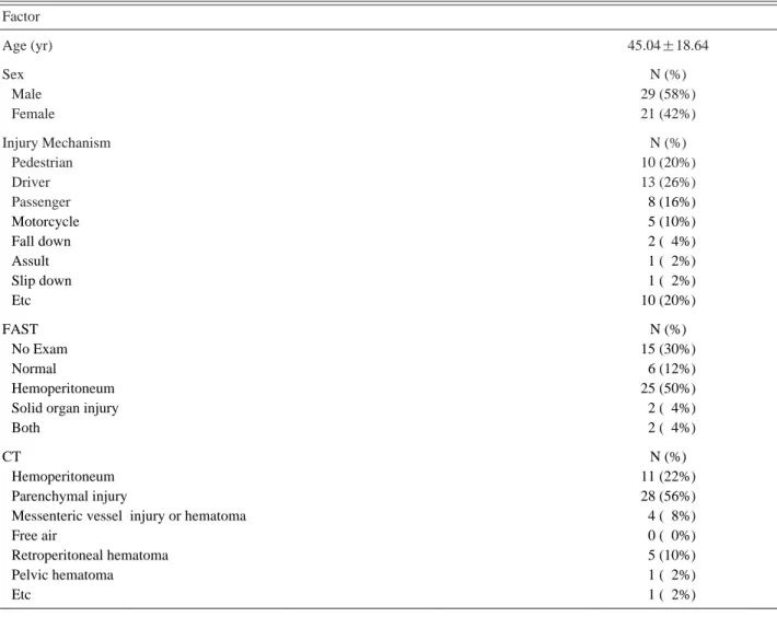
Table 1. The baseline characteristics and findings on *FAST and �CT scan Factor
Age (yr) 45.04±18.64
Sex N (%)
Male 29 (58%)
Female 21 (42%)
Injury Mechanism N (%)
Pedestrian 10 (20%)
Driver 13 (26%)
Passenger 08 (16%)
Motorcycle 05 (10%)
Fall down 02 (04%)
Assult 01 (02%)
Slip down 01 (02%)
Etc 10 (20%)
FAST N (%)
No Exam 15 (30%)
Normal 06 (12%)
Hemoperitoneum 25 (50%)
Solid organ injury 02 (04%)
Both 02 (04%)
CT N (%)
Hemoperitoneum 11 (22%)
Parenchymal injury 28 (56%)
Messenteric vessel injury or hematoma 04 (08%)
Free air 00 (00%)
Retroperitoneal hematoma 05 (10%)
Pelvic hematoma 01 (02%)
Etc 01 (02%)
* FAST : focussed assessment of sonography for trauma
�CT : computed tomography
후 조영제의 혈관 외 유출이 발생할 때 그 발생부위가 환 자의 치료와 예후에 어떤 영향을 미치는지 알아보고자 하 는데 있다.
II. 대상 및 방법
1. 대상 환자
2004년 1월부터 2006년 9월까지 복부 둔상을 입은 후 원 주기독병원 강원영서권역 응급의료센터로 내원하여 조기 평가상 혈역학적으로 안정적이어서 복부 및 골반 전산화 단층촬영을 시행한 환자 중 조영제의 혈관 외 유출이 관 찰된 환자를 대상으로 하였다. 혈역학적으로 불안정하더라 도 소생술 후 안정적이 되어 전산화 단층촬영을 시행한 환자도 대상에 포함시켰다. 먼저 응급의학과 전공의 및 전 문의에 의해 전산화 단층 촬영상 조영제 혈관 외 유출을 의심하고, 이후 영상의학과 전문의에 의한 판독소견에 근 거하여 혈관 외 유출을 확진 한 환자만 연구대상에 포함 시켰다. 나이 18세 이하, 임신, 화학요법을 받고 있는 암환 자, 면역억제상태, 심폐소생술거부상태는 제외기준으로서 대상에서 제외하였다.
2. 연구방법
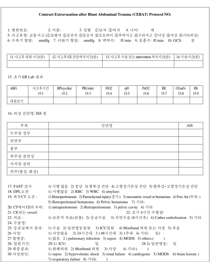
대상환자의 수집은 미리 계획된 연구 지침(Fig 1.)에 따
라 전향적으로 선정하였다. 각 환자의 임상적 소견 및 검 사실 소견을 먼저 조사하고 각 환자의 응급초음파소견을 기록하였다. 신체 6 부위의 외상진단에 따라 AIS (abbrevi- ated injury scale)를 측정하고 이를 통해 ISS (Injury severi- ty score)를 계산하여 객관적인 외상 정도를 비교하였다.
복부 및 골반 전산화 단층 촬영소견 등의 자료를 분석 하여 혈복강, 고형장기 손상, 장간막 혈관의 손상, 유리공 기(free air), 후복막강 혈종, 골반강 혈종여부를 파악하였 다. 조영제의 혈관 외 유출을 파악하여 유출되는 혈관을 기록하고 이를 복강 내, 후복막강, 골반강으로 군을 나누고 각 군에 따른 치료방법, 치료 후 합병증 및 사망률, 이환 율, 입원기간 등을 비교하였다. 치료방법은 응급실에서 초 기에 진단적 개복술이나 혈관색전술을 한 경우를 침습적 수술적 치료로, 보존적 치료를 한 경우와 입원 후에 지연 수술을 한 경우는 비수술적 치료로 구분하였다.
3. 통계분석
조사항목의 통계분석은 SPSS for WindowTM 13.0 을 이용하였다. 범주형 변수의 분석에는 카이제곱검정을 사용 하였고, 연속형 변수의 분석에는 one way ANOVA를 이 용하였다. 모든 통계분석에서 p<0.05 인 경우를 의미 있는 것으로 분석하였다.
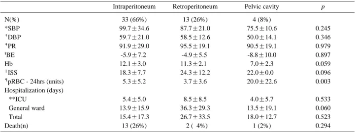
Table 2. The comparison of the characteristics according to CE site on CT scan
Intraperitoneum Retroperitoneum Pelvic cavity p
N(%) 33 (66%) 13 (26%) 4 (8%)
*SBP 99.7±34.6 87.7±21.0 75.5±10.6 0.245
�DBP 59.7±21.0 58.5±12.6 50.0±14.1 0.346
�PR 91.9±29.0 95.5±19.1 90.5±19.1 0.979
§BE -5.9±7.2. -4.9±5.50 -8.8±10.0 0.897
Hb 12.1±3.00 11.3±2.10 7.0±2.3 0.059
‖ISS 18.3±7.70 24.3±12.2 22.0±0.00 0.096
¶pRBC - 24hrs (units) 5.3±5.2 3.7±3.6 20.0±22.6 0.003
Hospitalization (days)
**ICU 5.4±5.0 8.5±8.5 4.0±5.7 0.533
General ward 13.9±15.9 36.3±29.3 13.5±19.1 0.060
Total 15.4±17.3 26.7±33.5 18.0±12.7 0.523
Death(n) 13 (26%) 02 (04%) 1 (2%) 0.294
* SBP : systolic blood pressure
�DBP : diastolic blood pressure
�PR : pulse rate
§BE : base excess
‖ISS : injury severity score
¶pRBC-24hrs : amount of packed RBC transfusion for 24hrs
** ICU : intensive care unit
III. 결 과
대상 환자는 모두50명으로 45.04±18세 (남자 29명, 여자 21명) 이었다(Table. 1). 전체 사망은 16명(32%)이었고 각 군에 따른 사망률의 차이는 없었다(p=0.294). 사고유형은 운전자 교통사고가 13명(26.5%)으로 가장 많았으며 전체 사고 중 교통사고가 73.5%로 많은 비중을 차지하였다. 내 원 시 측정한 활력징후와 검사실 소견은 각군에 따라 특 별한 차이를 보이지 않았으며 외상진단에 따라 계산된 ISS는 평균 19.66이었고 각군에 따른 차이는 없었다 (Table 2). 내원 초기 혈역학적으로 불안정한 환자의 경우 응급실에서 먼저 외상환자용 복부초음파로 혈복강이나 고 형장기의 손상이 진단된 경우는 29예(혈복강 25예, 고형장 기 손상 2예, 혈복강+고형장기 손상 2예)였으며, 진단적 복강 세척술은 연구지침에는 계획하였으나 대상환자 모두 에서 시행하지 않아 분석에서 제외하였다.
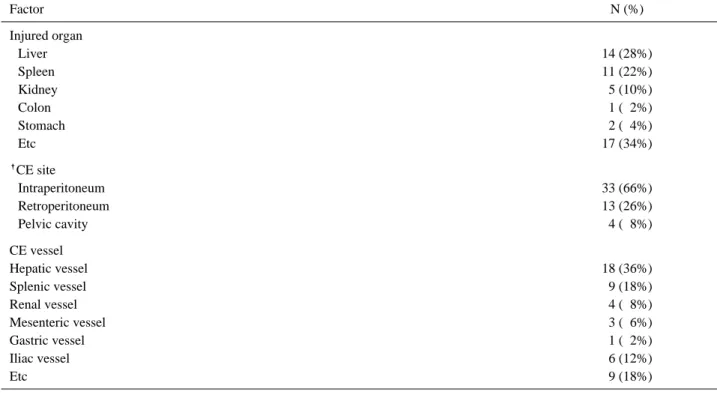
복부 및 골반 전산화 단층촬영상(Table. 1, 3) 고형장기 의 손상은 28예(56%), 혈복강은 11예(22%), 후복막강 혈 종 5예(10%), 장간막 손상 4예(8%), 골반강 혈종 1예 (2%)였으며, 조영제 혈관 외 유출은 복강내군이(Fig. 2) 33예(66%), 후복막강군(Fig. 3) 13예(26%), 골반강군 4예 (8%)였다. 유출혈관은 간 혈관이 18예(36%), 비장혈관 9 예(18%), 장골혈관 6예(12%), 신혈관 4예(8%), 장간막혈 관 3예(6%)등의 순서로 많았다.
각 군에 따른 치료방법(Table. 4)에서는 복강내군은 수술 적 치료22예(진단개복술 16예, 혈관색전술 6예), 비수술적 치료 11예(보존적 치료 9예, 입원 후 지연수술 2예) 였고, 후복막강군은 수술적 치료 3예(진단개복술 1예, 혈관색전술 2예), 비수술적 치료10예(보존적 치료 9예, 입원 후 지연수 술 1예)였으며, 골반강군은 수술적 치료 1예(혈관색전술 1 예), 비수술적 치료 3예(보존적 치료 1예, 입원 후 지연수술 2예)로 각 군에 따른 치료방법의 차이를 보였다(p=0.025).
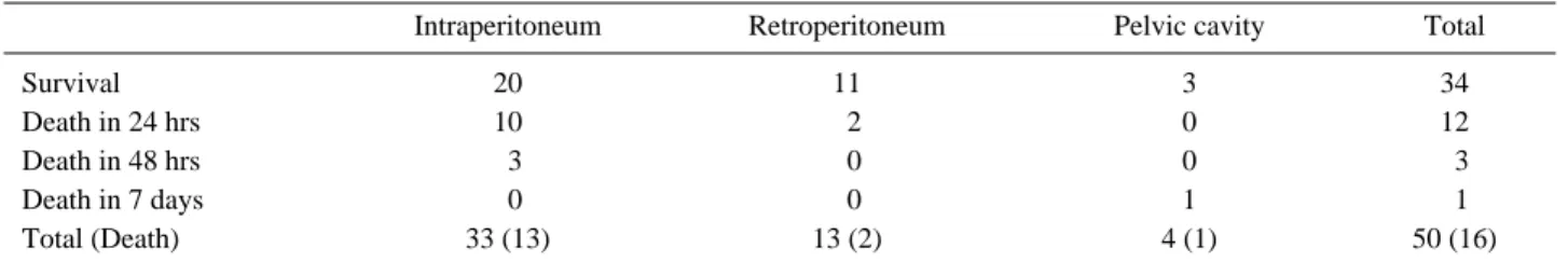
초기 24시간 내 농축적혈구 수혈양은 골반강군 4예에서 평균 20 unit으로 다른 군보다 많았다(p=0.003). 조영제 유 출부위에 따른 예후(Table 2, 5)에 대하여는 중환자실 및 총 입원기간 각 군에 따른 사망률에는 차이가 없었으나 (p=0.553, p=0.523, p=0.294), 총 16명의 사망 환자 중 복 강 내 조영제 혈관 외 유출이 13명(39%)으로 가장 많았으 며, 이중 24시간 내 조기사망이 10예(76%)로서 복강 내 군에서 조기사망률이 높았다(p=0.001).
IV. 고 찰
복부 둔상환자에 있어 혈역학적으로 안정된 경우는 조 기 평가를 위한 방법으로 정맥 혹은 경구 투여 조영제를 이용한 복부 및 골반 전산화 단층촬영이 매우 유용하게 이용되고 있다.(13,14) 전산화 단층촬영상 정맥투여 조영제 의 혈관 외 유출은 넓게 퍼지거나(diffuse), 국소적인(focal)
Table 3. The Specific findings on *CT scan of patients
Factor N (%)
Injured organ
Liver 14 (28%)
Spleen 11 (22%)
Kidney 05 (10%)
Colon 01 (02%)
Stomach 02 (04%)
Etc 17 (34%)
�CE site
Intraperitoneum 33 (66%)
Retroperitoneum 13 (26%)
Pelvic cavity 04 (08%)
CE vessel
Hepatic vessel 18 (36%)
Splenic vessel 09 (18%)
Renal vessel 04 (08%)
Mesenteric vessel 03 (06%)
Gastric vessel 01 (02%)
Iliac vessel 06 (12%)
Etc 09 (18%)
* CT : computed tomography
�CE : contrast extravasation on CT scan
고농도의 한 부분으로 표현되며, 주변 혈관조직들과 비교 했을 때 비슷한 농도로 나타나 동맥 혹은 정맥의 파열에 의한 출혈을 의미한다.(4) 이는 대부분 침습적인 중재시술
의 적응증이 된다.(5-12) 수많은 연구자들이 이런 소견을 보이는 환자들에 대한 경험을 연구하여, 대부분이 조영제 의 혈관 외 유출이 있을 때 중재시술을 권장하였는데, 과
Contrast Extravasation after Blunt Abdominal Trauma (CEBAT) Protocol NO:
1. 병원번호: 2. 이름: 3. 성별 ①남자 ②여자 4. 나이: 세
5. 사고유형: 교통사고 (①보행자 ②운전자 ③동승자 ④오토바이 ⑤추락사고 ⑥구타사고 ⑦낙상 ⑧자상 ⑨기타외상) 6. 수축기 혈압: mmHg 7. 이완기 혈압: mmHg 8. 맥박수: 회/min 9. 호흡수: 회/min 10. GCS: 점
15. 초기 ER Lab 결과
16. 외상 진단명: ISS 점
17. FAST 검사 1) 시행 않음 2) 정상 3) 혈복강 진단 4) 고형장기손상 진단 5) 혈복강+고형장기손상 진단
18. DPL소견: 1) 시행않음 2) RBC 3) WBC 4) amylase:
19. 복부CT 소견 : 1) Hemoperitoneum 2) Parenchymal injury(장기:) 3) mesenteric vessel or hematoma 4) Free Air (부위: ) 5) Retroperitoneal hematoma 6) Pelvic hematoma 7) 기타:
20. CT에서 CE의 부위: 1) intraperitoneum 2) Retroperitoneum 3) pelvic cavity 4) 기타
21. CE되는 vessel: 22. 초기 4시간 수혈량:
23. 치료 : 1) 보존적 치료(관찰) 2) 응급수술 3) 지연수술 (8시간후) 4) Cather embolization 5) 기타 24. 수술명:
25. 응급실에서 결과: 1) 수술 2) 일반병동입원 3) ICU입원 4) Moribund 퇴원 또는 사망 5) 후송
26 사망: 1) 사망않음 2) 24시간내 3 ) 48시간내 3) 1주내 4) 기타: 일)
27 합병증: 1) 없음 2 ) pulmonary infection 3) sepsis 4) MODS 5) others:( )
28. 입원기간: 28-1) ICU: 일 28-2) 일반병동: 일
29 최종결과: 1) 완쾌퇴원 2) Moribund 퇴원 3) 사망 4) 기타:( )
30 사망원인: 1) sepsis 2) hypovolemic shock 3) renal failure 4) cardiogenic 5) MODS 6) brain lesion( ) 7) respiratory failure 8) 기타( )
11. 사고후 내원 시간(분) 12. 사고후 CE 진단까지시간(분) 13. 사고후 수술 또는 intervention 까지시간(분) 14. 수술시간(분)
ABG 사고후시간 BP(sys/dia) PR(/min) FiO2 pH PaO2 BE O2sat% Hb
15-1 15-2 15-3 15-4 15-5 15-6 15-7 15-8 15-9
내원초기
부위 진단명 AIS
두부및 경부 안면부 흉부
복부및 골반강 사지및 골반 외부(열상, 화상)
Fig. 1. Contrast Extravasation after Blunt Abdominal Trauma (CEBAT) Protocol.
거에는 대부분 진단적 개복술을 권장하였으나 최근 영상 의학적 중재시술의 발달로 혈관색전술의 사용이 폭발적으 로 증가하고 있다. Fang 등(15)은 혈역학적으로 안정적인 외상성 간 손상환자 212명을 연구하였는데 이들 중 15명이 조영제의 혈관 외 유출을 나타내었다. 유출은 복강 내와 장기 내 유출로 구분하였으며 복강 내 유출은 진단적 개 복술을 필요로 한 반면 장기 내 유출은 혈관색전술을 권 장하였다. Wong 등(16)은 32명의 복강 내 조영제 유출환
자를 조사하였고 이중 85%가 중재시술이 필요했다고 발 표하였다. 또한 중재시술의 필요성을 결정짓는 요소로는 수축기 혈압이 100 mmHg이하, 편평한 대정맥소견(a flat vena cava), 복강 내 유출에 반하는 복강 외 유출로 결정 지었다. Ivan 등(17)은 1435명의 복부 둔상환자를 후향적으 로 연구하였는데 이중 70명(4.8%)의 환자에서 조영제 유 출이 관찰되었다. 조영제 유출부위에 따라 복강내유출군이 25명, 골반강-후복막강유출군이 39명, 두 곳 모두 유출된
Fig. 2. Computed tomography (CT) scan showing extravasa- tion of contrast within the mesentery. The patient underwent laparotomy and was found to have ileal mesentery rupture, ileal perforation and jejunal tear, which required segmental resection of ileum and primary repair of ileum and sigmoid mesocolon.
Fig. 3 . Computed tomography (CT) scan showing extravasa- tion of contrast from left renal artery. The patient underwent simple left nephrectomy at the time of laparotomy.
Table 4. The comparison of the treatment according to *CE site on �CT scan
Treatment Intraperitoneum Retroperitoneum Pelvic cavity Total
Operative 22 03 1 26
Nonoperative 11 10 3 24
Total 33 13 4 50
p=0.025
* CE : contrast extravasation on CT scan
�CT : computed tomography
Table 5. The comparison of the prognosis according to CE* site on CT�scan
Intraperitoneum Retroperitoneum Pelvic cavity Total
Survival 20 11 3 34
Death in 24 hrs 10 02 0 12
Death in 48 hrs 03 00 0 03
Death in 7 days 00 00 1 01
Total (Death) 33 (13) 13 (2) 4 (1) 50 (16)
p=0.001
* CE : contrast extravasation on CT scan
�CT : computed tomography
경우가 3명이 있었으며 이들 전체환자 중 30명(47%)이 진 단적 개복술 혹은 혈관조영술 같은 중재시술을 받았고, 34 명(53%)이 중재시술을 받지 않았다. 비수술적 치료는 골 반강-후복막강유출군이 복강내유출군에서보다 성공적이었 으며 결론적으로 조영제 혈관 외 유출은 모든 환자에서 침습적 중재시술의 적응증은 아니라고 발표했다.
본 연구에서는 전산화 단층촬영상 조영제의 혈관 외 유 출이 관찰된 50명의 환자를 대상으로 전향적으로 연구를 진행하였으며 유출부위를 복강 내, 후복막강, 골반강 세부 위로 나누어 각각의 치료방법과 예후를 비교하였다. 이중 복강내군은 다른 군에 비해 침습적인 수술적 치료가 선호 되었으나 후복막강, 골반강군은 비수술적 치료가 더 많이 시행되었음을 알 수 있었다(p=0.025). 그러나 각 군에 따른 예후비교에서 입원기간(p=0.523)과 사망률(p=0.294)은 유 의한 차이가 없는 것으로 나타나 해석에 주의를 요할 필요 가 있다. 이는 각군에 따라 다른 기준과 가설로 치료방법을 선정한 것이 아니므로, 치료방법의 경향은 파악할 수 있겠 으나 치료방법선정의 성공여부와는 관련성이 없다고 하겠 다. 오히려 침습적인 수술적 치료를 시행했던 복강 내 군에 서 24시간 이내 조기사망률이 높은 것으로 나타나 (p=0.001), 복강 내 조영제 혈관 외 유출소견 그 자체가 침 습적 중재시술의 절대적 적응증은 아닌 것으로 생각한다.
본 연구에서는 또한 농축적혈구 수혈 양이 골반강군에 서 의미 있게 많은 것으로 나타났는데, 이는 골반골 골절 등과 같은 과다 출혈을 초래하는 상황에서 혈관색전술 또 는 보존적 치료를 하면서 수혈을 많이 가하여 그런 결과 가 나온 것으로 생각한다. 본 연구의 제한점으로는 첫째, 단일병원 연구로 대상환자수가 적었던 점이며, 둘째, 연구 지침에 따라 전향적으로 환자를 선정하여 자료를 분석하 였으나, 특정한 기준과 가설에 근거하여 환자의 치료방법 을 적용하지 못하고, 해당 외과의의 결정에 근거하여 치료 방법이 선정되고 이를 분석했던 점이다. 향후 보다 많은 환자를 선정하고, 미리 합의된 기준에 근거 외상팀의 리더 에 의한 치료방법선정 등을 적용하여 전향적으로 치료를 시행하고 그 결과를 비교하면 더욱 의미 있는 결과를 얻 을 수 있을 것이다.
V. 결 론
복부 둔상을 입은 환자의 복부 및 골반 전산화 단층촬 영상 혈관 외 유출이 관찰되는 경우, 복강 내로 유출된 환 자에서 침습적인 수술적 치료를 선호하며 후복막강 또는 골반강 내 유출보다 24시간 내 조기 사망률이 높았다. 또 한 골반강 내로 유출된 환자에서 초기 24시간 수혈 양이 많았다. 그러나 유출부위에 따른 입원기간, 이환율, 최종사 망률에서는 차이가 없었다.
REFERENCES
01) Cho YD, Hong YS, Lee SW, Choi SH, Yoon YH, Lim SI, et al. Impact of Initial Helical Abdominal Computed Tomography on the Diagnosis of Hollow Viscus Injury and Blunt Abdominal Trauma. J Korean Soc Traumatol 2008;21:28-35
02) Kim HS, Hwang SY, Won HS, Kim SE, Park CW, Lee K. A Clinical Study of Abdominal CT in Blunt Abdominal Trauma, J Korean Soc Emerg Med 1994;5:100-6
03) Viscomi GN, Gonzalez R, Taylor KJ, Crade M.
Ultrasonic evaluation of hepatic and splenic trauma.
Arch Surg. 1980;115:320-1.
04) Yao DC, Jeffrey RB Jr, Mirvis SE, Weekes A, Federle MP, Kim C, et al. Using contrast-enhanced helical CT to visualize arterial extravasation after blunt abdominal trauma: incidence and organ distribution. AJR Am J Roentgenol. 2002;178:17-20.
05) Willmann JK, Roos JE, Platz A, Pfammatter T, Hilfiker PR, Marincek B, et al. Multidetector CT:
detection of active hemorrhage in patients with blunt abdominal trauma. AJR Am J Roentgenol.
2002;179:437-44.
06) Stephen DJ, Kreder HJ, Day AC, McKee MD, Schemitsch EH, ElMaraghy A, et al. Early detection of arterial bleeding in acute pelvic trauma. J Trauma.
1999;47:638-42.
07) Fang JF, Chen RJ, Wong YC, Lin BC, Hsu YB, Kao JL, et al. Pooling of contrast material on computed tomography mandates aggressive management of blunt hepatic injury. Am J Surg. 1998;176:315-9.
08) Jeffrey RB Jr, Cardoza JD, Olcott EW. Detection of active intraabdominal arterial hemorrhage: value of dynamic contrastenhanced CT. AJR Am J Roentgenol.
1991;156:725-9.
09) Taylor GA, Kaufman RA, Sivit CJ. Active hemorrhage in children after thoracoabdominal trauma: clinical and CT features. AJR Am J Roentgenol. 1994;162:401-4.
10) DiGiacomo JC, McGonigal MD, Haskal ZJ, Audu PB, Schwab CW. Arterial bleeding diagnosed by CT in hemodynamically stable victims of blunt trauma. J Trauma. 1996;40:249-52.
11) Federle MP, Courcoulas AP, Powell M, Ferris JV, Peitzman AB. Blunt splenic injury in adults: clinical and CT criteria for management, with emphasis on active extravasation. Radiology. 1998;206:137-42.
12) Pereira SJ, O’Brien DP, Luchette FA, Choe KA, Lim E, Davis Jr K, et al. Dynamic helical computed tomography scan accurately detects hemorrhage in patients with pelvic fracture. Surgery. 2000;128:678- 85.
13) Novelline RA, Rhea JT, Rao PM, Stuk JL. Helical CT in emergency radiology. Radiology. 1999;213:321-39.
14) Fang JF, Wong YC, Lin BC, Hsu YP, Chen MF.
Usefulness of multidetector computed tomography for the initial assessment of blunt abdominal trauma patients. World J Surg. 2006;30:176-82.
15) Fang JF, Chen RJ, Wong YC, Lin BC, Hsu YB, Kao JL, et al. Classification and treatment of pooling of contrast material on computed tomographic scan of blunt hepatic trauma. J Trauma. 2000;49:1083-8.
16) Wong YC, Wang LJ, See LC, Fang JF, Ng CJ, Chen
CJ. Contrast material extravasation on contrast-enhanced helical computed tomographic scan of blunt abdominal trauma: its significance on the choice, time, and out- come of treatment. J Trauma. 2003;54:164-70.
17) Diamond IR, Hamilton PA, Garber AB, Tien HC, Chughtai T, Rizoli SB, et al. Extravasation of Intravenous Computed Tomography Scan Contrast in Blunt Abdominal and Pelvic Trauma. J Trauma.
2009;66:1102-7.