─ 192 ─
대 한 외 상 학 회 지
Vol. 19, No. 2, December, 2006 � 증 례 �
복부 외상 후 발생한 동맥-집뇨계 간 연결
한일병원 비뇨기과
이창욱∙권오정∙방성학∙최낙영∙이창섭∙안승현
─ Abstract ─
Artery to Collecting System Communication after Abdominal Trauma
Chang Ug Lee, M.D., O Jung Kwon, M.D., Sung Hak Bang, M.D., Nak Young Choi, M.D., Chang Sub Lee, M.D., Seung Hyun Ahn, M.D.
Department of Urology, Hanil General Hospital, Seoul, Korea
Degenerative vascular disease, previous arterial surgery, long-term ureteral stenting, pelvis surgery, and radiotheraphy are reported as causes of artery-to-collecting-system communication.. Artery-to-collecting-sys- tem-communication associated with blunt trauma is rare, but potentially fatal. The diagnosis is very difficult and requires a high degree of suspicion. We were able to make the diagnosis based on the characteristic finding of contrast-enhanced computed tomography (CT) obtained in the early phase, equivalent to the finding obtained in the corticomedullary phase of the kidney. We report a case of artery to collecting system communi- cation due to blunt abdominal trauma following a fall, which was treated by embolization. (J Korean Soc Traumatol 2006;19:192-195)
Key Words: Artery and collecting system, Renal injury, Computed tomography
� Address for Correspondence : Seung Hyun Ahn, M.D.
Department of Urology, Hanil General Hospital 388-1 Ssangmun-dong, Dobong-gu, Seoul, Korea
Tel : 82-2-901-3961, Fax : 82-2-901-3158, E-mail : [email protected]
접수일: 2006년 10월 27일, 심사일: 2006년 10월 30일, 수정일: 2006년 11월 8일, 승인일: 2006년 11월 13일 외상으로 인한 동맥-집뇨계 간의 연결은 매우 드물게 발
생하며 치명적이다. 1, 2 저자들은 낙상으로 인한 신손상 환자에서 전산화 단층촬영의 조영 후 특징적인 동맥상 소 견을 통해 진단되어 신동맥 색전술을 시행한 동맥-집뇨계 간 연결증례 1례를 치험하였기에 문헌고찰과 함께 보고하 고자 한다.
Ⅰ. 증 례
40세 여자환자가 2층에서 떨어진 후 우측 측복부 동통
과 혈뇨를 주소로 응급실로 내원하였다. 과거력 상 우울증 으로 진단받은 후 수차례 자살기도를 했었으나 그 외에 특 이소견은 없었다. 이학적 소견 상 활력 징후는 혈압이 60/30 mmHg으로 떨어져 있었고, 맥박은 138회, 호흡은 18회, 체온은 36.5�C였다. 복부는 단단하고 팽만되어 있 었으며, 우측 늑골척추각압통이 있었고, 육안적 혈뇨를 보 여 방광 카테터를 삽입하였으며 혈뇨가 지속되었다. 내원 시 혈액학 검사에서는 혈색소 수치가 11.0 g/dl 등 특이 소견을 보이지 않았으나 2시간 후 시행한 검사에서는 혈색 소 수치가 3.6 g/dl로 감소하였다.
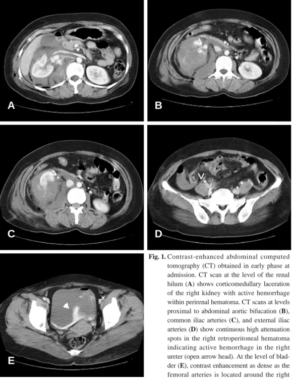
내원 당시 시행한 복부 전산화 단층촬영에서 우측신의 다발성 신손상과 신주위 혈종을 보였고(Fig. 1A), 혈종으 로의 조영제의 누출이 확인되었다. 혈종의 내부로 대동맥 이나 동맥과 같은 고음영 농도를 보이는 요관이 관찰되었 으며(Fig. 1B, C, D), 조영제가 방광내 우측 요관구 주 변으로 배출되었다(Fig. 1E). 수혈과 수액투여에도 혈역 학적으로 안정되지 않아 시행한 신동맥조영술에서 명확하 게 동맥과 요관의 누출이 확인되지는 않았으나 가성동맥류 가 관찰되었다(Fig. 2). 가성동맥류와 손상받은 부위에 대
하여 코일색전술을 시행한 뒤 혈역학적으로 안정되었으며, 6주 후 다른 시술없이 퇴원하였고 추후 복부 전산화 단층 촬영으로 추적관찰하기로 하였다.
Ⅱ. 고 찰
외상에 의한 비뇨생식기 손상 중에서 신장 및 방광파열 은 흔히 발생하는데 비하여 동맥과 집뇨계 간 연결은 매우 드물지만 치명적이며 진단과 치료가 쉽지 않은 질환으로
─ 193 ─
— 이창욱 외: 복부 외상 후 발생한 동맥-집뇨계 간 연결 —
Fig. 1. Contrast-enhanced abdominal computed tomography (CT) obtained in early phase at admission. CT scan at the level of the renal hilum (A) shows corticomedullary laceration of the right kidney with active hemorrhage within perirenal hematoma. CT scans at levels proximal to abdominal aortic bifucation (B), common iliac arteries (C), and external iliac arteries (D) show continuous high attenuation spots in the right retroperitoneal hematoma indicating active hemorrhage in the right ureter (open arrow head). At the level of blad- der (E), contrast enhancement as dense as the femoral arteries is located around the right ureteral orifice in the bladder (arrow head).
A B
C
E
D
1908년에 Moschowitz가 처음으로 발표한 이래로 60례 정도가 보고되고 있다.(3,4)
동맥과 집뇨계 간 연결의 발생부위로는 총장골동맥과 요 관이 교차하는 부위에 일어나는 경우가 대부분이며, 신동 맥과 신우, 신배 등은 서로 근접하게 위치하고 있으나 서 로 간의 연결은 매우 드물다. 또한 외장골동맥과 내장골동 맥보다 총장골동맥에서 일어난 예가 많이 보고되고 있다.
원인으로는 동맥류, 외상, 감염, 수술 및 방사선조사로 인한 부작용 등이 있다.(1,2,5) 또한 1차성과 2차성으로 구분할 수 있는데 1차성의 경우 대부분 둔상, 관통상 등의 외상이나 동맥류가 원인이며,(1,6,7) 2차성의 경우는 방사 선 요법이나 종양의 광범위 수술 등 의인성 원인이 대부분 을 차지한다.(8)
동맥과 집뇨계의 연결은 심한 출혈과 저혈압을 유발하여 치명적인 결과를 초래할 수 있다.(2) 그에 대하여 대부분 문헌에서는 동맥과 집뇨계 간 연결을 동맥조영술을 통하여 발표하고 있다.(1,6)
전산화 단층촬영은 복부둔상의 경우 대부분 시행되는 검 사이지만 전산화 단층촬영에서의 신동맥과 집뇨계 간 연결 소견이 발표된 예가 드물다. 본 증례에서는 내원 직후 시 행한 전산화 단층촬영에 의해 우측 요관의 특징적인 조영 증강과 방광내 조영제 배출이 확인되어 진단에 도움을 주 었다. 동맥조영술은 30%의 경우에서만 진단적이라고 Quillin 등(9)은 발표하고 있으며 특히 지속적으로 출혈되
는 경우에만 확인된다고 하였다. 본 증례에서는 동맥조영 술에 의한 동맥과 집뇨계의 직접적인 연결이 확인되지 않 았으나, 혈종으로 인한 종괴효과와 혈역학적으로 안정되지 않아 동맥조영술을 시행한 당시에는 일시적인 지혈이 이루 어진 것으로 생각된다. 그러나 전산화 단층촬영으로 동맥 과 집뇨계간 연결의 특징적인 소견을 확인하여 신속하게 진단할 수 있었고 적절한 치료를 시행할 수 있었다.
동맥과 요관 간 연결을 치료하는 방법으로는 개복술과 혈관내 시술이 행해지고 있으며, 신 절제술, 혈관치환술, 혈관결찰술, 요관 결찰술, 자가이식 등과 신동맥색전술, 스텐트 삽입술 등이 보고되고 있다. 본 증례에서는 O’
Donnell 등(10)이 발표한 바와 같이 신동맥에 대한 코일 색전술을 시행하였다. 그 후에 혈역학적으로 안정되었고 육안적 혈뇨가 감소하여 적절한 시술이 이루어 졌음을 확 인할 수 있었다.
복부 둔상 후 발생하는 동맥과 집뇨계 간 연결은 치명적 일 수 있다. 복부 둔상시 일반적으로 시행하는 전산화 단 층촬영을 이용하여 동맥과 집뇨계간 연결의 진단을 신속하 게 할 수 있어, 그에 따른 빠르고 적절한 시술이 가능할 수 있을 것으로 생각한다.
─ 194 ─
— 대한외상학회지 제 19 권 제 2 호 —
Fig. 2. Arterial (A) and parenchymal (B) phases of right renal arteriograms. A pseudoaneurysm is noted at the middle pole of the right kidney (open arrow head).
A B
─ 195 ─
— 이창욱 외: 복부 외상 후 발생한 동맥-집뇨계 간 연결 —
REFERENCES
01) Shinojima H, Seki T, Kumagai A, Sakurai Y, Shinno Y. Non-traumatic renal arteriopelvic fistula.
Int J Urol 1999;6:260-3.
02) Fay R, Brosman SA, Lindstrom R, Cohen A. Renal arterial-pelvic fistula. Urology 1973;2:292-5.
03) Moschowitz A. Simultaneous ligation of both exter- nal iliac arteries for secondary hemorrhage follow- ing bilateral ureterolithotomy. Ann Surg 1908;48:
872-5.
04) Vandersteen DR, Saxon RR, Fuchs E, Keller FS, Taylor LM Jr, Barry JM. Diagnosis and manage- ment of ureteroiliac artery fistula: value of provocative arteriography followed by common iliac artery embolization and extraanatomic arterial bypass grafting. J Urol 1997;158:754-8.
05) Batter SJ, McGovern FJ, Cambria RP.
Ureteroarterial fistula: case report and review of
the literature. Urology 1996;48:481-9.
06) Benoit G, Charpentier B, Roche A, Bellamy J, Mohamedi D, Fries D. Arteriocalyceal fistula after grafted kidney biopsy. Successful management by selective catheter embolization. Urology 1984;24:
487-90.
07) Giordanengo F, Vandone PL, Trimarchi S, Zaniboni N, Miani S. Ruptured aneurysm of the internal iliac artery. Panminerva Med 1995;37:150-4.
08) Bergqvist D, Parsson H, Sherif A. Arterio-ureteral fistula, a systematic review. Eur J Vasc Endovasc Surg 2001;22:191-6.
09) Quillin SP, Darcy MD, Picus D. Angiographic eval- uation and therapy of ureteroarterial fistulas. AJR Am J Roentgenol 1994;162:873-8.
10) O’Donnell M, Fakhry S, Chambers T. Traumatic left renal arterioureteral fistula. J Trauma 2002;
53:395.