대한외과학회지:제 74 권 제 2 호
□ 원 저 □
Vol. 74, No. 2, February, 2008
140
책임저자:남영수, 경기도 구리시 교문동 249-1번지
471-701, 한양대학교 구리병원 외과 Tel: 031-560-2292, Fax: 031-566-4409 E-mail: ysnam@hanyang.ac.kr
접수일:2007년 7월 10일, 게재승인일:2007년 11월 1일 중심 단어: 재발성 모소동, Limberg 피판술, 변형 마름모꼴 절제
재발성 모소동의 치료에 비대칭 마름모꼴 절제와 Limberg 피판술의 유용성
한양대학교 의과대학 구리병원 외과학교실 남 영 수
Effect of Modified Rhomboid Excision and Lim- berg Flap for the Treatment of Recurrent Pilono- dal Sinus
Young Soo Nam, M.D.
Department of Surgery, Guri Hospital, College of Medicine, Hanyang University, Guri, Korea
Purpose: Recurrence of a pilonidal sinus after surgery is well known. Many surgical techniques have been developed but there is no efficient method available. This study evaluated the results of a Modified Rhomboid excision and Limberg flap of a pilonidal sinus, and examined the value of this method.
Methods: Five patients, who had been treated with a modi- fied rhomboid excision and Limberg flap procedure for re- current pilonidal sinus, were evaluated. The patient's age, gender, duration of symptoms, length of hospital stay, compli- cations, time required for the return to normal activity, and prior history of surgery were evaluated.
Results: The mean age of the 5 patients (4 males and 1 female) was 22.7 years, and all had a history of previous surgery. The mean duration of symptoms was 4.2 years.
Only one patient developed seroma. The mean hospital stay was 7.2 days, and the mean time to normal activity was 14.4 days. There was no recurrence.
Conclusion: Modified Rhomboid excision and Limberg flap procedure is the optimal method for treating recurrent piloni- dal sinus with low complication and recurrence rates. (J Korean Surg Soc 2008;74:140-142)
Key Words: Modified Rhomboid excision, Limberg flap, Recurrent pilonidal sinus
서 론
모소낭은 청소년기에서 청년기에 주로 남성에서 호발하 는 천미부의 감염성과 재발성이 많은 질환으로 알려져 있 다.(1-3) 이 질환의 원인은 확실하지는 않으나, 선천적이라 고 주장하는 학자(4,5)와 후천적으로 털이 밀려들어가서 지 속적인 자극과 마찰에 의해 발생한다고 주장하는 학자(6,7) 도 있으나, 현재는 후자가 우세하다. 수많은 치료법이 보고 되고 있으나, 치료의 원칙은 모소낭의 광범위 절제이다. 그 러나 심한 재발성 모소동은 병변 부위가 광범위하여 광범 위 절제술을 하면 봉합의 문제가 발생하게 된다. 이런 경우 에 광범위 절제술 후 단순 피부 봉합이 불가능하다고 판단 되면 표판 이동술을 하여 피부의 긴장도 완화하고 봉합을 쉽게 하여, 수술 후 합병증 및 재발률을 최소화할 수 있다.
이에 저자는 재발성 모소동 환자의 치료로 광범위 비대 칭 마름모꼴 절제와 피판 이동술을 시행하여 임상 고찰과 함께 보고하고자 한다.
방 법
2003년 12월부터 2006년 12월까지 한양대학교병원에서 재발성 모소동으로 광범위 비대칭 마름모꼴 절제와 피판 이동술을 시행 받은 5명을 대상으로 환자의 나이와 성별, 병력 기간, 같은 부위의 수술 경력, 수술 후 합병증 및 재발 률, 입원 기간, 일상 생활로의 복귀 기간, 수술법 등에 대하 여 환자의 병력지 검토 및 인터뷰를 통하여 조사하였다. 추 적 기간은 평균 3.2년이었다.
수술 방법은 병변을 충분히 포함한 마름모꼴 절개를 하 는데, 마름모꼴 하변의 절개는 중심선에서 좌측으로 1.5 cm 외측으로 하였고, 우측으로는 이동시킬 피판을 도안하였다.
마름모꼴절개 안의 모소동은 광범위 절제를 시행하고, 피 판은 둔부건을 포함하여 충분히 박리하여 조직 결손부위로 이동하여 피하층을 바이크릴 3/0로 봉합하고, 피부는 피부 봉합기를 이용하여 봉합하고, Hemovac을 장치하였다. He- movac은 수술 후 5일째에 제거하였고, 피부 봉합은 수술 후 14일에 제거하였다(Fig. 1).
Young Soo Nam:Modified Rhomboid Excision and Limberg Flap for Recurrent Pilonodal Sinus 141
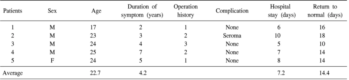
Table 1. Patients' demographics and results of each patients
Duration of Operation Hospital Return to
Patients Sex Age Complication
symptom (years) history stay (days) normal (days)
1 M 17 2 1 None 6 16
2 M 23 3 2 Seroma 10 18
3 M 24 4 3 None 5 10
4 M 25 7 2 None 7 14
5 F 24 5 1 None 8 14
Average 22.7 4.2 7.2 14.4
Fig. 1. Diagram of modified Rhomboid excision and Limberg flap.
결 과
5명의 환자 중에 남자가 4명이었고, 1명은 여자였다. 나 이 분포는 17세부터 25세까지로 평균 22.7세였다. 병력 기 간은 2년부터 7년까지로 평균 4.2년이었다. 5명 모두에서 같은 부위 수술이 1회에서 3회까지 있었는데 이중에 1명은 단순 배농술을 한 경우이고, 4명은 절제술을 받은 병력을 갖고 있었다.
5명의 환자 중에 1명에서 seroma의 축적이 있었고, 그 외 의 합병증은 없었다. 입원 기간은 5일부터 10일까지로 평균 7.2일이었으며 일상 생활로의 복귀는 평균 14.4일이었고, 추적기간 동안의 재발은 없었다(Table 1).
추적 기간은 평균 3.2년이었다.
고 찰
모소동의 발생 기전에 대하여는 선천적인 원인으로 주장 하는 학자와 후천적인 원인으로 주장하는 학자들로 나눠진 다. 후천적인 인자로 운전 여부, 개인 위생 상태 및 비만이 관계가 있다고 보고되기도 한다.(8) Karydakis는 후천적인 원인으로 세 가지의 중요한 인자들이 털의 침습 과정에 관 여한다고 하였다.(6) 첫째로 느슨해진 털로 이루어진 침습
군, 둘째로 털의 침습을 유발하는 힘, 셋째는 피부의 유연성 이다. 그러므로 모소낭의 치료와 예방은 이 세 가지 원인을 없애면 되는 것이다.
모소동의 치료는 첫 번째 인자를 없애기 위해서는 개인 위생을 깨끗이 해야 하고, 둘째와 셋째의 인자를 없애기 위 해서는 모든 염증성 조직과 모소낭 또는 모소동을 광범위 하게 완전히 제거해야만 한다. 이러한 광범위한 염증 조직 제거 후에는 피부 봉합을 하여야 한다. 방법으로는 단순봉 합술,(3,9,10) Z-성형술,(11) W-성형술,(12) V-Y 성형술,(13) 마름모꼴 제거 및 Limberg 피판술,(14-16) 대둔근 회전 피판 술, Bascom 수술법(17) 등이 있다.
모소동이 심하지 않는 경우에는 광범위 절제와 단순봉합 술로 완치될 수 있으나, 염증이 심하거나 재발한 모소동의 치료시에는 광범위 절제를 하면 봉합할 피부의 긴장이 심 하여서 봉합이 어렵고, 봉합을 하여도 피부의 긴장으로 상 처의 분리와 상처 감염 등의 합병증이 증가되고 재발률이 높아진다. 저자는 이런 문제점을 해결하기 위하여 병변이 심한 재발성 모소동의 치료로서 광범위한 비대칭 마름모꼴 제거와 Limberg 피판술을 적용하였다. 결과는 매우 양호하 여 한명에서 소량의 seroma의 축적이 있었고, 그 외의 합병 증은 발견되지 않았다.
Karydakis(18,19)는 단순 봉합술로 1%의 재발률을 보고하 였으나, 정중앙에 수술 반흔을 남길 때는 재발률이 높다고 보고하는 저자도 있었다.(15,20) 마름모꼴 제거와 Limberg 피판술을 시행한 경우는 합병증과 재발률을 0%로 보고하 는 경우도 있고,(21,22) 감염률을 17%로 보고하기도 하였으 나(20) 본 저자의 경우에는 아직 수술 인원이 많지 않으나 한명에서 소량의 seroma가 생겼고 감염률은 없었다. 재발률 도 각각 5%(23)와 7%(24)로 보고하는 저자도 있으나 본 저 자의 경우에는 0%이었다. 이는 마름모꼴의 하변의 절개선 을 정중앙에 하지 않고 좌측으로 1.5 cm 외측으로 하여 피 판의 이동 봉합시에 봉합선이 중앙에 오지 않게 한 것이 중요한 역할을 하지 않았나 생각한다.
입원 기간은 Akinci 등(3)은 2.6일, Mustafa 등(16)은 3.7일 로 보고하였으나 본 연구에서는 7.2일이었다. 이는 합병증 등의 원인때문이 아니라 각 나라마다의 관례에 따른 차이
142 J Korean Surg Soc. Vol. 74, No. 2
로 여겨진다. 일상 생활로의 복귀도 Akinci 등(3)은 12.4일, Mustafa 등(16)은 7일로 보고하였으나, 본 연구에서는 14.4 일 이었다.
재발률의 판단에 추적 기간이 영향을 미칠 수 있다는 보 고도 있다. Allen Mersh(7)는 비대칭 일차 봉합술을 이용한 수술 결과 1년 내에는 1%의 재발률을 보였으나, 장기적인 관찰에서는 3%로 증가하였다고 보고하였다. 본 연구에서 는 평균 3.2년의 추적 기간 동안에 재발률은 없었다.
재발성 모소동은 염증이 심하고 병변이 넓어서 치료 방 법을 결정하기도 어렵고, 수술 후에도 합병증과 재발률이 높아서 외과의사들의 비선호 질환이기도 하다. 이러한 문 제점을 보완하기 위하여 여러 가지의 수술법이 개발되었 다. 재발성 모소동 수술의 가장 큰 관건은 병소의 광범위 절제후의 피부의 무긴장 봉합이다. 이를 충족시키는 가장 이상적인 방법이 피판술이라고 생각한다. 단점으로는 수술 후 반흔이 많이 남으나, 최소한의 수술 후 합병증 및 재발률 의 장점이 있다. 외과의사의 피판술 수술 경험 부족이 문제 점으로 제기될 수 있으나 피판의 혈액 순환만 고려하면 해 결되리라 생각한다.
결 론
재발성 모소동의 치료로 염증 부위의 광범위 비대칭 마 름모꼴 절제와 피판술은 수술 후 합병증과 재발률을 최소 화 할 수 있는 안전한 수술법으로 생각한다.
REFERENCES
1) Chintapatla S, Safarani N, Kumar S, Haboubi N. Sacrococcy- geal pilonidal sinus: historical review, pathological insight and surgical options. Tech Coloproctol 2003;7:3-8.
2) Hegele A, Strombach FJ, Schonbach F. Reconstructive surgi- cal therapy ofinfected pilonidal sinus. Chirurg 2003;74:749-52.
3) Akinci OF, Coskun A, Uzunkov A. A simple and effective surgical treatment ofpilonidal sinus. Dis Colon Rectum 2000;
43:701-7.
4) da Silva JH. Pilonidal cyst: cause and treatment. Dis Colon Rectum 2000;43:1146-56.
5) Davage ON. The origin of sacrococcygeal pilonidal sinuses:
based on an analysisof four hundred sixty-three cases. Am J Pathol 1954;30:1191-205.
6) Karydakis GE. The etiology of pilonidal sinus. Hellenic Arm
Forc Med Rev 1975;7:411-6.
7) Allen Mersh TG. Pilinidal sinus: finding the right tract for treatment. Br J Surg 1990;77:123-32.
8) Buie LA. Jeep disease. South Med J 1944;37:103-9.
9) Al-Jaberi TM. Excision and simple primary closure of chronic pilonidal sinus. Eur J Surg 2001;167:133-5.
10) Gasten DF, Tan BY, Ayuyao A. A technique of radical ex- cision of pilonidaldisease with primary closure. Surgery 1973;
73:109-14.
11) Bose B, Candy T. Radical cure of pilonidal sinus by Z-plasty.
Am J Surg 1970;120:783-5.
12) Sood SC, Green UJ, Perni R. Results of various operations for sacrococcygealpilonidal disease. Plast Reconstr Surg 1975;
56:559-66.
13) Khatri VP, Espinosa MH, Amin AK. Management of recurrent pilonidal sinus bysimple V-Y fasciocutaneous flap. Dis Colon Rectum 1994;37:1232-5.
14) Eryilmaz R, Sahin M, Alimoglu O, Dasiran F. Surgical treat- ment of sacrococcygeal pilonidal sinus with Limberg trans- position flap. Surgery 2003;134:745-9.
15) Alper C, Bulent HU, Mustafa C, Ali C. Superiority of asym- metric modified Limberg flap for surgical treatment of piloni- dal disease. Dis Colon Rectum 2005;49:244-9.
16) Mustafa KU, Fikri K, Koray T, Ilter O. Rhomboid excision and Limberg flap formanaging pilonidal sinus. Dis Colon Rectum 2002;45:656-9.
17) Bascom JU. Pilonidal sinus. Curr Pract Surg 1994;6:175-80.
18) Karydakis GE. Easy and successful treatment of pilonidal si- nus after explanationof its causative process. Aust N Z J Surg 1992;62:385-9.
19) Karydakis GE. New approach to the problem of pilonidal sinus. Lancet 1973;2:1414-5.
20) Azab AS, Kamal MS, Saad RA, Abou al Atta KA, Ali NA.
Radical cure ofpilonidal sinus by a transposition rhomboid flap. Br J Surg 1984;71:154-5.
21) Manterola C, Barroso M, Araya JC, Fonseca L. Pilonidal dis- ease: 25 casestreated by the Dufourmentel technique. Dis Colon Rectum 1991;34:649-52.
22) Bozkurt MK, Tezel E. Management of pilonidal sinus with the Limberg flap. Dis Colon Rectum 1998;41:775-7.
23) Gwynn BR. Use of the rhomboid flap in pilonidal sinus. Ann R Coll Surg Eng 1986;68:40-1.
24) Hoehn JG, Elliott RA, Stayman JW. The use of Limberg flaps for repairongsmall decubitus ulcers. Plat Reconstr Surg 1977;
60:548-57.