JCS
Journal of Chest SurgeryClinical Research Association between Cumulative Fluid Balance and Outcomes in Acute Respiratory Distress Syndrome Patients Treated with Extracorporeal Membrane Oxygenation
Jun Hee Lee, M.D., Jong Yun Won, M.D., Ji Eon Kim, M.D., Hee Jung Kim, M.D., Ph.D., Jae Seung Jung, M.D., Ph.D., Ho Sung Son, M.D., Ph.D.
Department of Thoracic and Cardiovascular Surgery, Korea University Anam Hospital, Korea University College of Medicine, Seoul, Korea
ARTICLE INFO
Received September 24, 2020 Revised November 6, 2020 Accepted November 10, 2020 Corresponding author Ho Sung Son
Tel 82-2-920-5448 Fax 82-2-927-1478 E-mail [email protected] ORCID
https://orcid.org/0000-0003-2766-1186
Background: Extracorporeal membrane oxygenation (ECMO) has become increasingly accepted as a life-saving procedure for patients with severe acute respiratory distress syn- drome (ARDS). This study investigated the relationship between cumulative fluid balance (CFB) and outcomes in adult ARDS patients treated with ECMO.
Methods: We retrospectively analyzed the data of adult ARDS patients who received ECMO between December 2009 and December 2019 at Korea University Anam Hospital.
CFB was calculated during the first 7 days after ECMO initiation. The primary endpoint was 28-day mortality.
Results: The 74 patients were divided into survivor (n=33) and non-survivor (n=41) groups based on 28-day survival. Non-survivors showed a significantly higher CFB at 1–7 days (p<0.05). Cox multivariable proportional hazard regression revealed a relationship be- tween CFB on day 3 and 28-day mortality (hazard ratio, 3.366; 95% confidence interval, 1.528–7.417; p=0.003).
Conclusion: In adult ARDS patients treated with ECMO, a higher positive CFB on day 3 was associated with increased 28-day mortality. Based on our findings, we suggest a re- strictive fluid strategy in ARDS patients treated with ECMO. CFB may be a useful predictor of survival in ARDS patients treated with ECMO.
Keywords: Acute respiratory distress syndrome, Extracorporeal membrane oxygenation, Fluid balance
Copyright©The Korean Society for Thoracic and Cardiovascular Surgery. 2021. All right reserved.
This is an Open Access article distributed under the terms of the Creative Commons Attribution Non-Commercial License (http://creativecommons.org/licenses/
Introduction
In recent years, extracorporeal membrane oxygenation (ECMO) has made remarkable progress to become an ac- cepted treatment option for patients refractory to conven- tional therapy [1]. Depending on the organ being support- ed, there are 3 indications for ECMO: (1) cardiac support, (2) respiratory support, and (3) both cardiac and respirato- ry support. Although both venovenous (VV) ECMO and venoarterial (VA) ECMO can be used for respiratory sup- port, VV ECMO is generally used for respiratory support [2].Acute respiratory distress syndrome (ARDS) is an acute inflammatory form of lung injury that is associated with significant mortality and morbidity. The incidence of
ARDS was reported to be 78.9 per 100,000 person-years in the United States; furthermore, ARDS was observed in 10.4% of all intensive care unit (ICU) admissions and in 23.4% of mechanically ventilated patients [3,4]. Although the mortality of ARDS has decreased over time, recent studies have still reported high mortality rates in patients with ARDS (30%–43%) [5-7]. According to the Berlin defi- nition, ARDS is an acute-onset (within 1 week) condition characterized by bilateral lung opacities on chest radiogra- phy, with no evidence of cardiac failure-related hydrostatic edema on echocardiography and moderate to severe im- pairment of oxygenation [8].
The principle of treatment for ARDS patients is to treat the underlying cause, while providing supportive therapy to prevent further lung injury through lung-protective ven-
https://doi.org/10.5090/kjtcs.20.123 pISSN: 2765-1606 eISSN: 2765-1614 J Chest Surg. 2021;54(1):36-44
Jun Hee Lee, et al. Cumulative Fluid Balance in Acute Respiratory Distress Syndrome
JCS
tilator management and conservative fluid therapy [4,9]. In patients with severe ARDS, defined as a ratio of the partial pressure of oxygen to the fraction of inspired oxygen (PaO2/FiO2) <100 mm Hg with a positive end-expiratory pressure ≥5 cmH2O [10], ECMO has become increasingly accepted as a rescue therapy to avoid the potentially injuri- ous aspects of mechanical ventilation [11,12], since the 2009 H1N1 influenza pandemic [13] and the “Conventional Ventilatory Support versus Extracorporeal Membrane Ox- ygenation for Severe Adult Respiratory Failure” trial [14].
The benefits of ECMO for severe ARDS was once again proven in the “ECMO to Rescue Lung Injury in Severe ARDS” study [15].
Daily fluid balance is the daily sum of all intake and out- put, and cumulative fluid balance (CFB) is the sum total of fluid accumulation within a set period of time. A higher CFB leads to an increased risk of death, longer time on me- chanical ventilation, and longer length of ICU stay in ARDS patients [16-18]. Although restrictive fluid therapy in ARDS patients reduces mortality and shortens the dura- tion of mechanical ventilation and the length of ICU stay [19], an inappropriate fluid restriction strategy can lead to hemodynamic aggravation and dysfunction of multiple or- gans [20].
Adequate fluid resuscitation is essential for initiating and achieving sufficient extracorporeal blood flow in patients treated with ECMO, and fluid overload commonly occurs.
Although restrictive fluid therapy in patients with ARDS can reduce mortality, the most appropriate fluid balance in ARDS patients treated with ECMO remains controversial.
CFB is widely used as a surrogate marker of intravenous fluid management. The present study investigated the as- sociation between CFB and outcomes in adult ARDS pa- tients treated with ECMO. To the best of our knowledge, this is the first study to analyze the association between CFB and outcomes in ARDS patients treated with ECMO.
Methods
Patients and data collection
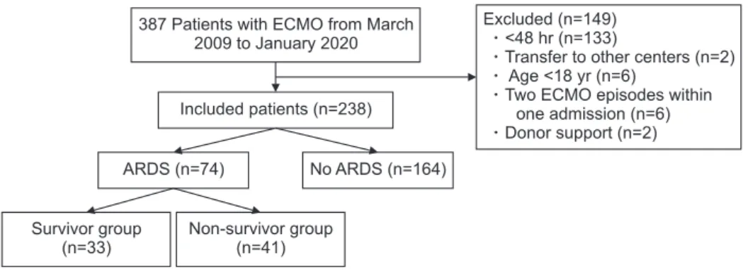
The data were collected retrospectively, and this study included ARDS patients treated with ECMO between De- cember 2009 and December 2019 at Korea University Anam Hospital, Korea University College of Medicine. The exclusion criteria for this study were as follows: (1) age <18 years old; (2) ECMO support for less than 48 hours; (3) re- peated ECMO applied within 1 admission; (4) transfer to other centers after ECMO application; and (5) ECMO sup- port for organ donation (Fig. 1). Initially, 387 adult patients treated during the study period were included in the analy- sis, of whom 149 were excluded (133 received ECMO for less than 48 hours, 2 were transferred to other centers, 6 were younger than 18 years old, 6 had 2 ECMO episodes within 1 admission, and 2 were organ donors). ECMO- treated patients who did not have ARDS (i.e., those who received ECMO for cardiac support and extracorporeal cardiopulmonary resuscitation) were also excluded (n=164).
The remaining 74 patients were included in the final sam- ple and subdivided into survivor (n=33) and non-survivor (n=41) groups based on mortality within 28 days of ECMO initiation.
Patient data included demographic characteristics, hos- pital course, daily input, daily output, basic blood testing, pre-ECMO ventilator settings, pre-ECMO arterial blood gas analysis (ABGA), post-ECMO ventilator settings, post-ECMO ABGA, ECMO complications, survival to hos- pital discharge, successful ECMO weaning, and ICU scor- ing systems, such as Acute Physiology And Chronic Health Evaluation II (APACHE II), Sequential Organ Failure As- sessment (SOFA), and Respiratory ECMO Survival Predic- tion (RESP), which were calculated on the basis of patients’
medical records. Screening of the underlying disease was mostly based on the patients’ previous medical history.
Fig. 1. Flowchart of participant en- rollment. ECMO, extracorporeal mem- brane oxygenation; ARDS, acute re- spiratory distress syndrome.
387 Patients with ECMO from March 2009 to January 2020
Survivor group (n=33)
Non-survivor group (n=41)
ARDS (n=74) No ARDS (n=164) Included patients (n=238)
Excluded (n=149)
<48 hr (n=133)
Transfer to other centers (n=2) Age <18 yr (n=6)
Two ECMO episodes within one admission (n=6) Donor support (n=2)
https://doi.org/10.5090/kjtcs.20.123
JCS
Monitoring lung function is essential for evaluating chang- es in a patient’s condition and includes regular assessments of the patient’s clinical status (vital signs; particularly the respiratory rate, respiratory muscle effort, and level of con- sciousness), lung mechanics, periodic ABGA, and lung im- aging. Bleeding complications were defined according to the Extracorporeal Life Support Organization definition.
Bleeding was defined as clinically overt bleeding recorded in the patient’s medical charts associated with a decrease in hemoglobin of at least 2 g/dL in 24 hours, or a transfu- sion requirement of 1 or more 10-mL/kg red blood cell transfusions over the same time period.
The primary endpoint was 28-day mortality. The sec- ondary endpoints included successful ECMO weaning, the duration of ECMO, the length of ICU stay, and complica- tions of ECMO, such as acute kidney injury (AKI), bleed- ing, and neurological complications.
Extracorporeal membrane oxygenation protocol
ECMO is indicated for patients with severe respiratory failure that is unresponsive to optimal mechanical ventila-
tion and medical treatment. The specific indications of ECMO are severe hypoxia (PaO2/FiO2 ratio <70 mm Hg), uncompensated respiratory acidosis (pH <7.15), or high plateau pressure (≥35 cmH2O). Patients on mechanical ventilation for over 7 days prior to ECMO initiation are relatively contraindicated for ECMO.
For respiratory support, VV ECMO is commonly used, while VA ECMO is used to provide respiratory and circu- latory support. The decision of which patients with ARDS should be initiated on ECMO was made by the ECMO team, which consisted of cardiologists, pulmonologists, and cardiovascular surgeons. In addition to routine ICU monitoring, monitoring of the ECMO device and potential risk was also performed. At our center, the target oxygen saturation (SaO2) was >85% and the target PaO2 was >50 mm Hg. If a Swan-Ganz catheter was inserted, a target of mixed venous saturation was >75%. To reach adequate ox- ygenation, the initial ECMO flow was 3–5 L/min, corre- sponding to 100% of the cardiac index (2.4 L/min/m2) in VA ECMO and about 80% of the cardiac index (2.4 L/min/m2) in VV ECMO. The initial sweep gas flow rate was set to 3 L/min, and was then changed according to the PaCO2 level
Table 1. Patient characteristics in the survivor and non-survivor groups
Characteristic Survivors (n=33) Non-survivors (n=41) p-value
Age (yr) 53.24±2.633 59.32±2.490 0.100
Sex (male) 16 (48.5) 17 (41.5) 0.546
Body mass index (kg/m2) 25.3±0.645 24.44±0.654 0.356
Comorbidities
Hypertension 14 (42.4) 20 (48.8) 0.585
Diabetes mellitus 6 (18.2) 9 (22.0) 0.688
Chronic kidney disease 3 (9.1) 4 (9.8) 1.000
Malignancy 5 (15.2) 10 (24.4) 0.326
Immunosuppression 8 (24.2) 13 (31.7) 0.479
Intensive care unit score
APACHE II 13.27±5.642 14.83±5.603 0.240
SOFA 6.48±3.938 6.63±2.300 0.848
RESP -0.55±3.133 -1.51±3.107 0.189
ECMO mode (venovenous mode) 31 (93.9) 34 (82.9) 0.283
Duration of MV support prior to ECMO initiation (day) 4.42±7.018 2.68±3.745 0.176
Cause of ARDS
Sepsis 2 (6.1) 6 (14.6) 0.286
Pneumonia 27 (81.8) 32 (78.0) 0.688
Aspiration 1 (3.0) 0 0.446
Massive transfusion 3 (9.1) 3 (7.3) 1.000
Trauma 0 1 (2.4) 1.000
Already on CRRT before ECMO initiation 2 (6.1) 7 (17.1) 0.283
New start of CRRT 12 (36.4) 17 (42.5) 0.594
Values are presented as mean±standard deviation or number (%).
APACHE, Acute Physiology And Chronic Health Evaluation; SOFA, Sequential Organ Failure Assessment; RESP, Respiratory Extracorporeal Membrane Oxygenation Survival Prediction; ECMO, extracorporeal membrane oxygenation; MV, mechanical ventilation; ARDS, acute respiratory distress syndrome; CRRT, continuous renal replacement therapy.
Jun Hee Lee, et al. Cumulative Fluid Balance in Acute Respiratory Distress Syndrome
JCS
on ABGA to maintain a PaCO2 of 45–50 mm Hg. Unfrac- tionated heparin (UFH) was given as a bolus injection (50 U/kg) before cannulation, and then UFH was continu- ously infused. The activated partial thromboplastin time (aPTT), which was used to monitor the patient’s response to heparin therapy, was checked every 6 hours for a 3-day period and every 12 hours thereafter. The aPTT target range was 45 to 65 seconds in VV ECMO and 60 to 80 sec- onds in VA ECMO.
Transthoracic echocardiography (TTE) prior to ECMO initiation was not routinely performed in all patients. VA ECMO was indicated when the ejection fraction (EF) was below 30%. At our center, TTE was routinely performed after 7 days after ECMO initiation to detect right ventricu- lar (RV) failure. If RV failure was confirmed, conversion to veno-arterio-venous mode was actively considered. The target mean arterial pressure (MAP) was 60 mm Hg to maintain optimal organ perfusion. Vasopressors were of- ten used to maintain the MAP above 60 mm Hg; norepi- nephrine was the first drug of choice and continuously in- fused (0.02–0.2 µg/kg/min). If the EF was reduced, dobutamine was continuously infused (2–20 µg/kg/min).
If hemodynamics and oxygen delivery were adequate on zero sweep gas and a FiO2 of 21%, weaning was considered.
Either the Emergency Bypass System (Terumo, Tokyo, Ja- pan) or the Permanent Life Support system (Getinge, Go- thenberg, Sweden) was used.
Cumulative fluid balance
CFB was calculated as the sum of total fluid accumula- tion during the first 7 days after ECMO initiation based on daily fluid balance (i.e., the daily sum of all intake and out- put). Fluid intake included oral intake, tube feeding, intra- venous fluids, medications, blood products, parenteral nu- trition, and dialysis influent-dialysate fluid. Fluid output included urine output, drainage from drains or chest tubes, stools, and dialysis effluent-dialysate from continuous re- nal replacement therapy (CRRT).
Statistical analyses
Categorical variables were analyzed using the chi-square test or the Fisher test. Continuous variables were represent- ed as mean values and standard deviations, and the t-test was used for between-group comparisons. Receiver operat- ing characteristic (ROC) curves were used to determine the cut-off value for the maximum sensitivity and specific- ity to evaluate CFB as a predictive factor of 28-day mortal- Table 2.
Ventilation parameters during ECMO support in the survivor and non-survivor groups VariableBefore ECMO initiationAfter ECMO initiation (immediate)After 24 hr Survivors (n=33)Non-survivors (n=41)p-valueSurvivors (n=33)Non-survivors (n=41)p-valueSurvivors (n=33)Non-survivors (n=41)p-value Vital signs MAP (mm Hg)81.67±20.90774.66±17.1870.11877.21±17.453108.36±21.6270.51669.42±24.02164.10±27.3000.382 Heart rate (/min)108.45±27.602115.20±25.4170.279108.36±21.627111.58±22.1910.53293.21±21.177102.32±19.7570.060 Respiratory rate (/min)22.94±6.51922.34±5.4480.66919.03±5.45720.02±5.8750.45815.55±4.02416.93±4.8910.196 Arterial blood gases pH7.26±0.1267.24±0.1730.5327.40±0.0887.38±0.1290.3757.42±0.0637.39±0.1080.198 PaO2 (mm Hg)63.34±13.15589.34±101.5510.149112.13±89.362109.69±73.1280.897102.22±61.68691.03±93.6100.348 PaCO2 (mm Hg)58.38±39.80462.09±40.0380.69233.91±12.96635.13±9.5000.64439.04±12.12938.40±8.1370.786 HCO3 (mEq/L)24.53±8.49126.51±14.2420.48421.09±5.80920.81±7.6360.86524.30±4.44323.69±5.4970.607 SaO2 (%)86.43±8.89985.73±13.5760.79995.75±4.43594.78±4.7440.37196.16±6.10293.29±8.4710.068 Ventilation parameters FiO2 (%)97.12±7.18492.8±15.2500.11445.90±13.37252.439±18.6780.09642.27±8.57744.43±17.0330.508 PEEP (cmH2O)9.70±2.7679.32±2.8320.5648.39±2.3708.39±2.5740.9458.21±1.6918.37±2.1290.717 PIP (cmH2O)26.24±4.93126.80±5.4600.64722.03±5.32323.37±4.9300.26321.51±4.05522.35±4.8940.432 Values are presented as mean±standard deviation. ECMO, extracorporeal membrane oxygenation; MAP, mean arterial pressure; Pa, partial pressure; SaO2, oxygen saturation; FiO2, fraction of inspired oxygen; PEEP, positive end-expiratory pressure; PIP, peak inspiratory pressure.
https://doi.org/10.5090/kjtcs.20.123
JCS
ity. Patients were subdivided into 2 groups using the cut-off value for CFB at 3 days. In order to analyze the relationship between outcomes and CFB, a Cox proportional hazards model was used, and the data were expressed as hazard ra- tios (HRs) with 95% confidence intervals (CIs). The data were analyzed with IBM SPSS ver. 20.0 (IBM Corp., Ar- monk, NY, USA) and p-values <0.05 were considered to in- dicate statistical significance.
The study was approved by the Institutional Review Board of Korea University (IRB approval no., 2020AN0424). The requirement for informed consent from individual patients was omitted because of the retrospective design of this study.
Results
The baseline characteristics of survivors and non-survi- vors are summarized in Table 1. No statistically significant differences were found in patient characteristics between the 2 groups. There were no significant differences in the APACHE II score (p=0.240), SOFA score (p=0.848), or RESP score (p=0.189) between the 2 groups. VV ECMO was performed in 93.9% of survivors and 82.9% of non- survivors (p=0.283), and the mean duration of mechanical ventilation support prior to ECMO initiation was 4.42±
7.018 days in survivors and 2.68±3.745 days in non-survi- vors (p=0.176). Pneumonia was the most common cause of ARDS in both groups (81.8% in survivors, 78% in non-sur-
vivors, p=0.688). The ventilation parameters during ECMO support are shown in Table 2. No statistically significant differences were found in ventilation parameters between the 2 groups.
Table 3 compares the clinical outcomes of survivors and non-survivors. The rate of successful ECMO weaning was 63.6% (n=21) in survivors and 24.3% (n=10) in non-survi- vors. Survivors had a significantly longer length of ICU stay (p=0.001), length of hospital stay (p=0.001), duration of ECMO support (p=0.001), and duration of mechanical ventilation support (p=0.001). The most common cause of death within 28 days after ECMO initiation was sepsis (n=21, 51.2%). One patient died with respiratory failure, as pneumonia worsened after successful ECMO weaning. No statistically significant differences were observed in the complications of ECMO, except for AKI (p=0.032). The causes of in-hospital death are shown in Supplementary Table 1. No statistically significant differences were ob- served in sepsis (p=0.201), bleeding/disseminated intravas- cular coagulation (p=0.373), respiratory failure (p=1.000), or anoxic brain injury (p=0.499). Non-survivors had a sig- nificantly higher rate of multiple organ failure (MOF) than survivors as the cause of in-hospital death (p=0.006). In this study, 9 patients received VA ECMO, of whom 2 pa- tients survived until 28 days after ECMO initiation and 1 patient was discharged alive from the hospital.
The mean CFB in both groups during 7 days after
Table 3. Clinical outcomes of patients in the survivor and non-survivor groups
Variable Survivors (n=33) Non-survivors (n=41) p-value
Successful ECMO weaning 21 (63.6) 10 (24.3) 0.001
Survival to hospital discharge 17 (51.5) -
Length of intensive care unit stay (day) 47.12±28.178 17.46±11.272 0.001
Length of hospital stay (day) 71.61±51.701 27.02±27.432 0.001
Duration of ECMO support (day) 28.67±24.441 9.27±6.527 0.001
ECMO mode change (veno-arterio-venous mode) 2 (6.0) 3 (7.3) 1.000
Duration of mechanical ventilation support 39.64±23.231 17.76±14.896 0.001
Cause of death (within 28 days of ECMO initiation)a)
Sepsis - 21 (51.2)
Multiple organ failure - 13 (31.7)
Bleeding/disseminated intravascular coagulation - 4 (9.7)
Respiratory failure - 1 (2.4)
Anoxic brain injury - 2 (4.8)
Complications of ECMO (in hospital)
Bleeding 15 (45.4) 10 (24.3) 0.057
Acute kidney injury 3 (9.0) 12 (29.2) 0.032
Brain hemorrhage 3 (9.0) 1 (2.4) 0.208
Leg ischemia 1 (3.0) 2 (4.8) 1.000
Values are presented as number (%) or mean±standard deviation.
ECMO, extracorporeal membrane oxygenation.
a)Causes of in-hospital death are shown in Supplementary Table 1.
Jun Hee Lee, et al. Cumulative Fluid Balance in Acute Respiratory Distress Syndrome
JCS
ECMO initiation was calculated (Fig. 2). Non-survivors had a higher CFB than survivors during 7 days after ECMO initiation. The mean CFB in survivors plateaued between 3 and 6 days, while the CFB in non-survivors con- tinued to increase throughout the 7-day period. Statistical- ly significant differences in CFB between non-survivors and survivors were found on days 1–7 (p<0.05) (Table 4).
The sample size of CFB on day 3 was 74 patients, which was the largest sample size. In addition, the p-value of CFB on day 3 was 0.015, which was the smallest value. There- fore, we used CFB on day 3 for Cox regression analysis.
The ROC curve of CFB on day 3 associated with 28-day mortality was used to determine the cut-off value for CFB on day 3 as a predictor of 28-day mortality (n=74) (Fig. 3).
The optimal cut-off value was 2629 mL, which showed sensitivity and specificity of 70.7% and 66.7%, respectively (Fig. 3). In order to obtain a clear evaluation in terms of HRs, the patients were subdivided into a low-CFB group (CFB on day 3 <the cut-off value) and a high-CFB group (CFB on day 3 ≥the cut-off value). The 28-day cumulative
survival curve for the low-CFB group and high-CFB group showed that the 28-day mortality rate was 37% in the low- CFB group and 72% in the high-CFB group (p<0.05) (Fig.
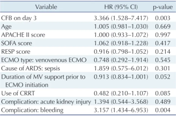
4).To determine the HR for 28-day mortality, CFB on day 3 (low versus high) and other confounding variables (age, ICU scoring systems [APACHE II, SOFA, and RESP], ECMO type, duration of mechanical ventilation support prior to ECMO initiation, use of CRRT, and complications [AKI and bleeding]) were analyzed using Cox multivari- able proportional hazard regression. In the Cox multivari- able proportional hazard regression model of factors associ- ated with 28-day mortality (Table 5), the factors in dependently related to 28-day mortality were high CFB on day 3 (HR, 3.366; 95% CI, 1.528–7.417; p=0.003), and bleeding (HR, 3.157; 95% CI, 1.434–6.953; p=0.004). Higher CFB on day 3 was an independent predictor of 28-day mortality.
10,000
8,000
6,000
4,000
2,000
7
Cumulativefluidbalance(mL)
Time (day)
0 1 2 3 4 5 6
Non-survivors Survivors
Fig. 2. Comparison of mean cumulative fluid balance between survivors and non-survivors.
100
80
60
40
20
100
20 40 60 80
Sensitivity
100-Specificity 0
AUC=0.674 p=0.006 ROC curve (n=74)
Fig. 3. ROC curve of cumulative fluid balance on day 3 associated with 28-day mortality. The optimal cut-off value was 2,632 mL, which showed sensitivity and specificity of 70.7% and 66.7%, respectively. ROC, receiver operating characteristic; AUC, area under the curve.
Table 4. Cumulative fluid balance during 7 days after extracorporeal membrane oxygenation initiation in the survivor and non-survivor groups
Variable Overall
no.
Survivors Non-survivors
p-value
No. Mean±SD No. Mean±SD
CFB on day 1 (mL) 74 33 1,433.82±1,869.363 41 2,792.59±3,439.949 0.045
CFB on day 2 (mL) 74 33 1,918.03±2,979.214 41 4,290.46±5,196.105 0.016
CFB on day 3 (mL) 74 33 2,558.58±3,992.746 41 5,843.49±7,113.176 0.015
CFB on day 4 (mL) 65 31 2,372.52±4,667.463 34 6,280.12±9,038.247 0.035
CFB on day 5 (mL) 62 31 2,502.71±5,126.720 31 6,913.61±10,474.581 0.039
CFB on day 6 (mL) 58 30 2,344.10±5,806.386 28 8,507.54±12,427.716 0.022
CFB on day 7 (mL) 55 29 3,123.07±6,107.004 26 9,843.54±13,966.551 0.030
SD, standard deviation; CFB, cumulative fluid balance.
https://doi.org/10.5090/kjtcs.20.123
JCS
Discussion
Appropriate fluid management to maintain adequate or- gan perfusion is a controversial issue that remains chal- lenging. The relationship of a negative fluid balance with clinical outcomes in critically ill patients has been de- scribed in several studies, and fluid overload has been re- ported to be strongly associated with negative outcomes [21-23]. A restricted fluid strategy has been widely adopted in clinical practice if a patient has stable hemodynamics.
ECMO has become widespread and its outcomes have improved in recent years, as advances have been made both in ECMO devices themselves and in the management of patients treated with ECMO. However, excessive fluid overload still remains a common characteristic of ECMO patients. Despite the short history of ECMO, the potential impact of fluid balance on outcomes in ECMO patients has been recognized. For instance, Kim et al. [24] reported a significantly increased risk of 90-day mortality in patients with higher CFB. Furthermore, Schmidt et al. [25] reported that survivors had a lower fluid balance on ECMO days 3–5.
In our study, we showed that CFB on day 3 was associat- ed with 28-day mortality. However, the mechanism by which positive fluid balance causes negative outcomes in ARDS patients remains unknown. A possible explanation is that positive fluid balance causes aggravation of extra- vascular and interstitial edema in the lungs and other or-
gans. Aggravation of interstitial edema in organs such as the lungs and kidneys leads to impaired oxygenation and perfusion, which cause MOF. Therefore, fluid restriction may help patients with ARDS by reducing edema [16,26].
Some factors other than fluid balance have been shown to be associated with mortality. Specifically, while previous studies showed that positive CFB was associated with mor- tality in ARDS patients, other potential confounding fac- tors, such AKI and sepsis, can also play a role in mortality [17,27-29]. The combination of ECMO and CRRT can ef- fectively improve fluid balance and electrolyte disturbanc- es. A systematic review showed that in ECMO survivors receiving CRRT, the overall fluid balance was lower than that in non-CRRT survivors [27]. In our study, the use of CRRT was not statistically significantly associated with 28- day mortality (HR, 0.482; 95% CI, 0.210–1.107; p=0.085).
This result may be due to the small sample size. A previous study reported that red blood cell transfusions were associ- ated with increased mortality in ARDS patients [30].
Bleeding is a common complication of ECMO and some- times requires massive transfusion. In our results, bleeding had a positive association with 28-day mortality (HR, 3.157; 95% CI, 1.434–6.953; p=0.005).
Limitations
The present study has several limitations. First, this was a retrospective single-center study. Second, the sample size of patients was small. Therefore, a larger-scale study would
100
80
60
40
28
5 10 15 20
Cumulativesurvivalrate(%)
Time (day) 0
Low CFB High CFB
35 39
31 30
27 22
24 17
22 13
21 9 CFB on day 3
Low CFB High CFB
No. at risk
Fig. 4. Cox regression model for 28-day survival between the low- CFB and high-CFB groups. Low-CFB group: CFB on day 3 <the cut-off value (2,629 mL), n=35; high-CFB group: CFB on day 3
≥the cut-off value (2,629 mL), n=39. The 28-day mortality rate was 37% in the low-CFB group and 72% in the high-CFB group (p=0.002). CFB, cumulative fluid balance.
Table 5. Cox multivariable proportional hazard regression of factors associated with 28-day mortality (n=74)
Variable HR (95% CI) p-value
CFB on day 3 3.366 (1.528–7.417) 0.003
Age 1.005 (0.981–1.030) 0.669
APACHE II score 1.000 (0.933–1.072) 0.997
SOFA score 1.062 (0.918–1.228) 0.417
RESP score 0.916 (0.798–1.052) 0.214
ECMO type: venovenous ECMO 0.748 (0.292–1.914) 0.545 Cause of ARDS: sepsis 1.859 (0.575–6.012) 0.301 Duration of MV support prior to
ECMO initiation
0.913 (0.834–1.001) 0.052
Use of CRRT 0.482 (0.210–1.107) 0.085
Complication: acute kidney injury 1.394 (0.544–3.568) 0.489 Complication: bleeding 3.157 (1.434–6.953) 0.004 HR, hazard ratio; CI, confidence interval; CFB, cumulative fluid balance;
APACHE II, Acute Physiology And Chronic Health Evaluation II; SOFA, Sequential Organ Failure Assessment; RESP, Respiratory Extracorporeal Membrane Oxygenation Survival Prediction; ECMO, extracorporeal membrane oxygenation; ARDS, acute respiratory distress syndrome; MV, mechanical ventilation; CRRT, continuous renal replacement therapy.
Jun Hee Lee, et al. Cumulative Fluid Balance in Acute Respiratory Distress Syndrome
JCS
be needed to more appropriately evaluate the 3-day CFB cut-off value and to find significant relationships in the data. Third, cardiac failure was not validated, as TTE was not routinely performed in all patients.
Conclusions
In adult ARDS patients treated with ECMO, a higher positive CFB on day 3 was found to be associated with a higher 28-day mortality risk. Based on our findings, we suggest restrictive fluid management in ARDS patients treated with ECMO. CFB may be a useful predictor of sur- vival in ARDS patients treated with ECMO.
Conflict of interest
No potential conflict of interest relevant to this article was reported.
ORCID
Jun Hee Lee: https://orcid.org/0000-0002-6592-6483 Jong Yun Won: https://orcid.org/0000-0002-9944-7261 Ji Eon Kim: https://orcid.org/0000-0002-1938-6412 Hee Jung Kim: https://orcid.org/0000-0001-5254-1405 Jae Seung Jung: https://orcid.org/0000-0002-8848-4112 Ho Sung Son: https://orcid.org/0000-0003-2766-1186
Supplementary materials
Supplementary materials can be found via https://doi.
org/10.5090/kjtcs.20.123. Supplementary Table 1. Causes of in-hospital death.
References
1. Makdisi G, Wang IW. Extra Corporeal Membrane Oxygenation (ECMO) review of a lifesaving technology. J Thorac Dis 2015;7:
E166-76.
2. Sorokin V, MacLaren G, Vidanapathirana PC, Delnoij T, Lorusso R.
Choosing the appropriate configuration and cannulation strategies for extracorporeal membrane oxygenation: the potential dynamic process of organ support and importance of hybrid modes. Eur J Heart Fail 2017;19 Suppl 2:75-83.
3. Bellani G, Laffey JG, Pham T, et al. Epidemiology, patterns of care, and mortality for patients with acute respiratory distress syndrome in intensive care units in 50 countries. JAMA 2016;315:788-800.
4. Diamond M, Peniston Feliciano HL, Sanghavi, D, Mahapatra S. Acute respiratory distress syndrome. Treasure Island (FL): StatPearls Pub-
lishing; 2020.
5. Zambon M, Vincent JL. Mortality rates for patients with acute lung injury/ARDS have decreased over time. Chest 2008;133:1120-7.
6. Davidson TA, Caldwell ES, Curtis JR, Hudson LD, Steinberg KP.
Reduced quality of life in survivors of acute respiratory distress syn- drome compared with critically ill control patients. JAMA 1999;281:
354-60.
7. Erickson SE, Martin GS, Davis JL, Matthay MA, Eisner MD; NIH NHLBI ARDS Network. Recent trends in acute lung injury mortali- ty: 1996-2005. Crit Care Med 2009;37:1574-9.
8. ARDS Definition Task Force, Ranieri VM, Rubenfeld GD, et al.
Acute respiratory distress syndrome: the Berlin definition. JAMA 2012;307:2526-33.
9. Thompson BT, Chambers RC, Liu KD. Acute respiratory distress syndrome. N Engl J Med 2017;377:562-72.
10. Villar J, Perez-Mendez L, Blanco J, et al. A universal definition of ARDS: the PaO2/FiO2 ratio under a standard ventilatory setting: a prospective, multicenter validation study. Intensive Care Med 2013;
39:583-92.
11. Hardin CC, Hibbert K. ECMO for severe ARDS. N Engl J Med 2018;378:2032-4.
12. Ma DS, Kim JB, Jung SH, Choo SJ, Chung CH, Lee JW. Outcomes of venovenous extracorporeal membrane oxygenation support for acute respiratory distress syndrome in adults. Korean J Thorac Car- diovasc Surg 2012;45:91-4.
13. Australia and New Zealand Extracorporeal Membrane Oxygenation (ANZ ECMO) Influenza Investigators, Davies A, Jones D, et al. Ex- tracorporeal membrane oxygenation for 2009 influenza A(H1N1) acute respiratory distress syndrome. JAMA 2009;302:1888-95.
14. Peek GJ, Clemens F, Elbourne D, et al. CESAR: conventional venti- latory support vs extracorporeal membrane oxygenation for severe adult respiratory failure. BMC Health Serv Res 2006;6:163.
15. Combes A, Hajage D, Capellier G, et al. Extracorporeal membrane oxygenation for severe acute respiratory distress syndrome. N Engl J Med 2018;378:1965-75.
16. Van Mourik N, Metske HA, Hofstra JJ, et al. Cumulative fluid bal- ance predicts mortality and increases time on mechanical ventilation in ARDS patients: an observational cohort study. PLoS One 2019;14:
e0224563.
17. Rosenberg AL, Dechert RE, Park PK, Bartlett RH; NIH NHLBI ARDS Network. Review of a large clinical series: association of cu- mulative fluid balance on outcome in acute lung injury: a retrospec- tive review of the ARDSnet tidal volume study cohort. J Intensive Care Med 2009;24:35-46.
18. Zinter MS, Spicer AC, Liu KD, et al. Positive cumulative fluid bal- ance is associated with mortality in pediatric acute respiratory dis- tress syndrome in the setting of acute kidney injury. Pediatr Crit Care Med 2019;20:323-31.
19. National Heart, Lung, and Blood Institute Acute Respiratory Distress
https://doi.org/10.5090/kjtcs.20.123
JCS
Syndrome (ARDS) Clinical Trials Network, Wiedemann HP, Wheel- er AP, et al. Comparison of two fluid-management strategies in acute lung injury. N Engl J Med 2006;354:2564-75.
20. Finfer S, Liu B, Taylor C, et al. Resuscitation fluid use in critically ill adults: an international cross-sectional study in 391 intensive care units. Crit Care 2010;14:R185.
21. Shen Y, Huang X, Zhang W. Association between fluid intake and mortality in critically ill patients with negative fluid balance: a retro- spective cohort study. Crit Care 2017;21:104.
22. Boyd JH, Forbes J, Nakada TA, Walley KR, Russell JA. Fluid resus- citation in septic shock: a positive fluid balance and elevated central venous pressure are associated with increased mortality. Crit Care Med 2011;39:259-65.
23. Malbrain ML, Marik PE, Witters I, et al. Fluid overload, de-resusci- tation, and outcomes in critically ill or injured patients: a systematic review with suggestions for clinical practice. Anaesthesiol Intensive Ther 2014;46:361-80.
24. Kim H, Paek JH, Song JH, et al. Permissive fluid volume in adult patients undergoing extracorporeal membrane oxygenation treatment.
Crit Care 2018;22:270.
25. Schmidt M, Bailey M, Kelly J, et al. Impact of fluid balance on out- come of adult patients treated with extracorporeal membrane oxy- genation. Intensive Care Med 2014;40:1256-66.
26. Roch A, Guervilly C, Papazian L. Fluid management in acute lung injury and ARDS. Ann Intensive Care 2011;1:16.
27. Chen H, Yu RG, Yin NN, Zhou JX. Combination of extracorporeal membrane oxygenation and continuous renal replacement therapy in critically ill patients: a systematic review. Crit Care 2014;18:675.
28. Vlaar AP, Binnekade JM, Prins D, et al. Risk factors and outcome of transfusion-related acute lung injury in the critically ill: a nested case-control study. Crit Care Med 2010;38:771-8.
29. Murphy CV, Schramm GE, Doherty JA, et al. The importance of flu- id management in acute lung injury secondary to septic shock. Chest 2009;136:102-9.
30. Gong MN, Thompson BT, Williams P, Pothier L, Boyce PD, Chris- tiani DC. Clinical predictors of and mortality in acute respiratory distress syndrome: potential role of red cell transfusion. Crit Care Med 2005;33:1191-8.