INTRODUCTION
Recent studies suggest that sphingolipids, in addition to their function as structural cell membrane constituents, play a key role as signaling molecules. In this regard, sphingolipid metabolites, including ceramide, sphingosine, and sphingo-sine 1-phosphate (S1P), have been identified as a new class of lipid messengers, which regulate cell proliferation, differ-entiation, and survival (1-3). S1P generated by sphingosine kinase, an intracellular enzyme and has been associated with the regulation of cell proliferation, differentiation, survival, and motility (1-5). In contrast to the general association of ceramide and sphingosine with cell growth arrest and apop-tosis, S1P that is a downstream metabolite of ceramide, gen-erally known to exert mitogenic and antiapoptotic effects (1-4). One of the better-known sources of S1P are human pla-telets, from which S1P is released upon activation by physio-logical stimuli, thereby suggesting that S1P is a factor signi-ficantly involved in endothelial injury, inflammation, throm-bosis, angiogenesis, and wound healing via an induced increase in the migration and proliferation of endothelial cells (6, 7). S1P reaches levels of up to 0.2 M in normal plasma, and may be as high as 10 M in normal serum, presumably
reflect-ing major contributions from platelets (8).
More recently, S1P has been identified as a first messenger, owing to its ability to bind and activate a series of G-protein coupled receptors (GPCRs) located on the cell surface (9), which have been designated S1P1(endothelial differentiation gene, Edg 1), S1P2(Edg 5), S1P3(Edg 3), S1P4(Edg 6), S1P5 (Edg 8), by a nomenclature subcommittee of the International Union of Pharmacologists (10, 11). S1P can function in either an autocrine or paracrine fashion via Edg receptors (9). The coupling of Edg receptors to diverse G proteins results in the activation of a number of downstream signaling pathways. Intracellular signaling pathways activated by Edg receptors include adenyl cyclase, phospholipase C (PLC), Ras, mitogen-activated protein kinase (MAPK), Rho, and several protein tyrosine kinases (12-18). S1P also acts as an intracellular sec-ond messenger to regulate calcium homeostasis, survival and growth (12). Indeed, a variety of effects were observed in con-junction with S1P, apparently depending on the cell type. Recently, S1P has occasionally been associated with certain growth-inhibitory and apoptotic effects in human hepatoma cells, ovarian cancer cells, cultured hippocampal neurons, and others (19-24).
In the present study, we have identified S1P receptors (Edg-Jeong-Hyun Shin, Gwang-Seong Choi, Won-Hyung Kang*, Ki-Bum Myung�
Department of Dermatology, School of Medicine, Inha University, Incheon; Department of Dermatology*, School of Medicine, Ajou University, Suwon; Department of Dermatology�
, School of Medicine, Ewha Womans University, Seoul, Korea *Kang WH changed affiliation since this work was done: Department of Dermatology, Kwandong University, Koyang.
Address for correspondence
Ki-Bum Myung, M.D.
Department of Dermatology, Ewha Womans University Mokdong Hospital, 911-1 Mock-dong, Yangcheon-gu, Seoul 158-710, Korea
Tel : +82.2-2650-5159, Fax : +82.2-2650-6295 E-mail : [email protected]
*This work was supported by Inha University Research Grant (INHA-31442) to JH Shin.
298
Sphingosine 1-Phosphate Triggers Apoptotic Signal for B16 Melanoma
Cells via ERK and Caspase Activation
The bioactive sphingolipid metabolite sphingosine 1-phosphate (S1P), recently was reported to induce apoptosis of some cancer cells and neurons, although it gener-ally known to exert mitogenic and antiapoptotic effects. In this study, we investigat-ed the effects of S1P on the cell growth, melanogenesis, and apoptosis of culturinvestigat-ed B16 mouse melanoma cells. In results, S1P was found to induce apoptosis in B16 melanoma cells in a dose- and time-dependent manner, but exerted minimal effects on melanogenesis. Although receptors of sphingosine 1-phosphate (endothelial dif-ferentiation gene 1 [Edg]/S1P1, Edg5/S1P2, Edg3/S1P3) were expressed in B16 melanoma cells, they were shown not to be associated with S1P-induced apopto-sis. In addition, pertussis toxin did not block the apoptotic effects of S1P on B16 melanoma cells. S1P induced caspase-3 activation and the extracellular signal-reg-ulated kinase (ERK) activation. Interestingly, the ERK pathway inhibitor, UO126, reversed the apoptotic effects of S1P on B16 melanoma cells. These results sug-gest that S1P induced apoptosis of B16 melanoma cells via an Edg receptor-inde-pendent, pertussis toxin-insensitive pathway, and appears to be associated with the ERK and caspase-3 activation.
Key Words : Sphingosine 1-Phosphate; Melanoma; Apoptosis; Caspases; Extracellular Signal-Regulated Kinase
Received : 28 June 2006
1, -3, and -5) in B16 mouse melanoma cells, and investigat-ed the actions of S1P on cell growth. S1P evidencinvestigat-ed an anti-proliferative effect on B16 mouse melanoma cells. S1P induced apoptosis via caspase-3 activation, in an Edg receptor-inde-pendent fashion. S1P activated the extracellular signal-reg-ulated protein kinase (ERK) and UO126, the ERK pathway inhibitor, reversed the apoptotic effects of S1P.
MATERIALS AND METHODS Materials
Sphingosine 1-phosphate and dihydrosphingosine 1-phos-phate were obtained from Calbiochem (SanDiego, CA, U.S.A.). Stock sphingolipid solutions were dissolved in methanol (S1P and dihydro-S1P). All stock solutions were maintained at -80℃until use. Dilutions of S1P and dihydro-S1P were freshly prepared after the evaporation of methanol via resus-pension in 0.4% fatty acid-free bovine serum albumin (BSA) and sonication. UO126 and pertussis toxin were purchased from the Sigma Co (Yongin, Korea). Stock UO126 solutions were dissolved in Me2SO and maintained at -80℃until use. Culture media and reagents were obtained from Gibco (LA, CA, U.S.A.), and fetal bovine serum (FBS) was obtained from Omega (Tarzana, CA, U.S.A.). Mouse monoclonal antibody against Edg-3, Edg-5 and rabbit polyclonal antibody against Edg-1 were from Merk Korea (Seoul, Korea). Rabbit mono-clonal anti-cleaved caspase-3 (Asp175) antibody was pur-chased from Cell Signaling Technology, Inc (Danvers, MA, U.S.A.). The ApopTag Kit (S7101) was purchased from the Intergen Company (Norcross, Eeorgia, U.S.A.).
Cell culture
B16 mouse melanoma cells (2×10⁴) were seeded into 30-mm culture dishes in maintenance medium (RPMI 1640 supplemented with 10% FBS, 1% streptomycin and peni-cillin), and permitted to attach overnight. The medium was then exchanged with serum-starved medium (0.25% FBS) for 48 hr, and treated with S1P or vehicle (PBS/BSA) for the indi-cated time courses. Every 2 days, the medium was changed. MTT dye-reduction assay
Cells (1×10⁴ cells/well), seeded into 12-well plates for 24 hr in maintenance media and 48 hr for serum-starvation media, and were incubated with the test substances for 24 hr at 37℃in 5%CO2. After the addition of 100 L/well of MTT solution (5 mg/mL), the plates were incubated for an additional 4 hr. The supernatants were then removed, and the formazan crystals were solubilized in 1 mL of dimethyl-sulfoxide. Optical density was determined at a wavelength of 540 nm, using an ELISA reader.
Melanin assay
For the quantitative analysis of melanin content, cells were harvested and counted (2×10⁵), and then centrifuged for 4 min with 100,000 r.p.m. After vortex, cells were incubated in 1N of NaOH to lysis overnight at 37℃and then optical density was determined at 470 nm using an ELISA reader. Immunocytochemistry for Edg receptors
Cells were grown for 24 hr on Lab-Tek chamber slides (Nunc Inc., Naperville, IL, U.S.A.). The cells were then fixed in 4% paraformaldehyde containing 0.1% Triton X-100 overnight at 4℃. Nonspecific binding sites were blocked with blocking solution (20% normal goat serum, 0.1% BSA, and 0.1% Triton X-100 in phosphate buffered solution [PBS]) for 10 min at room temperature. The cells were then incu-bated for 25 min at 45℃with mouse monoclonal antibody against Edg-3, Edg-5 diluted to 1:50, and rabbit polyclonal antibody against Edg-1 diluted to 1:100, in PBS. After wash-ing with PBS, the cells were incubated with biotin-labeled mouse anti-IgG antibody (1:200, DAKO, Carpinteria, CA, U.S.A.) and streptavidin alkaline phosphate anti-alkaline phosphate, for 15 min each at room temperature. The sub-strate chromogen New fuchsin (ScyTek, Logan, UT, U.S.A.) was also applied for 20 min. The cells were examined with a Zeiss microscope. PBS instead of a primary antibody was administered as a negative control.
In situ analysis of DNA integrity (TUNEL assay)
Cells were grown on Lab-Tek chamber slides (Nunc Inc.) for 24 hr in maintenance medium to attach, after which the medium was changed to fresh maintenance medium or serum-starved medium (0.5% FBS) for 48 hr, then treated for 24 hr with S1P or a vehicle. The nick-end-labeling (terminal deoxy-nucleotidyl transferase-mediated dUTP TUNEL) technique was conducted as described. In brief, the slides were fixed for 10 min in 1% paraformaldehyde in PBS, and then washed twice in PBS. The cells were preincubated for 2 min at room temperature in equilibration buffer, and incubated for 1 hr in a moisture chamber at 37℃with a working strength (55 L/5 cm2) formulation of TdT enzyme (77 L of reaction buffer with 33 L of TdT enzyme). The reaction was halted via the transference of the slides to stop buffer (1 mL buffer with 34 mL D/W) in a coplin jar, with 15 sec of agitation, incubated for 10 min at room temperature, and then washed three times in PBS. The cells were incubated for 30 min in a moisture chamber at room temperature in anti-dioxigenin peroxidase conjugate, and then washed four times in PBS. DAB peroxidase substrate was applied to the stains for 10 min at room temperature, followed by washing in distilled water. The cells were counterstained with 0.5% methyl green in a coplin jar for 10 min at room temperature, then
mount-ed in an aqueous mounting mmount-edium (Niommount-eda, Foster City, CA, U.S.A.).
We utilized computerized image analysis to quantify the number of apoptotic cells in each sample. A CCD camera (CCD-IRIS, Sony, Tokyo, Japan) mounted on a microscope (Olympus BX50F, Olympus Optical Co., Tokyo) was con-nected to an IBM personal computer (PC). The image sig-nals acquired by the PC were then evaluated using Image Pro Plus, Version 3.0 (Media Cybernetics Co., Silver Spring, MD, U.S.A.). Image analyses were conducted on four repre-sentative areas of the slides, under constant magnification (×200), after which the percentage of apoptotic cells was determined.
Western blot analysis
Cells were grown for 24 hr in 100 mm culture dishes, sub-jected to serum starvation for 48 hr, and treated with S1P or vehicle for the indicated times. They were then harvested and lysed in radioimmunoprecipitation (RIPA) buffer (150 mM NaCl, 1% NP-40, 0.5% deoxycholate, 0.1% SDS, and 50 nM Tris [pH 8.0]) containing the phosphatase inhibitor Na3VO4(10 mM), and the protease inhibitors phenylmethyl-sulfonyl fluoride (200 mM) and aprotinin (10 g/mL). Ten
to 20 micrograms of protein per lane was separated via SDS-polyacrylamide gel electrophoresis, and blotted onto nitro-cellulose membranes, which were then saturated with 2% bovine serum albumin in Tris-buffered saline containing 0.05% Tween 20. The blots were then incubated with the designated primary antibodies at a 1:1,000 dilution, and then incubated further with horseradish peroxidase-conjugated secondary antibody. The bound antibodies were detected using a Renaissance chemiluminescence kit (Dupont NEN, Boston, MA, U.S.A.).
Statistical analysis
All experiments were independently repeated at least twice. Statistics were determined via Student’s t-test (SPSS 12.0 for Windows); p values <0.05 as compared with control cells were considered to be statistically significant.
RESULTS
Sphingosine 1-phosphate shows proliferation inhibitory effects in B16 melanoma cells
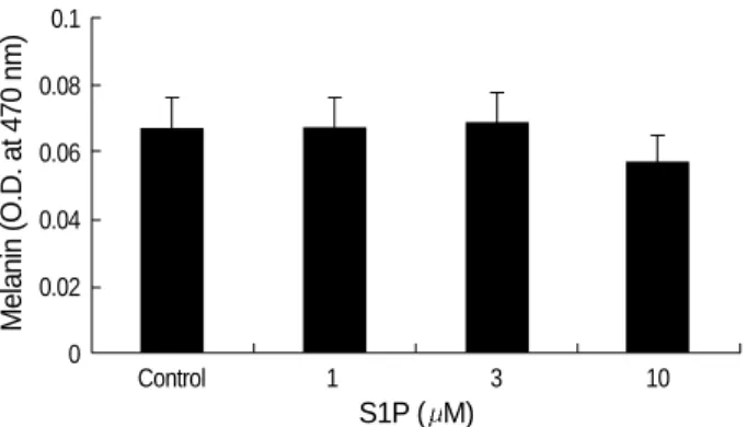
S1P was found to inhibit the proliferation of B16 melanoma cells (Fig. 1). After 24 hr, S1P significantly inhibited cell growth at concentrations in excess of 5 M (p<0.05). Indeed, after 24 hr, S1P induced the shrinkage, rounding and detach-ment of cells, whereas serum deprivation alone had no detec-table effects on cell morphology (data not shown). To assess the effect of S1P on melanogenesis in B16 melanoma cells, melanin content was measured as described in method. The melanin synthesis of B16 melanoma cells was not signifi-cantly affected by S1P (p>0.05, Fig. 2).
Sphingosine 1-phosphate induces apoptosis of B16 melanoma cells via caspase-3 activation
In order to identify more precisely the inhibitory effects of S1P on the growth of B16 melanoma cells, we attempted to determine whether S1P induced apoptosis in B16 melanoma
Cell viability (O.D.)
3.5 3 2.5 2 1.5 1 0.5 0 0 1 5 10 20 30 * S1P ( M) 12 hr
Fig. 1.Treatment with more than 5 M inhibited the growth of B16 melanoma cells (*: p<0.05). The control was treated with vehicle instead of S1P for 24 hr. Optical density was determined at a wave-length of 540 nm. The experiment was repeated five times.
24 hr Melanin (O.D. at 470 nm) 0.1 0.08 0.06 0.04 0.02 0 Control 1 3 10 S1P ( M)
Fig. 2.Melanin content was not significantly affected by S1P (p> 0.05).
Fig. 3.The number of apoptotic B16 melanoma cells increased in the S1P treatment group (10 M for 24 hr) (B) as compared to the vehicle-treated control group (A). (TUNEL, ×200), (Inset: apop-totic cells, ×400).
cells. Using TUNEL assays, the numbers of positively stained cells were found to have increased after 72 hr of treatment with 10 M of S1P, whereas the majority of serum-deprived control cells evidenced negative findings (Fig. 3). This was quantitatively analyzed as described in the methods, and the percentage of apoptotic cells after 24 hr of treatment with 10 M of S1P was found to have been increased significantly as compared with the control group (p<0.001) (Table 1).
In order to further characterize the apoptotic effects of S1P on B16 melanoma cells, we conducted Western blot analy-sis for caspase-3 activation. As shown in Fig. 4, 10 M of S1P induced time-dependent caspase-3 activation.
Receptors of sphingosine 1-phosphate (Edg1/S1P1, Edg5/ S1P2, Edg3/S1P3) are expressed in B16 melanoma cells
To identify the receptors of sphingosine 1-phosphate on the surfaces of B16 melanoma cells, we conducted immuno-cytochemical staining with anti Edg-1, -3, and -5, as des-cribed in the methods. The plasma membrane of B16 mela-noma cells stained positively for Edg-1, Edg-3, and Edg-5 (Fig. 5A). These expressions were verified via Western blot analysis (Fig. 5B).
The apoptotic effect of sphinosine 1-phosphate is Edg-independent
In order to determine whether S1P triggers apoptosis via Edg receptors, we employed dihydro-S1P (Di-S1P) as the method of Van Brocklyn et al. (15), a structural analogue of
*, p<0.001; control group, vehicle treated; S1P group, 10 M of S1P treatment for 24 hr.
TUNEL positive cells (%) (mean±SD) Groups
Control group 8.80±1.96
S1P group 80.92±11.19 *
Table 1.The comparison of the TUNEL positive cells
Viable cells (% of control)
140 120 100 80 60 40 20 0 Control Di-S1P S1P
Fig. 6.MTT dye reduction assays were conducted as described in the methods section (Optical density at 540 nm). 24 hr of treat-ment with dihydro-S1P (10 M), a structural analogue of S1P that binds and activates only Edg receptors, dose not induce cell death in contrast to the effect of S1P (*, p<0.05). This result suggested that the apoptotic effects of S1P were Edg-independent. This ex-periment was repeated three times.
Fig. 4.Western blot analysis of the cell extracts from B16 cells treat-ed with 10 M S1P using caspase-3 antibody showtreat-ed decreastreat-ed inactive casapse-3 (35 kDa) and increased active cleaved caspase-3 (17, 19 kDa).
Fig. 5.(A) Cell surface receptors of S1P are expressed abundantly in B16 melanoma cells (Immunocytochemistry, ×400). (B) West-ern blot analysis of cell extracts from B16 melanoma cells using specific anti-Edg antibodies shows 45 kDa proteins.
35 kDa
45 kDa
-actin 19 kDa
Cont 12 hr 24 hr
Edg-1 Edg-3 Edg-5
Edg-1 Edg-3 Edg-5
17 kDa
ERK
A
B
*
Cell viability (O.D.)
3.5 3 2.5 2 1.5 1 0.5 0 Control S1P PTX S1P+PTX
Fig. 7.Serum-deprived cells were preincubated for 24 hr with 10 ng/mL of pertussis toxin (PTX) or vehicle, and incubated further in the presence of PTX or vehicle, together with S1P (10 M). The MTT dye reduction assays were conducted as was described in the methods section. After time, cell viability was found to have been significantly decreased after treatment with S1P, with or with-out PTX (*: p<0.05). PTX failed to reverse the apoptotic effects of S1P. This experiment was repeated three times.
* *
12 hr 24 hr
S1P that binds and activates Edg receptors, and exerts only receptor-associated signals. 10 M of Di-S1P induced a slight increase in B16 melanoma cell viability, but exhibited no significant effects in the MTT dye reduction assay (p>0.05) (Fig. 6). This result was contrasted with the apoptotic effects evidenced by S1P. Furthermore, pertussis toxin (PTX), which inhibits cAMP pathway, did not block the apoptotic effects of S1P on B16 melanoma cells (p>0.05) (Fig. 7). These results indicated that the apoptotic effects of S1P occurred via a PTX-insensitive, Edg-independent pathway.
Sphingosine 1-phosphate induces the ERK activation and its inhibition reverses cell death
The antiapoptotic functions of ERK, which are known to be activated via Edg receptors, have been well established, and S1P was reported to exert protective effects against apop-tosis via ERK and Akt activation in the melanocytes (25). In order to characterize the survival mechanisms by which S1P counteracts its own apoptotic effects, we investigated the activation of ERK by S1P and its responsive role, using UO126, the MEK inhibitor that is a kinase upstream of ERK. According to our results, S1P induced more intense ERK activation than control or Di-S1P until 12 hr, and UO126 blocked this effect (Fig. 8). Interestingly, UO126 pretreat-ment reversed S1P-induced cell death (Fig. 9, p<0.05). How-ever, UO126 did not work at 24 hr. These results suggested that S1P induced cell death via ERK activation, at least dur-ing the early stages.
DISCUSSION
We determined that sphingosine 1-phosphate induced apoptosis in B16 melanoma cells. The evidence for
S1P-in-duced apoptosis are as follows; first, 24 hr of treatment with more than 5 M of S1P resulted in a significant inhibition of B16 melanoma cell growth, second, S1P-treated B16 mela-noma cells exhibited round, fragmented and condensed nuclei when stained with TUNEL method, and finally, S1P treat-ment resulted in a stimulation of caspase-3 cleavage, followed by a loss of cell viability. We observed that B16 melanoma cells expressed the Edg receptors of S1P, but the apoptotic effects of S1P were found not to be dependent on the activa-tion of the receptors, and were not blocked by treatment with pertussis toxin. S1P was shown to induce activation of the ERK, and UO126, the MEK inhibitor that is a kinase ups-tream of the ERK, reversed B16 melanoma cell death induced by S1P. Collectively, our results suggest that S1P induces apoptosis involving the activation of the ERK and caspase, via an Edg receptor-independent and pertussis toxin-insensi-tive pathway.
This is, to our knowledge, the first study to report that S1P induces apoptosis in B16 melanoma cells. The exact mecha-nism underlying the apoptotic effects of S1P remains to be elucidated. According to our results, the pathway is Edg-independent, and this is consistent with other reports that reveal the proapoptotic effects of S1P. Recent research into the apoptotic effects of S1P in mesangial cells showed that they were Edg-independent, and the results of this research essentially suggested that the conversion of S1P into sphin-gosine constituted the relevant mechanism (24). However, the conversion of S1P into sphingosine was not relevant in our study, because S1P-induced apoptosis was caspase-depen-dent, whereas the cytotoxic effects of sphingosine occurred in a caspase-independent manner (26). In addition, both sph-ingolipids required similarly high concentrations for the in-duction of cell death, which militates against the possibility that micromolar concentrations of S1P are being converted completely to micromolar concentrations of sphingosine (27). Fig. 8.Serum-deprived cells were preincubated for 1 hr with 10
M of UO126 or vehicle, and incubated further in the presence of UO126 (the MAP kinase inhibitor) or vehicle, together with 10 M of S1P or dihydro-S1P, for the indicated time periods. Western blot analysis was conducted as described in the methods sec-tion. This figure indicates that S1P activates ERK, and UO126 in-hibits the activation of ERK.
P-ERK1/2 0 hr 2 hr 6 hr 12 hr Control S1P S1P+UO126 Di-S1P Di-S1P+UO126 UO126 0 hr 2 hr 6 hr 12 hr ERK1/2
Cell viability (O.D.)
3.5 3 2.5 2 1.5 1 0.5 0
Control S1P UO126 S1P+UO126
Fig. 9.Serum-deprived cells were preincubated for 1 hr with 10 M of UO126 or vehicle, and incubated further in the presence of UO126 or vehicle, together with 10 M of S1P, for the indicat-ed time periods. MTT dye rindicat-eduction assays were conductindicat-ed as described in the methods section. After 12 hr, S1P significantly induced cell death, and UO126 reversed S1P-induced cell death (*, p< 0.05). This result indicated that S1P induced cell death via ERK activation in early stages. This experiment was repeated three times.
*
12 hr 24 hr
Davaille et al. (27) suggested that the effects of S1P were con-centration-dependent. Whereas micromolar concentrations of S1P exerted proapoptotic effects, which were attributed to an Edg-independent mechanism, submicromolar S1P con-centrations evidenced antiapoptotic, proliferative effects via Edg-receptors, and ERK and PI3K/Akt stimulation in hep-atic myofibroblasts. Liu et al. (28) showed that sphingosine kinase type 2 was a putative BH3-only protein that induced apoptosis, whereas sphingosine kinase type 1 stimulated both growth and survival. Sphingosine kinase type 2-induced apop-tosis was found to be preceded by caspase-3 activation, and occurred independently of Edg receptor activation. This en-zyme was also over-expressed under serum starvation condi-tions. Down regulation of gene of S1P phosphates 1, which dephosphorylates S1P selectively, was detected in stress con-ditions in NIH3T3 cells (29). Indeed, S1P itself is known to stimulate sphingosine kinase (9). In hippocampal neurons or ovarian cancer cells, S1P-triggered apoptosis relies on a calcium-dependent pathway (20, 21). S1P is capable of mobi-lizing calcium in the ER, in the absence of plasma membrane S1P receptors (30). Collecting the above findings, S1P is shown to exert proapoptotic signal via Edg-independent intracellu-lar pathway, which may activate calcium or sphingosin kinase type 2 and caspase cascade. It is possible that sphingosin kinase type 2 was over expressed in our study, because we deprived serum from culture media to devoid the effect of S1P exist-ing serum.
By the way, it is interesting the role of the ERK in apop-tosis in our study, since the ERK is well known to mediate mitogenic and cell survival effects. However, there are reports that ERK can be associated with apoptosis in certain condi-tions. In neuronal cells, stimuli that induce prolonged ERK activation, most notably oxidative stress or ischemia, also trigger cell death (31, 32). Transient ERK activation stimu-lates cell proliferation, but prolonged ERK pathway activa-tion results in cell cycle arrest. However, the mediators of cell death remain enigmatic. Recently, Cagnol et al. (33) showed that caspase-8 was the substrate of prolonged activated ERK in HEK293 cells. In our study, S1P was shown to induce the ERK activation proceeding to the activation of caspase-3 in B16 melanoma cells. This suggested that S1P activates the ERK and then caspases, leading cell death. We are current-ly undertaking a series of S1P-induced caspases cascade and Bcl-2/Bax protein changes. In melanoma cell lines, Bcl-2 protein is highly expressed to resist apoptosis. Recent study showed Bcl-2 over expression stimulated sphingosine kinase type 1 in human melanoma cell line (34).
In conclusion, sphingosine 1-phosphate exerts apoptotic effects on B16 melanoma cells, an effect which is mediated by ERK and caspase-3 activation. It is a PTX-insensitive and Edg receptor-independent pathway. The downstream path-ways associated with the apoptotic effects of S1P should be further investigated. Furthermore, the finding that high dose of S1P can be proapoptotic should be proven also in a couple
of other (human) melanoma cell lines. Then it would become clear, whether S1P-induced apoptosis is general for melanoma cells or it is a special feature of B16 melanoma cells.
ACKNOWLEDGEMENT
We thank Mr. Young-Bae Kim (Ajou University Hospi-tal, Suwon, Korea) and Ms. Myung-Hee Cha (Inha Univer-sity Hospital, Incheon, Korea) for technical assistances.
REFERENCES
1. Sigal YJ, McDermott MI, Morris AJ. Integral membrane lipid
phos-phatases/phosphotransferases: common structure and diverse func-tions. Biochem J 2005; 387: 281-93.
2. Spiegel S, Milstein S. Sphingosine 1-phosphate, signaling inside and
out. FEBS Lett 2000; 476: 55-7.
3. Pyne S, Pyne NJ. Sphingosine 1-phosphate signalling in mammalian
cells. Biochem J 2000; 349: 385-402.
4. Spiegel S, Cuvillier O, Edsall LC, Kohama T, Menzeleev R, Olah Z, Olivera A, Pirianov G, Thomas DM, Tu Z, Van Brocklyn JR, Wang F. Sphingosine 1-phosphate in cell growth and cell death. Ann N Y
Acad Sci 1998; 845: 11-8.
5. Goetzl EJ, An S. Diversity of cellular receptors and functions for the
lysophospholipid growth factors lysophosphatidic acid and sphingo-sine 1-phosphate. FASEB J 1998; 12: 1589-98.
6. Yatomi Y, Igarashi Y, Yang L, Hisano N, Qi R, Asazuma N, Satoh K, Ozaki Y, Kume S. Sphingosine 1-phosphate, a bioactive
sphin-golipid abundantly stored in platelets, is a normal constituent of hu-man plasma and serum. J Biochem (Tokyo) 1997; 121: 969-73.
7. Ruwisch L, Schafer-Korting M, Kleuser B. An improved
high-per-formance liquid chromatographic method for the determination of sphingosine-1-phosphate in complex biological materials. Naunyn Schmiedebergs Arch Pharmacol 2001; 363: 358-63.
8. Tokumura A. A family of phospholipid autacoids: occurrence,
meta-bolism, and bioactions. Proc Lipid Res 1995; 34: 151-64.
9. Spiegel S, Milstein S. Sphingosine 1-phosphate, a key cell signaling
molecule. J Biol Chem 2002; 277: 25851-4.
10. Fukushima N, Ishii I, Contos JJ, Weiner JA, Chun J.
Lysophospho-lipid receptors. Annu Rev Pharmacol Toxicol 2001; 41: 507-34.
11. Chun J, Goetzl EJ, Hla T, Igarashi Y, Lynch KR, Moolenaar W, Pyne S, Tigyi G. International union of pharmacology. XXXIV.
Lysophos-pholipid receptor nomenclature. Pharmacol Rev 2002; 54: 265-9.
12. An S, Goetzl EJ, Lee H. Signaling mechanisms and molecular
char-acteristics of G protein-coupled receptors for lysophosphatidic acid and sphingosine-1-phosphate. J Cell Biochem Suppl 1998; 31: 147-57.
13. Lee MJ, Van Brocklyn JR, Thangada S, Liu CH, Hand AR, Menze-leev R, Spiegel S, Hla T. Sphingosine-1-phosphate as a ligand for the
G protein-coupled receptor EDG-1. Science 1998; 279: 1552-5.
14. Okamoto H, Takuwa N, Gonda K, Okazaki H, Chang K, Yatomi Y, Shigematsu H, Takuwa Y. EDG1 is a functional
sphingosine-1-phos-phate receptor that is linked via a Gi/oto multiple signaling pathways, including phospholipase C activation, Ca2+mobilization, Ras-mito-gen-activated protein kinase activation, and adenylate cyclase inhi-bition. J Biol Chem 1998; 273: 27104-10.
15. Van Brocklyn JR, Lee MJ, Menzeleev R, Olivera A, Edsall L, Cuvil-lie O, Thomas DM, Coopman PJ, Thangada S, Liu CH, Hla T, Spie-gel S. Dual actions of sphingosine-1-phosphate: extracellular through
the Gi-coupled receptor Edg-1 and intracellular to regulate prolifer-ation and survival. J Cell Biol 1998; 142: 229-40.
16. Zondag GC, Postma FR, Etten IV, Verlaan I, Moolenaar WH.
Sph-ingosine 1-phosphate signalling through the G-protein-coupled recep-tor Edg-1. Biochem J 1998; 330: 605-9.
17. Okamoto H, Takuwa N, Yatomi Y, Gonda K, Shigematsu H, Takuwa Y. EDG3 is a functional receptor specific for sphingosine
1-phos-phate and sphingosylphosphorylcholine with signaling characteris-tics distinct from EDG1 and AGR16. Biochem Biophys Res Com-mun 1999; 260: 203-8.
18. Gonda K, Okamoto H, Takuwa N, Yatomi Y, Okazaki H, Sakurai T, Kimura S, Sillard R, Harii K, Takuwa Y. The novel sphingosine
1-phosphate receptor AGR16 is coupled via pertussis toxin-sensitive and -insensitive G-proteins to multiple signalling pathways. Biochem J 1999; 337: 67-75.
19. Hung WC, Chuang LY. Induction of apoptosis by
sphingosine-1-phosphate in human hepatoma cells is associated with enhanced ex-pression of bax gene product. Biochem Biophys Res Commun 1996; 229: 11-5.
20. Hong G, Baudhuin LM, Xu Y. Sphingosine-1-phosphate modulates
growth and adhesion of ovarian cancer cells. FEBS Lett 1999; 460: 513-8.
21. Moore AN, Kampfl AW, Zhao X, Hayes RL, Dash PK.
Sphingosine-1-phosphate induces apoptosis of cultured hippocampal neurons that requires protein phosphatases and activator protein-1 complexes. Neuroscience 1999; 94: 405-15.
22. Van Brocklyn JR, Tu Z, Edsall L, Schmidt RR, Spiegel S.
Sphingo-sine 1-phosphate-induced cell rounding and neurite retraction are mediated by the G protein-coupled receptor H218. J Biol Chem 1999; 274: 4626-32.
23. Davaille J, Gallois C, Habib A, Li L, Mallat A, Tao J, Levade T, Lotersztajn S. Antiproliferative properties of sphingosine
1-phos-phate in human hepatic myofibroblasts. A cyclooxygenase-2 medi-ated pathway. J Biol Chem 2000; 275: 34628-33.
24. Gennero I, Fauvel J, Nieto M, Cariven C, Gaits F, Briand-Mesange
F, Chap H, Salles JP. Apoptotic effect of sphingosine 1-phosphate
and increased sphingosine 1-phosphate hydrolysis on mesangial cells cultured at low cell density. J Biol Chem 2002; 277: 12724-34.
25. Kim DS, Hwang ES, Lee JE, Kim SY, Park KC.
Sphingosine-1-phos-phate promotes mouse melanocyte survival via ERK and Akt activa-tion. Cell Signal 2003; 15: 919-26.
26. Daugas E, Nochy D, Ravagnan L, Loeffler M, Susin SA, Zamzami N, Kroemer G. Apoptosis-inducing factor (AIF): a ubiquitous
mito-chondrial oxidoreductase involved in apoptosis. FEBS Lett 2000; 476: 118-23.
27. Davaille J, Li L, Mallat A, Lotersztajn S. Sphingosine-1-phosphate
triggers both apoptotic and survival signals for human hepatic myofi-broblasts. J Biol Chem 2002; 277: 37323-30.
28. Liu H, Toman RE, Goparaju SK, Maceyka M, Nava VE, Sankala H, Payne SG, Bektas M, Ishii I, Chun J, Milstein S, Spiegel S.
Sph-ingosine kinase type 2 is a putative BH3-only protein that induces apoptosis. J Biol Chem 2003; 278: 40330-6.
29. Lee JS, Jung JH, Kim TH, Seo JS. Changes of gene expression in
NIH3T3 cells exposed to osmotic and oxidative stresses. Genomics Inform 2004; 2: 67-74.
30. Kim MY, Liang GH, Kim JA, Kim YJ, Oh S, Suh SH.
Sphingosine-1-phosphate activates BKCa channels independently of G protein-coupled receptor in human endothelial cells. Am J Physiol Cell Phys-iol 2006; 290: 1000-8.
31. Stanciu M, Wang Y, Kentor R, Burke N, Watkins S, Kress G, Rey-nolds I, Klann E, Angiolieri MR, Johnson JW, DeFranco DB.
Per-sistent activation of ERK contributes to glutamate-induced oxidative toxicity in a neuronal cell line and primary cortical neuron cultures. J Biol Chem 2000; 275: 12200-6.
32. Namura S, Iihara K, Takami S, Nagata I, Kikuchi H, Matsushita K, Moskowitz MA, Bonventre JV, Alessandrini A. Intravenous
admin-istration of MEK inhibitor U0126 affords brain protection against forebrain ischemia and focal cerebral ischemia. Proc Natl Acad Sci USA 2001; 98: 11569-74.
33. Cagnol S, Van Obberghen-Schilling E, Chambard JC. Prolonged
activation of ERK1,2 induces FADD-independent caspase 8 activa-tion and cell death. Apoptosis 2006; 11: 337-46.
34. Bektas M, Jolly PS, Muller C, Eberle J, Spiegel S, Geilen CC.
Sph-ingosine kinase activity counteracts ceramide-mediated cell death in human melanoma cells: role of Bcl-2 expression. Oncogene 2005; 24: 178-87.